Introduction
Pulpal and periapical pathologies are associated with microorganisms that are polymicrobial in nature. Ultimate aim of root canal treatment is to completely eliminate microorganisms from root canal system through mechanical and chemical debridement. Although mechanical debridement can reduce the bacteria from the root canal, chemical debridement is associated with high success rates in bacterial elimination from the root canal system.[1]
Most persistent root canal infection in root canal system is Enterococcus faecalis that has the ability to survive harsh environment and it possess a potential difficulty in removing from the root canal system.[2] Fungi have occasionally been found in primary root canal infections, but they appear to occur more often in filled root canals of teeth in which treatment has failed. Candida albicans is by far the fungal species most commonly isolated from infected root canals. Candida species is one of the commonly isolated microorganisms from root canals in 55% of the cases and been associated with necrotic pulp in 15.3% of the
ABSTRACT
The aim of this study is to compare the antimicrobial activity of Aloe vera nanoparticles to that of calcium hydroxide in persistent endodontic infections. Microorganisms that cause persistent apical periodontitis usually exhibit a high level of resistant to disinfection and are able to survive against harsh conditions of root canal system and organize a mature biofilm. Therefore, these microrganisms are not easily eliminated from the root canal by common irrigation solutions or intracanal medicaments. It may be stated that efforts to eliminate these bacteria may equate with achieving successful disinfection. The study consists of four groups of intracanal medicament, Group A - calcium hydroxide (positive control), Group B - sterile water (negative control), Group C - A. vera, and Group D - silver nanoparticles (Ag-NPs) of A. vera which was tested against Enterococcus faecalis, Streptococcus mutans, and Candida albicans that were common in persistent endodontic infections. Strains of these microorganisms were isolated and were subcultured. Disc diffusion was done for the primary evaluation of antimicrobial susceptibility, in which microorganisms were incubated in agar plates, following which 50 µL of each medicament was added in filter paper and placed on the plates. The plates were incubated, and zone of inhibition around each disc was measured. Ag-NPs of A. vera demonstrated antimicrobial activity; henceforth, research should be directed toward the use as an intracanal medicament in root canal treatment.
Keywords: Aloe vera, silver nanoparticles, antimicrobial efficacy
Antimicrobial efficacy of silver nanoparticles of
Aloe vera
S. Haripriya, P. Ajitha
Department of Conservative Dentistry and Endodontics, Saveetha Dental College, Saveetha University, Chennai, Tamil Nadu, India.
Correspondence: Dr. P. Ajitha, Department of Conservative Dentistry and Endodontics, Saveetha Dental College, Saveetha University, 162, Poonamallee High Road, Chennai - 600 077, Tamil Nadu, India. Tel.: +9444174551. E-mail: [email protected]
cases.[3] Streptococcus mutans though commonly associated with dental caries, it has also been reported to be associated with root canal infections such as apical periodontitis.[4]
Calcium hydroxide is the most commonly used intracanal medicament due to its high alkaline pH that has seem to have the inhibitory effect on bacterial growth. However, it is not proven to be effective against all endodontic pathogens.[5] Although synthetic chemical medicaments such as calcium hydroxide and chlorhexidine are commonly used, development of antibiotic-resistant strains and the side effects associated with it have directed the researchers to look for the use of herbal alternatives for root canal disinfection.[6]
Aloe barbadensis Mill is a short succulent herb resembling a cactus, with green dagger-shaped fleshy, spiny, and marginated leaves, filled with a clear viscous gel belong to family Liliaceae. It has been used in root canal disinfection due to its antimicrobial property both as an intracanal medicament and as an irrigant. The antimicrobial effects of Aloe vera have been attributed to the plant’s natural anthraquinones that have demonstrated in numerous vitro studies.[7-10]
Nanoparticles offer distinct properties such as particle size, increased chemical reactivity, and increased surface area/mass ratio compared to their bulk counterparts. These distinct properties of nanomaterials have made them an immensible tool in dentistry. Advantages of these nanoparticles are that they interact with at the subcellular and molecular level of the human body to achieve maximal therapeutic efficacy with minimal side effects.[11]
How to cite this article: Haripriya S, Ajitha P.Antimicrobial efficacy of silver nanoparticles of Aloe vera. JAdv Pharm Edu Res 2017;7(2):163-167.
Source of Support: Nil, Conflict of Interest: None declared.
Access this article online
Among all the nanomaterials, clusters of silver atoms, which are defined as silver nanoparticles (Ag-NPs), have the highest degree of commercialization, and it been used exponentially.[12] In endodontics, Ag-NPs have been used to inhibit microbial development and prevent infections; as a disinfectant in root canal treatments to eliminate bacteria, bacterial products, and debris from the root canal system, this rapid spread of Ag-NP use in endodontic materials was mainly because of its proven antimicrobial activity against nearly 650 different disease-causing microorganisms, including multidrug-resistant strains. Another reason that has encouraged the use of Ag-NPs for therapeutic and clinical purposes was information showing that silver, in the form of nanoparticles, would be less toxic to cells and tissues.[13-16]
Therefore, this study was conducted to assess the synergistic action of nanoparticles and A. vera in root canal disinfection.
Aim
The aim of this study was to evaluate the antimicrobial efficacy of Ag-NPs of A. vera against E. faecalis, S. mutans, and C. albicans.
Materials and Methods
Test materials used are as follows:
1. Calcium hydroxide (RC Cal; Prime Dental Products, India) - positive control
2. Normal saline - negative control 3. A. vera extract
4. Ag-NPs of A. vera.
Microorganisms:
• Candida albicans (ATCC 10556)
• Enterococcus faecalis (ATCC 29212)
• Streptococcus mutans
• Brain heart infusion (BHI) broth • Vernier callipers.
Preparation of A. vera extract
A. vera gel was extracted from the plant’s leaves. The leaves of the plants were washed with distilled water, and the surfaces of the leaves were disinfected with 70% ethyl alcohol. After cutting and opening the leaves, the fresh pulp was collected and homogenized.
Synthesis of Ag-NPs
30 g portion of A. vera leaves were thoroughly washed and finely cut. Leaves were boiled in 100 ml sterile distilled water. The resulting extract was used for the synthesis of nanoparticles.
25 ml of ammonia was added to the 50 ml of 0.01M silver nitrate solution followed by the addition of 50 ml leaf extract. The concentration of silver nitrate was adjusted to 0.001M by making up the final volume to 500 ml using distilled water. The observation of color change after 24 h incubation in dark place indicates the formation of Ag-NPs. These mixtures were centrifuged at 10,000 rpm
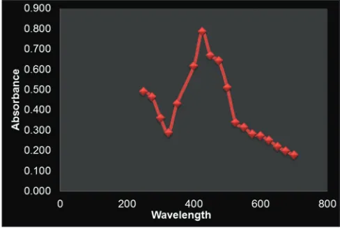
for 10 min at room temperature. The pellet was collected and dried in a hot plate. The dried powder was collected in Eppendorf tubes and stored in refrigerator and which was further characterized by spectrophotometer [Figure 1].
Preparation of experimental solutions
A. vera gel and Ag-NPs of A. vera were diluted with distilled water to obtain a concentration of 500 µg, 1000 µg, and 2000 µg.
Agar diffusion test
Microorganisms were subcultured, and they were streaked in Petri dish containing BHI agar. After drying the inoculums, sterile filter paper discs were applied. 0.1 ml of each solution was pipetted onto the sterile paper disks. Five replicates were prepared for each organism. The plates were incubated at 37°C for 48 h.
Microbial inhibition was measured around the papers disks, and the test was repeated under aseptic conditions to ensure consistency. The antimicrobial activity of each medicament was expressed in terms of the mean of the diameter of zone of inhibition (in mm).
Statistics
The normality tests Kolmogorov–Smirnov and Shapiro–Wilks tests results reveal that the variable follows a normal distribution. Therefore, to analyze the data, parametric methods are applied.
To compare the mean values between groups, one-way ANOVA is
applied. SPSS version 22.0 is used to analyze the data. Significance level is fixed as 5% (α =0.05).
Results
The mean values of growth inhibition produced by different test groups against the test microorganisms are given. Table 1 shows the mean zone of inhibition of calcium hydroxide, A. vera gel (500 µg), and Ag-NPs of A. vera (500 µg), and there was a statistical significant difference between the groups. Table 2 shows the mean zone of inhibition of calcium hydroxide, A. vera gel (1000 µg), and Ag-NPs of A. vera (1000 µg), and there was a statistical significant difference
between the groups. Table 3 shows the mean zone of inhibition of calcium hydroxide, A. vera gel (2000 µg), and Ag-NPs of A. vera (2000 µg), and there was a statistical significant difference between the groups.
Calcium hydroxide performed better than all the test groups, followed by Ag-NPs of A. vera (2000 µg >1000 µg >500 µg) and A. vera extract (2000 µg >1000 µg >500 µg) against all the tested microorganisms after 48 h incubation [Tables 1-3].
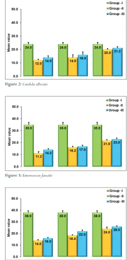
Mean zone of inhibition for C. albicans (Group I - calcium hydroxide, Group II - A. vera at a concentration of 500 µg, 1000 µg, and 1500 µg and Ag-NPs of A. vera at a concentration of 500 µg, 1000 µg, and 1500 µg) [Figure 2].
Mean zone of inhibition for E. faecalis (Group I - calcium hydroxide, Group II - A. vera at a concentration of 500 µg, 1000 µg, and 1500 µg and Ag-NPs of A. vera at a concentration of 500 µg, 1000 µg, and 1500 µg) [Figure 3].
Mean zone of inhibition for S. mutans (Group I - calcium hydroxide, Group II - A. vera at concentration of 500 µg, 1000 µg, and 1500 µg and Ag-NPs of A. vera at concentration of 500 µg, 1000 µg, and 1500 µg) [Figure 4].
Table 1: Mean zone of inhibition calcium hydroxide versus Aloe vera (500 µg) versus silver nanoparticles of Aloe vera (500 µg)
Variables Group Mean±SD F value P value
C. albicans Group I 24.00±1.225 177.143 <0.001 Group II 12.00±0.707
Group III 14.00±1.225
E. faecalis Group I 35.00±2.000 393.781 <0.001 Group II 11.20±1.304
Group III 14.20±0.837
S. mutans Group I 38.00±1.581 760.000 <0.001 Group II 14.00±0.707
Group III 16.00±0.707
E. faecalis: Enterococcus faecalis, S. mutans: Streptococcus mutans, C. albicans: Candida albicans, SD: Standard deviation
Table 2: Mean zone of inhibition calcium hydroxide versus Aloe vera (1000 µg) versus silver nanoparticles of Aloe vera (1000 µg)
Variables Group Mean±SD F value P value
C. albicans Group I 24.00±1.225 40.000 <0.001 Group II 14.00±2.121
Group III 16.00±2.121
E. faecalis Group I 35.00±2.000 297.404 <0.001 Group II 16.20±0.837
Group III 17.00±1.000
S. mutans Group I 38.00±1.581 429.684 <0.001 Group II 18.40±0.548
Group III 22.00±1.000
E. faecalis: Enterococcus faecalis, S. mutans: Streptococcus mutans, C. albicans: Candida albicans, SD: Standard deviation
Table 3: Mean zone of inhibition calcium hydroxide versus Aloe vera (2000 µg) versus silver nanoparticles of Aloe vera (2000 µg)
Variables Group Mean±SD F value P value
C. albicans Group I 24.00±1.225 23.407 <0.001 Group II 20.00±0.707
Group III 21.20±0.837
E. faecalis Group I 35.00±2.000 140.211 <0.001 Group II 21.80±0.837
Group III 23.00±1.000
S .mutans Group I 38.00±1.581 156.364 <0.001 Group II 24.00±1.581
Group III 26.00±0.707
E. faecalis: Enterococcus faecalis, S. mutans: Streptococcus mutans, C. albicans: Candida albicans, SD: Standard deviation
Figure 2:Candida albicans
Figure 3:Enterococcus faecalis
Discussion
A variety of laboratory methods are available to evaluate the in vitro antimicrobial activity of an extract or a pure compound. The most known and basic methods are the disc diffusion method. It is commonly used because of advantage of providing direct estimation of its antimicrobial activity against a specific microorganism and added advantages of simplicity, low cost, the ability to test enormous numbers of microorganisms and antimicrobial agents, and the ease of results interpretation. Although there are new technologies in the field of microbiology, disc diffusion is still one of the preliminary tests to assess the antimicrobial activity of a material.[17-19] To further study the antimicrobial effect of an agent in depth, time-kill test and flow cytofluorometric methods are recommended, which provide information on the nature of the inhibitory effect (bactericidal or bacteriostatic) (time-dependent or concentration-dependent). The incubation time of 48 h was chosen owing to the reason majority of these intracanal medicaments were placed for a period of 7–10 days in root canal system.
As expected, calcium hydroxide had better antimicrobial property when compared to other medicaments. Antimicrobial activity of Ca (OH)2 is related to the release of highly reactive hydroxyl ions in an aqueous environment, which mainly affects cytoplasmic membranes, proteins, and DNA.[20,21] Among the microbes, C. albicans was found to have a minimal zone of inhibition signifying that they require more amount of hydroxyl ions release to inhibit its growth.
Plants and other natural sources provide a huge range of complex and structurally diverse compounds. Recently, many researchers have focused on the investigation of plant and microbial extracts, essential oils, pure secondary metabolites, and new synthetized molecules as potential antimicrobial agents. The fact that a plant extract exhibits antimicrobial activity is of interest, A. vera had 75 potentially active constituents such as vitamins, enzymes, minerals, sugars, lignin, saponins, salicylic acids, and amino acids which were possible reasons for its antimicrobial action. It also contains curcumin, nimbidin, myristric acid, and anthraquinones, all of which can account for its antimicrobial activity.[22]
Biologically synthesized Ag-NPs are commonly used in the field of medicine. Extracellular biosynthesis of Ag-NPs was carried out using medicinal plant extracts. It has been reported that Ag-NPs are non-toxic to humans and most effective against bacteria, virus, and other eukaryotic microorganism at low concentrations and without any side effect.[23] Moreover, several salts of silver and their derivatives are commercially manufactured as antimicrobial agents. In small concentrations, silver is safe for human cells but lethal for microorganisms.[24] Silver is known to produce an antibacterial effect by acting on multiple targets starting from interaction with the sulfhydryl groups of proteins and DNA, alter the hydrogen bonding/respiratory chain, unwind DNA, and interfere with cell-wall synthesis/cell division. Ag-NPs are known to further destabilize the bacterial membrane and increase permeability, leading to leakage of cell constituents. This could be the possible reason for Ag-NPs of A. vera to demonstrate antimicrobial activity.
Conclusion
Within the limitations of the study, it can be concluded that Ag-NPs of A. vera demonstrated antimicrobial activity against E. faecalis, S. mutans, and C. albicans.
Henceforth, future research should be carried out for using Ag-NPs of A. vera in root canal disinfection by increasing its concentration and future research oriented toward testing its biocompatibility.
References
1. Bystrom A, Claesson R, Sundqvist G. The antibacterial effect of camphorated paramonochlorophenol, camphorated phenol and calcium hydroxide in the treatment of infected root canals. Endod Dent Traumatol 1985;1:170-5. 2. Siqueira JF Jr, Rôças IN. Diversity of endodontic microbiota revisited. J Dent
Res 2009;88:969-81.
3. Maekawa LE, Valera MC, Oliveira LD, Carvalho CA, Koga-Ito CY, Jorge AO.
In vitro evaluation of the action of irrigating solutions associated with intracanal medications on Escherichia coli and its endotoxin in root canals. J Appl Oral Sci 2011;19:106-12.
4. Gomes BP, Pinheiro ET, Gadê-Neto CR, Sousa EL, Ferraz CC, Zaia AA, et al.
Microbiological examination of infected dental root canals. Oral Microbiol Immunol 2004;19:71-6.
5. Gomes BP, Ferraz CC, Vianna ME, Rosalen PL, Zaia AA, Teixeira FB, et al. In vitro antimicrobial activity of calcium hydroxide pastes and their vehicles against selected microorganisms. Braz Dent J 2002;13:155-61.
6. Newell CA, Anderson LA, Phillipson JD. Herbal Medicines: A Guide For Health Care Professional. London: Pharmaceutical Press; 1996.
7. Bhardwaj A, Ballal S, Velmurugan N. Comparative evaluation of the
antimicrobial activity of natural extracts of Morinda citrifolia, papain and Aloe vera (all in gel formulation), 2% chlorhexidine gel and calcium hydroxide, against Enterococcus faecalis: An in vitro study. J Conserv Dent 2012;15:293-7.
8. Kurian B, Swapna DV, Nadig RR, Ranjini MA, Rashmi K, Bolar SR. Efficacy
of calcium hydroxide, mushroom, and Aloe vera as an intracanal medicament against Enterococcus faecalis: An in vitro study. Endodontology 2016:28:137-42. 9. Maguire H, Torabinejad M, Kettering J. The use of Aloe vera gel as an intracanal
medicament. J Endod 1996;22:193.
10. Priya S, Vundavalli RM, Reddy VK, Pradeep G, Babu P, Geetha V. Dentinal tubule disinfection with 2% Chlorhexidine Gel, Aloe Vera, Proplois, Septilin and Calcium Hydroxide. Int J Med Res Rev 2016;4:950-5.
11. Monteiro DR, Gorup LF, Takamiya AS, Ruvollo-Filho AC, de Camargo ER, Barbosa DB. The growing importance of materials that prevent microbial adhesion: Antimicrobial effect of medical devices containing silver. Int J Antimicrob Agents 2009;34:103-10.
12. Beer C, Foldbjerg R, Hayashi Y, Sutherland DS, Autrup H. Toxicity of silver nanoparticles - nanoparticle or silver ion? Toxicol Lett 2012;208:286-92. 13. Hobson DW. Commercialization of nanotechnology. Wiley Interdiscip Rev
Nanomed Nanobiotechnol 2009;1:189-202.
14. Gomes-Filho JE, Silva FO, Watanabe S, Cintra LT, Tendoro KV, Dalto LG, et al.
Tissue reaction to silver nanoparticles dispersion as an alternative irrigating solution. J Endod 2010;36:1698-702.
15. Bahador A, Pourakbari B, Bolhari B, Hashemi FB. In vitro evaluation of the antimicrobial activity of nanosilver-mineral trioxide aggregate against frequent anaerobic oral pathogens by a membrane-enclosed immersion test. Biomed J 2015;38:77-83.
purposes in Endodontics: A systematic review of in vitro studies. Mater Sci Eng C Mater Biol Appl 2016;58:1269-78.
18. Gomes BP, Pedroso JA, Jacinto RC, Vianna ME, Ferraz CC, Zaia AA, et al.In vitro evaluation of the antimicrobial activity of five root canal sealers. Braz Dent J 2004;15:30-5.
19. Gomes BP, Vianna ME, Sena NT, Zaia AA, Ferraz CC, de Souza Filho FJ. In vitro
evaluation of the antimicrobial activity of calcium hydroxide combined with chlorhexidine gel used as intracanal medicament. Oral Surg Oral Med Oral Pathol Oral Radiol Endod 2006;102:544-50.
20. Pizzo G, Giammanco GM, Cumbo E, Nicolosi G, Gallina G. In vitro antibacterial
activity of endodontic sealers. J Dent 2006;34:35-40.
21. Siqueira JF Jr, Lopes HP. Mechanisms of antimicrobial activity of calcium hydroxide: A critical review. Int Endod J 1999;32:361-9.
22. Alamdar S, Agaoglu JS. Investigation of in vitro antimicrobial activity of Aloevera
juice. Anim Vet Adv 2009;8:99-102.
23. Jeong SH, Yeo SY, Yi SC. The effect of filler particle size on the antibacterial properties of compounded polymer/silver fibers. J Mat Sci 2005;40:5407-11.
24. Krutyakov YA, Kudrynskiy A, Olenin AY, Lisichkin GV. Extracellur biosynthesis and antimicrobial activity of silver nanoparticles. Russ Chem Rev 2008;77:233.