Original Article
The Effect of Sintering Program on the Compressive Strength of Zirconia Copings
Amir Ali Reza Khaledi 1, Mahroo Vojdani 1, Mitra Farzin 2, Soudabeh Pirouzi 3
1
Dept. of Prosthodontic, Biomaterials Research Center, School of Dentistry, Shiraz University of Medical Sciences, Shiraz, Iran. 2 Dept. of Prosthodontic, School of Dentistry, Shiraz University of Medical Sciences, Shiraz, Iran.
3
Post Graduate, Dept. of Prosthodontic, School of Dentistry, Shiraz University of Medical Sciences, Shiraz, Iran.
KEY WORDS
Compressive strength; Zirconia;
Sintering;
Received July 2017;
Received in Revised form August 2017; Accepted September 2017;
ABSTRACT
Statement of the Problem: Considering the limitations of conventional sintering of zirconium oxide (ZrO2) copings, shortening the sintering time can be proposed as an
alternative method for making the copings.
Purpose: This study aimed to assess the effects of sintering time on compressive strength of Yttria Tetragonal Zirconia Polycrystal (Y-TZP) copings.
Materials and Method: Thirty copings of pre-sintered 3Y-TZP blanks were milled and sintered in a special furnace for three different durations (n=10 per group). The sintering time was 1 h 15 min for IPS e.max ZirCAD, 4 h 20 min for Speed ZrO2,
and 7 h 20 min for the conventional ZrO2 group. The specimens were cemented on
the brass dies by using conventional glass ionomer cement. The copings were verti-cally loaded until fracture by using a universal testing machine. The data were ana-lyzed through one-way analysis of variance (ANOVA) and post hoc test to compare the mean differences of compressive strength yielded in three study groups (α=0.05).
Results: The mean ± SD of compressive strength value was (3617 ± 543.54) N for IPS-e.maxZirCAD group, (2663 ± 508.11) N for Speed ZrO2 group, and (1662±
466.71 N) for conventional ZrO2 group. There were statistically significant
differ-ences among compressive strength values of the tested groups (p< 0.05). The highest compressive strength values were obtained from the IPS e.max ZirCAD group.
Conclusion: Within the limitations of this in vitro study, it can be concluded that compressive strength of the zirconia copings is affected by the sintering time. High compressive strength of zirconia copings can be obtained by shortening the sintering time.
Corresponding Author: Pirouzi S.,Dept. of Prosthodontic, School of Dentistry, Shiraz University of Medical Sciences, Shiraz, Iran. Tel: +98-7136263193-4 Email: [email protected]
Cite this article as: Khaledi AAR., Vojdani M., Farzin M., Pirouzi S. The Effect of Sintering Program on the Compressive Strength of Zirconia Copings. J Dent Shiraz Univ Med Sci.,
2018 September; 19(3):206-211.
Introduction
Zirconia is being increasingly used in dentistry due to the development in evolution of dental advanced ceram-ics. Its practical properties as well as high productivity in computer-aided design and computer aided manufac-turing (CAD-CAM) systems have promoted it to the category of top quality materials. [1]
Zirconia is a polycrystalline available in three al-lotropic forms. The monoclinic (M) phase remains
sta-ble up to 1170° C. At higher temperatures, it turns to tetragonal (T) phase, which is stable up to 2370°C. The cubic phase is formed in 2680°C, which equals the melting point of zirconia. [2] The tetragonal phase of partially stabilized zirconia (PZS) is metastable at room temperature due to the presence of stabilizers such as Yttrium oxide (Y2O3) (2-3 mol%). [3] Thereby, the
cracks and consequently inhibits their development. [4] This feature, called toughening transformation, im-proves the physical and mechanical properties of zirco-nia. [5]
The CAD-CAM technologies enable the milling of zirconia into reconstructions with complicated geom-etries. [1, 6] Currently, soft and hard milling processes are known for zirconia. [6-7] In the former, which is done on green and pre- sintered blanks, the zirconia frameworks are sintered after milling. In that condition, the zirconia surface is free of monoclinic phase unless the restoration is sandblasted. On the other hand, the hard-milled zirconia is done on fully sintered frame-works. Therefore, the milling process is lengthy and difficult, yielding restoration with a considerable mono-clinic phase on the surface. [7]
The zirconia-based ceramics are favorably em-ployed in dentistry because of their unique mechanical properties that cannot be found in any other ceramic system. [8] Being the most commonly used type of zir-conia ceramic material, Y-TZP has some exclusive properties such as high fracture strength, high thermal resistance, low thermal conductivity, and chemical sta-bility. [9] These properties make Y-TZP a quite suitable material for posterior prostheses. The fracture strength is affected by various factors such as size and distribu-tion of the crystalline phase, crown dimensions, geome-try, as well as other factors related to some properties of the supporting structure such as the modulus of elastici-ty. [10-12]
The relationship between the microstructure and mechanical properties of Y-TZP has been discussed in many studies. They all found that the transformation toughening in these ceramics depended on the grain size. [13-15] In the zirconia sintering process, alteration in sintering time and temperature can influence the grain size. [16] The higher the temperature and time is, the larger the grains size would be. [17] The larger tetrago-nal grains are prone to undergo stress-induced transfor-mation to a balanced structure, resulting in an increased toughness. The maximum toughness is quite close to the critical grain size (1µm). Larger grain size than this crit-ical size leads to spontaneous T-M transformation, which consequently decrease the material stability. [18] Accordingly, lower temperature and time reduce the grain size down to 0.2μm, at which T-M transform does
not occur, and consequently the toughness would be in the lowest level. [19] Contrary to the toughness, the strength is at the highest level in small grains since the fracture size increases in relation to the grain size. [20]
Gupta et al. [21] reported that fine grain ZrO2
(generally<0.5μm) with small concentrations of stabiliz-ing Y2O3 contained up to 98% of the post-sintering
met-astable T phase. High strength coincided with high te-tragonal phase content and low strength coincided with high monoclinic phase content.
Ruiz and Ready [16] reported that the grain size increased with increasing the sintering temperature, which in turn, led to increased fracture toughness owing to larger transformation zones. However, they observed no significant difference in biaxial flexural strength of zirconia ceramics with various grain sizes. In contrast, however, Casellas et al. [22] found that decreasing the grain sizes lightly increased the flexural strength.
Evidence is limited about the efficacy of short time sintering on the compressive strength of zirconia copings; thus, the objective of the present investigation was to describe the relationship between the sintering time and the compressive strength of zirconia copings. The null hypothesis was that there would be no differ-ence between the compressive strength of 3Y-TZP cop-ings sintered in conventional and short time condition.
Materials and Method
The designed copings were milled from three types of pre-sintered zirconia blanks (Ivoclar Vivadent, Germa-ny) in different sintering programs as displayed in Table 1.
The copings were inspected for any imperfection and rejected in case of any defect. Then, they were ce-mented conventionally with glass ionomer cement (GIC; Fuji I, GC, Japan) on the brass dies, which were already cleaned with steam and alcohol. All copings were filled with the luting cement and, then, loaded with a vertical force of 10 N for 10 minutes in a cementation device. The copings were clamped in the holder of a universal testing machine (Zwick Z2.5; Zwick, Ulm, Germany). They were vertically loaded on the occlusal surface at a crosshead speed of 0.5mm/m. The mini-mum force leading to fracture was recorded for each sample. The universal testing machine was controlled via a computer software system, which completed the stress-strain diagram.
Table 1: Classification of the copings to the groups accord-ing to sinteraccord-ing time
Group N Sintering
temperature
Sintering time Conventional ZrO2 10 1530 7 h 20 min
Speed ZrO2 10 1530 4 h 20 min
IPS-emaxZirCAD 10 1530 75 min
The statistical analyses were performed by using SPSS software, version 14(SPSS Inc.). The mean values and standard deviations (SD) were calculated for each group. One-way ANOVA and post hoc tests were used to compare the results between the groups. The signifi-cance level was set at 0.05.
Results
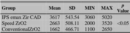
Table 2 represents the mean values and SD of the force leading to fracture in each group. The mean ± SD frac-ture load was 1662±466.71 in the conventional ZrO2 group, 2663±508.11 in the Speed ZrO2 group, and 3617±543.54 in IPS e.max ZirCAD group.
Table 2: The mean values and standard deviation of fracture load in Newton
Group Mean SD MIN MAX p
Value
IPS emax Zir CAD 3617 543.54 3060 5020 <0.05 Speed ZrO2 2663 508.11 2000 3520 ConventionalZrO2 1662 466.71 1100 2650
The results of post hoc test showed that compressive strength was significantly different among the test groups (df=2, F=17.488, p= 0.000)
The highest mean± SD of fracture load was ob-served in IPS e.max Zir CAD (3617± 543.54) which was sintered with in the shortest sintering time (75min). The results of ANOVA and post hoc indicated that the compressive strength was affected by the sintering time.
Discussion
This study aimed to evaluate the relationship between sintering time and compressive strength in 3Y-TZP cop-ings. The findings rejected the null hypothesis through proving that the sintering time significantly affected the in vitro compressive strength of cemented zirconia cop-ings (p< 0.05).
The mean compressive strength in the present study was found 3617, 2663, and 1662 N for groups 1 to 3, respectively. It is difficult to compare failure loads found in the literature to those found in this study, due to different experimental variables. A study reported a failure load of 381 N for zirconia coping of 0.6 mm thickness on non-carious incisor. [23] In another study, the failure load was 1670 N in zirconia coping with 0.4mm thickness on resin die. [24] Other studies sur-veyed the failure load in complete crowns and reported the fracture load values to range between 980 and 1400 N. [25-26] In the present study, the compressive strength was assessed on brass dies instead of natural teeth to control the accuracy and certainty of reposition and similarity of parameters such as margin and conver-gence coping. Thus, the copings geometries were iden-tical, although their elastic modulus and fracture strength were not similar to natural teeth. The results of some previous studies showed that increasing the elastic modulus of supporting structure led to an increase in the failure resistance of the crown. [27-30] It can be con-cluded that the higher range of failure load in this study was due to the high elastic modulus of the supporting structure, which may lead to an overestimation of the clinical values.
Zesewitz et al. [35] have shown that there was not a substantial difference in fracture load between zirconia crowns that were cemented with resin-bonded cements and metal dies that were cemented with glass ionomer cement. They concluded that the strength of zirconia might overcome against the influence of cement proper-ties and thickness. In the current study, the glass iono-mer cement was used with minor self-adhesive proper-ties. In the present study, we surveyed the zirconia cop-ings without veneering materials, because the recent studies reported that the presence and thickness of ve-neering porcelain affected the compressive load to fail-ure. [25-26, 36] Moreover, the copings were fabricated with flat occlusal morphology because the natural oc-clusal anatomy was likely to affect the outcome due to the lateral component of the loading force. [37]
The relationship between the grain size and the mechanical properties in 3Y-TZP was previously stud-ied. It was found that the grain size determined the transformation toughening effect, toughness, and strength. [38] Sintering condition directly influenced the grain size of zirconia. The strength of porous ceramics decreases significantly with the increase of porosity. The compressive strength is influenced by the porosity and pore size. [39] Kim et al. [40] evaluated the effects of sintering time on the grain size of zirconia. They re-ported that the density of all samples ranged from 6.06 to 6.07 g/cm. They noted no statistically significant dif-ference among the samples sintered for different time spans. However, Tekeli and Erdogan [41] noticed that the sintering time influenced the density and mechanical properties. They reported that high sintering tempera-ture and extended sintering time increased the grain size and consequently, the number of pore; and thereupon yielded a material with reduced mechanical properties. In contrast, Hjerppe et al. [42] showed that short sinter-ing time for zirconia decreased the grain size. Yet, the results were not statistically significant and had no ef-fect on the mechanical properties. Cottom et al. [19] reported that shorter sintering time yielded smaller grain size.
In the current study, different sintering time was applied to pre-sintered zirconia specimens in order to obtain dense sintered copings with different grain size, based on our finding the shorter sintering time resulted higher compressive strength. However, the strength of
ceramic material depends more upon the experiment condition rather than the materials properties. In con-trast, the fracture toughness is a more inherent property of ceramics, which is not affected by surface flaws and the initial crack size. [43] Based on Hall-Petch [44] re-gime, the fracture in large grain sizes is controlled by the intrinsic defects; where as in small grain sizes, fail-ure is governed by extrinsic processing defects such as preparation cracks and pores. In this case, the grain size is much smaller than the defects. Thus, the fracture strength depends on the grain size less than it does on the geometry of the fracture origin. Hoffman et al. [45-46] demonstrated that the critical stress for microcrack formation decreased with increasing the grain size. They also explained that the failure in grains smaller than a certain critical size occurred due to the stress concentra-tions around the pore before the formation of the mi-crocracks. The criterion for fracture changes from lim-ited crack extension to limlim-ited crack initiation. This variation increases the fracture strength in smaller grains. According to the result of the present study, shortening the sintering time could led to smaller grain size in the IPS e.max ZirCAD group, which in turn in-creased the compressive strength load value.
This study has some limitations; short time sinter-ing may also influence other properties of the zirconia ceramics but this study was limited to only one feature. Static in vitro tests were used in current study; however, dynamic fatigue tests are more representative of clinical masticatory forces and further in vitro and in vivo tests are required.
Conclusion
Within the limitations of this study, it can be concluded that the sintering protocol affects the compressive strength of zirconia copings, the shorter the sintering time, the higher the compressive strength.
Conflict of Interest
The authors declare that they have no conflict of inter-est.
References
[1] Denry I, Kelly JR. State of the art of zirconia for dental
applications. Dent Mater. 2008; 24: 299-307.
Tetragonal-Monoclinic Transformation in Zirconia:
Les-sons Learned and Future Trends. J Am Ceram Soc. 2009;
92: 1901–1920.
[3] Hannink RHJ, Kelly PM, Muddle BC. Transformation
toughening in zirconia containing ceramics. J Am Ceram
Soc. 2000; 83: 461–487.
[4] Kelly JR, Campbell SD, Bowen HK. Fracture-surface
analysis of dental ceramics. J Prosthet Dent. 1989; 62:
536-541.
[5] Anusavice KJ. Mechanical properties of dental materials.
In: Phillips' science of dental materials. Anusavice KJ,
Shen C, Rawls HR, editors. 12th ed. St. Louis, MO,
USA: Saunders; 2013. p. 48-68.
[6] Raigrodski AJ, Chiche GJ, Potiket N, Hochstedler JL,
Mohamed SE, Billiot S, et al. The efficacy of posterior
three-unit zirconium-oxide-based ceramic fixed partial
dentalprostheses: a prospective clinical pilot study. J
Prosthet Dent. 2006; 96: 237-244.
[7] Beuer F, Aggstaller H, Edelhoff D, Gernet W. Effect of
preparation design on the fracture resistance of zirconia
crown copings. Dent Mater J. 2008; 27: 362-367.
[8] Chen YM, Smales RJ, Yip KH, Sung WJ. Translucency
and biaxial flexural strength of four ceramic core
materi-als. Dent Mater. 2008; 24: 1506-1511.
[9] Hu L, Wang CA, Huang Y. Porous yttria-stabilized
zirconia ceramics with ultra-low thermal conductivity J
Mater Sci. 2010; 45: 3242-3246.
[10]Snyder MD, Hogg KD. Load-to-fracture value of
differ-ent all-ceramic crown systems. J Contemp Ddiffer-ent Pract.
2005; 6: 54-63.
[11]White SN, Miklus VG, McLaren EA, Lang LA, Caputo
AA. Flexural strength of a layered zirconia and porcelain
dental all-ceramic system. J Prosthet Dent. 2005; 94:
125-131.
[12]Deany IL. Recent advances in ceramics for dentistry. Crit
Rev Oral Biol Med. 1996; 7: 134-143.
[13]Lange FF. Transformation Toughening 1: Size Effects
Associated with the Thermodynamics of Constrained
Transformations. J Mater Sci. 1982; 17: 225–234.
[14] Lange F. Transformation toughening Part 3
Experimen-ta/ observations in the Zr02- Y203 system. J Mater Sci.
1982; 17: 240-246.
[15]Becher PF, Swain MV. Grain-size-dependent
transfor-mation behavior in polycrystalline tetragonal zirconia. J
Am Ceram. 1992; 75: 493–502.
[16]Ruiz L, Readey MJ. Effect of heat-treatment on grain size
phase assemblage, and mechanical properties of 3 mol%
Y-TZP. J Am Ceram Soc. 1996; 79: 2331–2340.
[17]Derafshi R, Khorshidi H, Kalantari M, Ghaffarlou I. Ef-
fect of mouthrinses on color stability of monolithic
zir-conia and feldspathic ceramic: an in vitro study. BMC
Oral Health. 2017; 17: 129.
[18]Bravo-Leon A, Morikawa Y, Kawahara M, Mayo MJ.
Fracture toughness of nanocrystalline tetragonal zirconia
with low yttria content. Acta Mater. 2002; 50: 4555–
4662.
[19]Cottom BA, Mayo MJ. Fracture toughness of
nanocrys-talline ZrO2–3mol% Y2O3 determined byVickers
inden-tation. Scripta Mater. 1996; 34: 809–814.
[20]Eichler J, Rodel J, Eisele U, Hoffman M. Effect of grain
size on mechanical properties of submicrometer 3Y-TZP:
Fracture strength and hydrothermal degradation. J Am
Ceram Soc. 2007; 90: 2830–2836.
[21]Gupta TK, Bechtold JH, Kuznicki RC, Cadoff LH,
Ros-sing BR. Stabilization of tetragonal phase in
polycrystal-line zirconia. J Mater Sci. 1977; 12: 2421–2426.
[22]Casellas D, Alcala J, Llanes L, Anglada M. Fracture
variability and R-curve behavior in yttria-stabilized
zir-conia ceramics. J Mater Sci. 2001; 36: 3011–3025.
[23]Potiket N, Chiche G, Finger IM. In vitro fracture strength
of teeth restored with different all-ceramic crownsystems.
J Prosthet Dent. 2004; 92: 491-495.
[24]Bindl A, Lüthy H, Mörmann WH. Thin-wall ceramic
CAD/CAM crown copings: strength and fracture pattern.
J Oral Rehabil. 2006; 33: 520-528.
[25]Leevailoj C, Platt JA, Cochran MA, Moore BK. In vitro
study of fracture incidence and compressive fracture load
of all-ceramiccrowns cemented with resin-modified glass
ionomer and other luting agents. J Prosthet Dent. 1998;
80: 699-707.
[26]Bernal G, Jones RM, Brown DT, Munoz CA, Goodacre
CJ. The effect of finish line form and luting agent on the
breaking strength of Dicor crowns. Int J Prosthodont.
1993; 6: 286–290.
[27]Al-Makramani BMA, Razak AAA, Abu-Hassan MI.
Evaluation of load at fracture of Procera AllCeram
cop-ings using different lutingcements. J Prosthodont. 2008;
17: 120-124.
[28]Akesson J, Sundh A, Sjögren G. Fracture resistance of
all-ceramic crowns placed on a preparation with a
slice-formedfinishing line. J Oral Rehabil. 2009; 36: 516-523.
Nagy WW. Resistance to fracture of two all-ceramic
crown materials following endodonticaccess. J Prosthet
Dent. 2006; 95: 33-41.
[30]Lee SK, Wilson PR. Fracture strength of all-ceramic
crowns with varying core elastic moduli. Aust Dent J.
2000; 45: 103-107.
[31]Manicone PF, Rossi Iommetti P, Raffaelli L. An
over-view of zirconia ceramics: basic properties and clinical
applications. J Dent. 2007; 35: 819-826.
[32]Proos KA, Swain MV, Ironside J, Steven GP. Influence
of core thickness on a restored crown of a first premolar
using finite elementanalysis. Int J Prosthodont. 2003; 16:
474-480.
[33]Kelly JR. Clinically relevant approach to failure testing
of all-ceramic restorations. J Prosthet Dent. 1999; 81:
652-661.
[34]Rekow ED, Harsono M, Janal M, Thompson VP, Zhang
G. Factorial analysis of variables influencing stress in
all-ceramic crowns. Dent Mater. 2006; 22: 125-132.
[35]Zesewitz TF, Knauber AW, Nothdurft FP. Fracture
re-sistance of a selection of full-contour all-ceramic crowns:
an in vitro study. Int J Prosthodont. 2014; 27: 264-266.
[36]Wakabayashi N, Anusavice KJ. Crack initiation modes in
bilayered alumina/porcelain disks as a function of
core/veneer thickness ratio and supporting substrate
stiff-ness. J Dent Res. 2000; 79: 1398-1404.
[37]Tsalouchou E, Cattell MJ, Knowles JC, Pittayachawan P,
McDonald A. Fatigue and fracture properties of yttria
partially stabilized zirconia crown systems. Dent Mater.
2008; 24: 308-318.
[38]Matsui K, Yoshida H, Ikuhara Y. Isothermal sintering
effect on phase separation and grain growth in
yttria-stabilized tetragonal zirconia polycrystal. J Am Ceram
Soc. 2009; 92: 467–475.
[39]Chen R, Wang CA, Huang Y, Ma L, Lin W. Ceramics
with special porous structures fabricated by
freeze-gelcasting: using tert-Butyl alcohol as a template. J Am
Ceram Soc. 2007; 90: 3478–3484.
[40]Kim MJ, Ahn JS, Kim JH, Kim HY, Kim WC. Effects of
the sintering conditions of dental zirconia ceramics on the
grain size and translucency. J Adv Prosthodont. 2013; 5:
161-166.
[41]Tekeli S, Erdogan M. A quantitative assessment of
cavi-ties in 3 mol% yttria-stabilized tetragonal zirconia
speci-mens containing various grain size. Ceram Int. 2002; 28:
785–789.
[42]Hjerppe J, Vallittu PK, Fröberg K, Lassila LV. Effect of
sintering time on biaxial strength of zirconium dioxide.
Dent Mater. 2009; 25: 166-171.
[43]Kelly JR. Dental ceramics: current thinking and trends.
Dent Clin N Am. 2004; 48: 513-530.
[44]Zimmermann A, Rödel J. Generalized Orowan-Petch plot
for brittle fracture. J Am Ceram Soc. 1998; 81: 2527–
2532.
[45]Hoffman M, Rodel J. Suggested mechanism of
strength-ening of. 'nanotoughened' ceramics. J Ceram Soc Jpn.
1997; 105:1086-1090.
[46]Merkert P, Hoffman M, Rödel J. Detection of prefracture
microcracking in Al2O3 by acoustic emission. Journal of