RESEARCH PAPER
Controlled Antibiotic Delivery by Gelatin Nanospheres:
Optimization, Characterization and Antibacterial Evaluation
Shahrzad Fathollahipour 1, Azadeh Ghaee 1*, Ali Abouei Mehrizi 1 and Mojtaba Koosha 21 Department of Life Science Engineering, Faculty of New Science and Technologies, University of Tehran,
Tehran, Iran.
2 Department of Pulp and Paper Energy and New Technologies, Engineering Faculty, Zirab Campus, Shahid
Beheshti University, Mazandaran, Iran
*Corresponding Author Email: [email protected] ARTICLE INFO
Article History:
Received 12 July 2016 Accepted 11 September 2016 Published 01 October 2016
Keywords:
Gelatin nanoparticles Nanoprecipitation
Pluronic based delivery systems Drug release
Antibacterial
ABSTRACT
How to cite this article
Fathollahipour S, Ghaee A, Abouei Mehrizia A, Koosha M. Controlled Antibiotic Delivery by Gelatin Nanospheres: Optimization, Characterization and Antibacterial Evaluation. J Nanostruct, 2016; 6(4): 285-292. DOI: 10.22052/ jns.2016.34330
The present work focuses on preparation and characterization of erythromycin loaded gelatin nanoparticles through nanoprecipitation method. The procedure consists of the addition of the aqueous gelatin solution to the non-solvent phase containing Lutherol F127. Three different measures of cross-linker and polymer concentration were also examined, and the optimum concentration was found. The morphology of gelatin nanoparticles was characterized by field emission scanning electron microscope. It was shown that the optimal morphology can be achieved at the concentration of 1.25 wt % of gelatin in aqueous phase by addition of 20 mL of glutaraldehyde 5%, as the crosslinking agent. Nanoparticle wet size determination was carried out using a dynamic light scattering system and found to be approximately 100 nm. Furthermore, Erythromycin release studies proved the suitability of these particles as a drug delivery system, at least in the studied 72 hours interval. As suggested by related measurements, these nanoparticles are good candidates for antibacterial agent release in any possible related application.
INTRODUCTION
In the past few decades, there has been substantial development in the use of biocompatible polymers as drug delivery systems [1-3]. Delivering therapeutic compounds to the target area has always been a major drawback in in treatment of many diseases. Conventional use of drugs is often described by their limited efficiency, poor bio-availability, and lack of selectivity. These limitations and drawbacks could be enhanced by controlling the process of drug delivery. In controlled drug delivery systems (DDS) the active material is transported directly to the place of action. This is counted as a unique way to minimize the unwanted side effects on surrounding tissues. On
the other hand, a suitable drug delivery system is able to protect the drug from degradation or clearance and increases the drug concentration in target tissues. As a result of this action, lower drug dosage would be required to treat a same disease, in contrast to other normal methods of drug application [4].
286 J Nanostruct 6(4): 285-292, Autumn 2016 Such unique carriers have so far been synthesized
from several biopolymers such as chitosan [8], alginate [9], guar gum [10], hydroxyethyl cellulose [11], gellan [12], carboxymethyl cellulose [13], dextrans [14], and gelatin [15]</EndNote>.
Of all the aforementioned biopolymers, gelatin-based carriers mark a major class in many different industries such as food production, pharmacology and medicine [5]. Gelatin is the product of collagen denaturation. Depending on the preparation method, the obtained gelatin are of two types, namely Type A and Type B [16].
Nanoprecipitation is a straight, rapid and easy method for fabricating nanoparticles. In this process, the addition of the polymer containing solvent to a miscible non solvent induces the production of nanoparticles with the help of a suitable surfactant [17]. Gelatin, as a natural protein, could be a safe choice for nanoprecipitation and drug release applications because of its numerous benefits [5, 18].
In the present study, followed by the optimization of processing parameters (gelatin and crosslinker content), erythromycin loaded gelatin nanoparticles (GNPs) were produced by a modified nanoprecipitation method. The primary focus point of the current survey is to evaluate the drug release behavior of the synthesized nanoparticles. Furthermore, we showed that these nanoparticles could be well used as antimicrobial agents in any related field.
MATERIALS AND METHODS
Gelatin of microbiology grade (porcine gelatin of type A) was purchased from Merck (Billerica, MA, USA). Pluronic F-127 was also the product of Sigma-Aldrich (Sigma-Aldrich, St. Louis, MO, USA). Glutaraldehyde (50% v/v), absolute ethanol, and all other reagents, including NaCl, KCl,
NaHCO3 and NaH2PO4, for fabrication of pseudo
extracellular fluid (PECF), were also purchased from Merck Chemicals (Merck, Billerica, MA, USA). Erythromycin was supplied by Tehran-Darou pharmaceuticals (Tehran, Iran). Deionized water was used in all experiments. All other chemicals were utilized without any further purification.
Synthesis of gelatin nanoparticles
Gelatin nanoparticles were prepared by nanoprecipitation method as described in our previous work [19]. Briefly, gelatin at the amount of 125 mg was dissolved in DI water, and put
on magnetic stirrer for 30 minutes, having the temperature of 60 ̊C. The prepared solution was then added to 20 ml alcoholic solution containing 0.4 gr of pluronic F-127. The next step was to add different amounts of glutaraldehyde (5% v/v, 16, 17 and 20 µl) after 30 minutes, with the aim of finding the minimum effective cross-linker concentration. After 24 hours of cross-linking the particles were centrifuged at 9000 rpm for 15 minutes. Re-dispersion in water, and shaking for 15 minutes in order to make sure of un-reacted glutaraldehyde removal were also done. Then the particles were re-centrifuged and the supernatant was collected. Gelatin nanoparticles were freeze-dried (FDB-5503, Operon, South Korea) after being frozen in liquid nitrogen.
Drug loading into gelatin nanoparticles
Erythromycin, as the model antibacterial agent, was loaded into GNPs through drug incubation method. Briefly, 500 mg of dried bare GNPs were put into dialysis membrane and allowed to swell for 1 hour under continues magnetic stirring at 37 ̊C in de-ionized water. After preparing saturated erythromycin solution, the dialysis tube containing GNPs were placed inside the solution for another 24 hours. The resulting drug loaded particles were then rinsed three times to remove surface drugs, collected by centrifugation and dried again for further use [20-22].
Characterization of gelatin nanoparticles
In order to evaluate the morphology and diameter of gelatin nanoparticles, field emission scanning electron microscopy (S4160, Hitachi, Japan), at accelerating voltage, of 10 kV was used. The particle diameters were measured using Image J software. The minimum of one hundred particles were measured in each statistical analysis.
The surface charge and z-average diameter of gelatin nanoparticles dispersed in de-ionized water at a concentration of (5 mg/ml) was measured using a Zetasizer and Dynamic Light Scattering system (Brookhaven, ZetaPals, USA). In this method, the extent of particle mobility is demonstrated as a result of applying an electrical charge to the nanoparticles. This method helps to predict the level of surface hydrophilicity [23].
The IR spectra of gelatin nanoparticles were recorded, via KBr method, using Fourier transform infrared spectrophotometer (PerkinElmer, USA).
287
J Nanostruct 6(4): 285-292, Autumn 2016
loaded cross-linked gelatin nanoparticles. Briefly, 100 mg of erythromycin loaded nanoparticles were put into a washed dialysis membrane and placed in the middle of release medium (PECF: pseudo extra cellular fluid). PECF is a mixture of
0.68 gr NaCl, 0.22 gr KCl, 2.5 gr NaHCO3, and 0.35
gr NaH2PO4 in 100 mL of distilled water, having the
pH value of 8.0±0.5.
The UV absorbance spectrum erythromycin over the range of 180 to 500 nm was recorded using a UV spectrophotometer (WPA, BiowaveT, England). The maximum absorption wavelength of erythromycin was shown to be 285 nm. The wavelength was used to prepare the linear calibration curve for the antibacterial drug.
Established along the calibration curve, the release profile of Erythromycin of gelatin nanoparticles in pseudo extracellular fluid was measured in triplicate for 72 hours in a handmade diffusion cell and plotted in cumulative drug release diagram versus time. In each sampling a volume of 2 ml was extracted out of the diffusion cell at determined time intervals for being measured spectrophotometrically, and instead 2 ml of fresh medium was poured into the cell in order to satisfy the sink conditions. The release medium temperature was held constant at 37 ̊C by a water bath and pump circulating system.
Antibacterial evaluation of gelatin nanoparticles was performed using the agar disk diffusion method. Agar disk diffusion is a commonly used method to examine the antibacterial activity as the active compound of the sample diffuses through the agar plate [24]. In order to assess the antibacterial activity, Mueller-Hinton agar solid was used as the culture medium for Staphylococcus aureus and Pseudomonas aeruginosa standard bacteria. The nutrient culture medium was poured into a Petri plate and a loopful of each bacterial
strain was spread on agar medium followed by incubation at 37 ̊C for 24 hours to create bacterial colonies. The created bacterial colony was picked off with a wire loop and put in pre-sterilized nutrient broth and then incubated overnight at 37 ̊ C for another 24 hours. By appropriately diluting with sterile distilled water and nutrient broth, the
cultures of bacteria containing ∼108 CFU/ml were
prepared. Then prepared bacteria were uniformly spread onto agar plate, and the powder solution was placed into a hole-shaped spot inside the culture medium. The incubation was continued for 24 hours at 37 ̊C and the diameter of inhibition zone was then measured [25]. Moreover, as the standard control antibiotic discs Cefoxitin and Ciprofloxacin were used.
RESULTS AND DISCUSSION
Morphological studies of gelatin nanoparticles as a function of gelatin concentration and amount of cross-linker
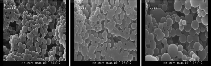
In order to find the best concentration of gelatin in aqueous phase from the morphological point of view, three different concentrations of 1.25 wt%, 2 wt% and 2.25 wt% were investigated. All other parameter including the cross-linker and surfactant concentrations are kept constant in this phase. SEM micrographs of gelatin nanoparticles in the three different concentrations are shown in Fig. 1 (a-c). Spherical, regular shaped particles with smooth surfaces and rather uniform size distribution of gelatin nanoparticles are observed at the concentration of 1.25 wt%. This non-agglomerated morphology observed at the concentration of 1.25 wt% could be related to appropriate proportion of surfactant to gelatin. In a previous case study, Lee et al. [26], reported the production of gelatin nanoparticles via nanoprecipitation method. They agreed that the surfactant
288 J Nanostruct 6(4): 285-292, Autumn 2016 concentration strongly affects the particle size and
morphology as the best emulsifier to gelatin mass ratio was reported to be 32:1.
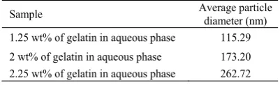
It is also seen that average diameters of nanoparticles are increased by increasing the gelatin content in the aqueous phase. However, the particles tend to agglomerate by changing the gelatin concentration from 1.25 wt% to 2 wt% and 2.25 wt%. The average nanoparticle diameters measured by Image J software for at least 100 particles are presented in Table 1. Increasing the average particle diameter of the produced gelatin nanoparticles might be described by the more tendency of gelatin molecules to each other rather than surfactant molecules which is predicted to increase with the addition of gelatin concentration, causing the growth of the total particle size. Moreover, increasing gelatin concentration in aqueous phase from 1.25 wt% to 2.25 wt% increased the amount of agglomeration. This may be due to the prevention of solvent diffusion through the non-solvent phase as a result of high viscosity. Which in turn results in the non-proper nanoprecipitation process, resulting in the creation of some visible agglomerates [7, 26].
Furthermore, the amount of cross-linker agent was studied to find out the minimum possible glutaraldehyde needed to crosslink the nanoparticles without forming any visible
agglomerates. In this regard three different glutaraldehyde (5% v/v) amounts of 16µl, 17µl and 20µl were added to the structure and after 24 hours of cross-linking the morphology of freeze-dried gelatin nanoparticles were studied by FE-SEM. Results are shown in Fig. 2 (a-c). Obviously, increasing the amount of cross-linker from 16µl to 17µl and finally 20µl has decreased the amount of agglomeration. In case of 16µl cross-linker agent, there are visible aggregates in gelatin nanoparticles, where there are no distinct borders between each two adjacent particles. This pattern visibly stops at the concentration of 20µl, where rather sharp borders and less agglomerated structure could be seen. As a result of these experiments, the formulation having 1.25 wt% of gelatin in aqueous phase and the amount of 20µl cross-linking agent was chosen to be optimal and used for all further experiments and characterizations.
Zeta potential measurement on gelatin nanoparticles Results of zeta potential for measuring the surface charge of gelatin nanoparticles are shown in Fig. 3. Table 2, shows the surface charge values calculated by zeta-sizer device for the nanoparticles in water as dispersant with pH value of 7±0.5.
As shown in Table 2, the average ξ-potential measured for the particles in three successive runs has been recorded to be -59.95 mv. Dynamic light
Sample Average particle diameter (nm) 1.25 wt% of gelatin in aqueous phase 115.29 2 wt% of gelatin in aqueous phase 173.20 2.25 wt% of gelatin in aqueous phase 262.72
Table 1. Average particle diameter of gelatin nanoparticles
Fig. 2. FE-SEM micrographs of gelatin nanoparticles with different amounts of cross-linker agent added, 16 µl (a); 17 µl (b) and 20 µl (c)
Runs Mobility Zeta Potential (mv)
1 -0.99 -49.78
2 -1.30 -65.75
3 -1.28 -64.34
289
J Nanostruct 6(4): 285-292, Autumn 2016
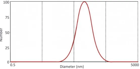
scattering (DLS) was used to calculate the particle size distribution of gelatin nanoparticles in optimal condition (Fig. 4).
The FOQELS software has recorded the mean wet particle diameter equal to 175.1 nm with the unimodal polydispersity of 0.350. In a previously reported nanoprecipitation method, Lee et al. [7], produced spherical, well-dispersed gelatin nanoparticles with an average size of around ~251 nm in wet condition, in which the preparation method is somehow comparable with the present study.
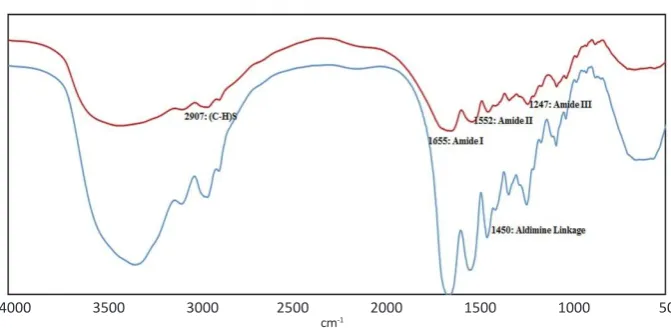
Fig. 5 (a, b) shows the FTIR spectra for cross-linked and un-crosslinked gelatin nanoparticles. The FTIR spectrum of un-crosslinked Gelatin nanoparticles exhibited an absorption peak in the range of
3308-3508 cm-1 which is related to (N-H) stretching
vibration [27]. The peak at 2907 cm-1 corresponds
to (C-H) stretching of methyl and methylene groups [28]. Besides, this spectrum showed the
two absorption characteristic peaks of 1655 cm-1,
1552 cm-1 and 1247 cm-1, respectively, for amide I
(C=O stretching), amide II ((N-H) bending and
(C-H) stretching) and amide III (C-N) stretching plus (N-H) bending) [7, 29]. Additionally, the characteristic peak at 1456 cm-1 is obviously related to (C=N) stretching vibration [28].
After crosslinking, in addition to the previously mentioned peaks, a strong absorption peak
at around 1450 cm-1 was observed in the FTIR
spectrum of the crosslinked gelatin nanoparticles which is certainly related to the aldimine linkage (CH=N) created as a result of bonding with glutaraldehyde as the crosslinking agent. The aldimine linkage is formed by the reaction of the aldehyde group of glutaraldehyde with the amino group of gelatin protein backbone [28].
In Vitro release profile
To evaluate the in vitro release properties of the drug delivery system, the Erythromycin release profile of gelatin nanoparticles was studied. Results of the 72 hours of release studies are shown in Fig. 6. As observed in Fig. 6, the amount of drug released in burst mode is equal to 10% of the initial erythromycin content for GNPs. This in turn
Fig. 3. Results of zeta potential measurement for gelatin nanoparticles
Fig. 4. Logarithmic size distribution of gelatin nanoparticles
Phase (Radians)
Time (Seconds)
Number
2.64
0.84 0.42
0.00 -2.11
100
75
50
25
00.5 5000
290 J Nanostruct 6(4): 285-292, Autumn 2016 shows the appropriate controlling characteristics
of the nanoparticles. The cumulative release of erythromycin after 72 hours was equal to 75.13% for GNPs.
Antibacterial testing
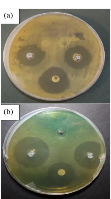
The restrictive effect of gelatin nanoparticles was studied against microbial strains of Staphylococcus aureus and Pseudomonas aeruginosa. This evaluation was carried out by measuring the clear zone of inhibition created at the surrounding area of the samples, which is shown in Fig. 7.
In this regard, when no clear zone has been created around a specific sample, there is no antibacterial activity assumed. Studies are carried out in triplicate and the results are reported as the
average diameter of the inhibition zone. The zone of inhibition diameter for gelatin nanoparticles loaded with erythromycin against Staphylococcus aureus and Pseudomonas aeruginosa has been 25.5 and 30.9 mm, respectively, which proves the antibacterial effect of the released erythromycin on these two bacterial strains.
Table 3 lists the of inhibitory zone diameters for all samples. The antimicrobial mechanism of the GNPs could be explained by the gradual release of erythromycin, as the antibiotic from the spherical structure. Therefore, a part of microbial membrane begins to absorb the released antibiotic, resulting in membrane disruption.
CONCLUSION
Gelatin nanoparticles were fabricated by Fig. 5. FTIR spectra of crosslinked and un-crosslinked gelatin nanoparticles
Fig. 6. Release behavior of GNPs
Cummula
tiv
e Drug R
elease (%)
Cross-linked GNP Uncross-linked GNP
4000 3500 3000 2500 2000 1500 1000 500 cm-1
80
70
60
50
40
30
20
10
0
291
J Nanostruct 6(4): 285-292, Autumn 2016
nanoprecipitation method with water and ethanol as solvent and non-solvent respectively. The SEM images show homogenous and round shape morphology in nanoparticles. Gelatin concentration and the extent of crosslinking were shown to have significant effect on agglomeration of particles. As it was proved that among the three tested concentrations for each of gelatin and glutaraldehyde, the optimum conditions could be obtained in 1.25 (wt%) and 20 µl, respectively. Either increasing the gelatin or decreasing the cross-linker concentrations would result in poor formed morphologies. FTIR analysis showed no harmful bonds created by each of the surfactant, or the cross-linker materials. When subjected to
the two bacterials strains of Staphylococcus aureus and Pseudomonas aeruginosa, the drug-loaded GNPs exhibited excellent antibacterial activity, which was shown by the large diameter zones on inhibition formed around samples. Drug release studies indicated appropriate results, which could be a proof to the fact that these particles could be further used as a release controlling system for different drug delivery purposes.
CONFLICT OF INTEREST
The authors declare that there is no conflict of interests regarding the publicaton of this manuscript.
REFERENCES
1. Joshi J, Patel RP. Role of biodegradable polymers in drug delivery. Int J Curr Pharm Res. 2012;4(4):74-81.
2. Kamaly N, Xiao Z, Valencia PM, Radovic-Moreno AF, Farokhzad OC. Targeted polymeric therapeutic nanoparticles: design, development and clinical translation. Chem. Soc. Rev.. 2012;41(7):2971-3010.
3. Vashist A, Vashist A, Gupta Y, Ahmad S. Recent advances in hydrogel based drug delivery systems for the human body. J. Mater. Chem. B. 2014;2(2):147-66.
4. Wilczewska AZ, Niemirowicz K, Markiewicz KH, Car H. Nanoparticles as drug delivery systems. Pharmacol Rep. 2012;64(5):1020-37.
5. Naidu B, Paulson AT. A new method for the preparation of gelatin nanoparticles: encapsulation and drug release characteristics. J. Appl. Polym. Sci. 2011;121(6):3495-500. 6. Parveen S, Misra R, Sahoo SK. Nanoparticles: a boon to
drug delivery, therapeutics, diagnostics and imaging. Nanomedicine: NANOMED NANOTECH BIOL MED. 2012;8(2):147-66.
7. Lee EJ, Khan SA, Park JK, Lim K-H. Studies on the characteristics of drug-loaded gelatin nanoparticles prepared by nanoprecipitation. Bioprocess and biosystems engineering. 2012;35(1-2):297-307.
8. Sharma K, Somavarapu S, Colombani A, Govind N, Taylor KM. Crosslinked chitosan nanoparticle formulations for delivery from pressurized metered dose inhalers. Eur. J. Pharm. Biopharm. 2012;81(1):74-81.
9. Manuja A, Kumar S, Dilbaghi N, Bhanjana G, Chopra M, Kaur H, et al. Quinapyramine sulfate-loaded sodium alginate nanoparticles show enhanced trypanocidal activity. Nanomed J. 2014;9(11):1625-34.
10. Kaur M, Malik B, Garg T, Rath G, Goyal AK. Development and characterization of guar gum nanoparticles for oral immunization against tuberculosis. J Drug Deliv.
2014(0):1-Sample Staphylococcus aureus Pseudomonas aeruginosa
Zone of Inhibition (mm) Zone of Inhibition (mm)
Standard disk of Cefoxitin 22.5 -
Standard disk of Ciprofloxacin - 25.8
Erythromycin loaded GNPs 25.5 30.9
Table 3. Inhibitory zone diameters
292 J Nanostruct 6(4): 285-292, Autumn 2016
7.
11. Pandey JK, Nakagaito AN, Takagi H. Fabrication and applications of cellulose nanoparticle-based polymer composites. Polym. Eng. Sci. 2013;53(1):1-8.
12. Prakash SJ, Santhiagu A, Jasemine S. Preparation, Characterization and In Vitro Evaluation of Novel Gellan Gum-Raloxifene HCl Nanoparticles. J Pharm BioSci. 2014;2:63-71.
13. Li M, Tang Z, Lin J, Zhang Y, Lv S, Song W, et al. Synergistic Antitumor Effects of Doxorubicin-Loaded Carboxymethyl Cellulose Nanoparticle in Combination with Endostar for Effective Treatment of Non-Small-Cell Lung Cancer. Adv Healthc Mater. 2014;3(11):1877-88.
14. Wang B, Liu P, Shi B, Gao J, Gong P. Preparation of pH-Sensitive Dextran Nanoparticle for Doxorubicin Delivery. JNN. 2015;15(4):2613-8.
15. Li W-M, Su C-W, Chen Y-W, Chen S-Y. In situ DOX-calcium phosphate mineralized CPT-amphiphilic gelatin nanoparticle for intracellular controlled sequential release of multiple drugs. Acta Biomater. 2015;15:191-9.
16. Elzoghby AO. Gelatin-based nanoparticles as drug and gene delivery systems: Reviewing three decades of research. Journal of Controlled Release. 2013;172(3):1075-91. 17. Khan SA, Schneider M. Improvement of nanoprecipitation
technique for preparation of gelatin nanoparticles and potential macromolecular drug loading. MACROMOL BIOSCI. 2013;13(4):455-63.
18. Mohanty B, Aswal V, Kohlbrecher J, Bohidar H. Synthesis of gelatin nanoparticles via simple coacervation. J. Surf. Sci. Technol.. 2005;21(3/4):149.
19. Fathollahipour S, Abouei Mehrizi A, Ghaee A, Koosha M. Electrospinning of PVA/chitosan nanocomposite nanofibers containing gelatin nanoparticles as a dual drug delivery system. J. Biomed. Mater. Res. Part A. 2015;103(12):3852-62. 20. Dinarvand R, Mahmoodi S, Farboud E, Salehi M, Atyabi
F. Preparation of gelatin microspheres containing lactic
acid–Effect of cross-linking on drug release. Acta pharm. 2005;55(1):57-67.
21. Adhirajan N, Shanmugasundaram N, Babu M. Gelatin microspheres cross-linked with EDC as a drug delivery system for doxycyline: development and characterization. J. Microencapsul. 2007; 24(7):659-71.
22. Kumbar S, Kulkarni A, Aminabhavi T. Crosslinked chitosan microspheres for encapsulation of diclofenac sodium: effect of crosslinking agent. J. Microencapsul.. 2002;19(2):173-80. 23. Soppimath KS, Aminabhavi TM, Kulkarni AR, Rudzinski WE.
Biodegradable polymeric nanoparticles as drug delivery devices. J. Control. Release. 2001;70(1):1-20.
24. Dickert H, Machka DBK, Braveny I. The uses and limitations of disc diffusion in the antibiotic sensitivity testing of bacteria. J. Infect. 1981;9(1):18-24.
25. Archana D, Dutta J, Dutta P. Evaluation of chitosan nano dressing for wound healing: Characterization, in vitro and in vivo studies. Int. J. Biol. Macromolec. 2013;57:193-203. 26. Lee E, Khan S, Lim K-H. Gelatin nanoparticle preparation
by nanoprecipitation. J Biomater Sci Polym Ed. 2011;22(4-6):753-71.
27. Gaihre B, Khil MS, Lee DR, Kim HY. Gelatin-coated magnetic iron oxide nanoparticles as carrier system: Drug loading and in vitro drug release study. Int. J. Pharm. 2009;365(1– 2):180-9.
28. Azimi B, Nourpanah P, Rabiee M, Arbab S. Producing Gelatin Nanoparticles as Delivery System for Bovine Serum Albumin. Iran Biomed J. 2014;18(1):34.