1-Methyl-3-phenylthiourea
Hou-xiang Su
Lvliang University, Lvliang, Shanxi 033001, People’s Republic of China Correspondence e-mail: [email protected]
Received 18 March 2014; accepted 3 April 2014
Key indicators: single-crystal X-ray study;T= 296 K; mean(C–C) = 0.004 A˚; Rfactor = 0.041;wRfactor = 0.114; data-to-parameter ratio = 18.6.
The title compound, C8H10N2S, was prepared by reaction of methylamine solution, KOH and phenyl-isothiocyanate in ethanol. It adopts asyn-Me andanti-Ph conformation relative to the C S double bond. The dihedral angle between the N— C( S)—N thiourea and phenyl planes is 67.83 (6). In the
crystal, the molecules centrosymmetrical dimers by pairs of N(Ph)—H S hydrogen bonds. The dimers are linked by N(Me)—H S hydrogen bonds into layers parallel to (100).
Related literature
For applications of thiourea derivatives, see: Madan & Taneja (1991); Xuet al.(2004); Borisovaet al.(2007). For the crystal structures of related compounds, see: Jiet al.(2002); Wenzelet al.(2011).
Experimental
Crystal data
C8H10N2S Mr= 166.24 Monoclinic,C2=c a= 17.348 (3) A˚
b= 8.6023 (13) A˚ c= 12.1672 (18) A˚
= 99.637 (3)
V= 1790.1 (5) A˚3
= 0.30 mm
Data collection
Bruker SMART CCD area-detector diffractometer
5444 measured reflections
2026 independent reflections 1424 reflections withI> 2(I) Rint= 0.033
Refinement
R[F2> 2(F2)] = 0.041 wR(F2) = 0.114 S= 1.03 2026 reflections 109 parameters
H atoms treated by a mixture of independent and constrained refinement
max= 0.24 e A˚
3
min=0.24 e A˚
3
Table 1
Hydrogen-bond geometry (A˚ ,).
D—H A D—H H A D A D—H A
N1—H1 S1i
0.81 (2) 2.55 (2) 3.351 (2) 169 (2)
N2—H2 S1ii
0.77 (2) 2.78 (2) 3.4229 (19) 142 (2)
Symmetry codes: (i)xþ1 2;yþ
5
2;zþ1; (ii)xþ 1 2;y
1 2;zþ
1 2.
Data collection:SMART(Bruker 1997); cell refinement:SAINT
(Bruker 1997); data reduction: SAINT; program(s) used to solve structure:SHELXS2013(Sheldrick, 2008); program(s) used to refine structure: SHELXL2013 (Sheldrick, 2008); molecular graphics:
SHELXTL(Sheldrick, 2008); software used to prepare material for publication:SHELXTL.
The diffraction data collection was carried by Hai-lian Xiao in the New Materials & Function Coordination Chemistry Laboratory, Qingdao University of Science & Technology.
Supporting information for this paper is available from the IUCr electronic archives (Reference: KQ2012).
References
Borisova, N. E., Reshetova, M. D. & Ustynyuk, Y. A. (2007).Chem. Rev.107, 46–79.
Bruker (1997).SMARTandSAINT. Bruker AXS, Inc., Madison, Wisconsin, USA.
Ji, B. M., Du, C. X., Zhu, Y. & Wang, Y. (2002).Chin. J. Struct. Chem.21, 252– 255.
Madan, V. K. & Taneja, A. D. (1991).J. Indian Chem. Soc.68, 162–163. Sheldrick, G. M. (2008).Acta Cryst.A64, 112–122.
Wenzel, M., Light, M. E., Davis, A. P. & Gale, P. A. (2011).Chem. Commun. 47, 7641–7643.
Xu, L., Jian, F., Qin, Y., Yu, G. & Jiao, K. (2004).Chem. Res. Chin. Univ.20, 305–307.
Online
supporting information
Acta Cryst. (2014). E70, o528 [doi:10.1107/S1600536814007442]
1-Methyl-3-phenylthiourea
Hou-xiang Su
S1. Comment
Thioureas have been studied for many years because of their broad antibiosis and sterilibzation properties. Recent years
study shows that thioureas not only can be used to kill insects and adjust plant growth but also have anti-viral activities
(Madan & Taneja, 1991; Borisova et al., 2007). From our early quantum study on these compounds we find that they
have several active centers and cart form polyligand complexes with metals easily (Xu et al., 2004). These complexes are
widely used as anti-medicines. Therefore study on thioureas has important impact on the future. In order to search for
new compounds with higher bioactivity, the title compound was synthesized.
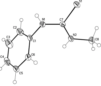
In the title compound, C8H10N2S (I), the bond lengths and angles are in a good agreement with those found in the related
compounds (Ji et al., 2002; Wenzel et al. 2011). Compound I adopts a cis-Me and trans-Ph conformation relative to the
C═S double bond (Figure 1). The dihedral angle between the N1—C7(═S1)—N1 thiourea and phenyl planes is
67.83 (6)°.
In the crystal, the molecules of I form centrosymmetrical dimers by the two intermolecular N1—H1···S1i hydrogen
bonds (Table 1, Figure 2). The dimers are further bound to each other by the intermolecular N2—H2···S1ii hydrogen
bonds (Table 1) into layers parallel to (100) (Figure 2). Symmetry codes: (i) –x + 1/2, –y + 5/2, –z + 1; (ii) –x + 1/2, y–
1/2, –z + 1/2.
S2. Experimental
The title compound was prepared by reaction of methylamine solution (40%, 0.05 mol, 5.5 ml), KOH (0.15 mol, 8.4 g)
and phenyl-isothiocyanate(0.05 mol, 4.65 g) in the ethanol solution (40 ml) at room temperature. Single-crystals of the
title compound suitable for X-ray measurements was obtained by recrystallization from ethanol/acetone (v/v=1:1) at room
temperature.
S3. Refinement
The hydrogen atoms of the amino groups were localized in the difference Fourier map and refined isotropically. The other
hydrogen atoms were placed in the calculated positions with C—H = 0.93 Å (aryl–H) and 0.96 Å (methyl–H) and refined
in the riding model with fixed isotropic displacement parameters: Uiso(H) = 1.5Ueq(C) for the CH3 group and 1.2Ueq(C) for
Figure 1
Molecular structure of I. Displacement ellipsoids are presented at the 40% probability level. H atoms are depicted as
Figure 2
A portion of the crystal structure of I demonstrating the H–bonded dimers and layers parallel to (100). The hydrogen
atoms participating in the formation of hydrogen bonds are shown only. The intermolecular N—H···S hydrogen bonds are
depicted by dashed lines.
1-Methyl-3-phenylthiourea
Crystal data
C8H10N2S Mr = 166.24 Monoclinic, C2/c a = 17.348 (3) Å b = 8.6023 (13) Å c = 12.1672 (18) Å β = 99.637 (3)° V = 1790.1 (5) Å3 Z = 8
F(000) = 704 Dx = 1.234 Mg m−3
Mo Kα radiation, λ = 0.71073 Å Cell parameters from 1286 reflections θ = 2.4–24.8°
µ = 0.30 mm−1 T = 296 K Bar, colorless
0.25 × 0.23 × 0.20 mm
Data collection
Bruker SMART CCD area-detector diffractometer
Radiation source: sealed tube phi and ω scans
5444 measured reflections 2026 independent reflections
1424 reflections with I > 2σ(I) Rint = 0.033
θmax = 27.5°, θmin = 2.4° h = −22→21
k = −8→11 l = −15→15
Refinement
Refinement on F2 Least-squares matrix: full R[F2 > 2σ(F2)] = 0.041 wR(F2) = 0.114 S = 1.03
2026 reflections 109 parameters 0 restraints
map
Hydrogen site location: difference Fourier map H atoms treated by a mixture of independent
and constrained refinement
where P = (Fo + 2Fc)/3 (Δ/σ)max < 0.001
Δρmax = 0.24 e Å−3 Δρmin = −0.24 e Å−3
Special details
Geometry. All e.s.d.'s (except the e.s.d. in the dihedral angle between two l.s. planes) are estimated using the full covariance matrix. The cell e.s.d.'s are taken into account individually in the estimation of e.s.d.'s in distances, angles and torsion angles; correlations between e.s.d.'s in cell parameters are only used when they are defined by crystal symmetry. An approximate (isotropic) treatment of cell e.s.d.'s is used for estimating e.s.d.'s involving l.s. planes.
Fractional atomic coordinates and isotropic or equivalent isotropic displacement parameters (Å2)
x y z Uiso*/Ueq
S1 0.16423 (3) 1.19173 (6) 0.35515 (4) 0.04468 (19)
N1 0.29456 (9) 1.0463 (2) 0.43958 (15) 0.0467 (5)
N2 0.23002 (10) 0.9493 (2) 0.27416 (15) 0.0485 (5)
C1 0.35645 (10) 0.9351 (2) 0.45446 (17) 0.0401 (5)
C2 0.36233 (13) 0.8311 (3) 0.5403 (2) 0.0648 (7)
H2A 0.3251 0.8303 0.5871 0.078*
C3 0.42442 (15) 0.7264 (3) 0.5572 (3) 0.0814 (9)
H3 0.4287 0.6555 0.6156 0.098*
C4 0.47899 (13) 0.7275 (3) 0.4884 (3) 0.0683 (7)
H4 0.5202 0.6569 0.4997 0.082*
C5 0.47334 (12) 0.8309 (3) 0.4037 (2) 0.0646 (7)
H5 0.5109 0.8317 0.3573 0.077*
C6 0.41193 (11) 0.9357 (3) 0.38576 (18) 0.0510 (5)
H6 0.4082 1.0064 0.3273 0.061*
C7 0.23382 (9) 1.0531 (2) 0.35543 (15) 0.0353 (4)
C8 0.16755 (12) 0.9440 (3) 0.1781 (2) 0.0685 (7)
H8A 0.1740 1.0278 0.1284 0.103*
H8B 0.1180 0.9540 0.2026 0.103*
H8C 0.1695 0.8467 0.1401 0.103*
H1 0.2993 (12) 1.117 (3) 0.4843 (19) 0.054 (7)*
H2 0.2646 (12) 0.893 (3) 0.2769 (18) 0.052 (7)*
Atomic displacement parameters (Å2)
U11 U22 U33 U12 U13 U23
S1 0.0346 (3) 0.0467 (3) 0.0524 (3) 0.0114 (2) 0.0062 (2) 0.0000 (2)
N1 0.0401 (9) 0.0497 (11) 0.0462 (11) 0.0157 (8) −0.0043 (8) −0.0147 (9)
N2 0.0365 (9) 0.0573 (12) 0.0482 (11) 0.0134 (8) −0.0034 (8) −0.0132 (9)
C1 0.0293 (8) 0.0418 (11) 0.0457 (11) 0.0061 (8) −0.0044 (8) −0.0085 (9)
C2 0.0512 (13) 0.0680 (17) 0.0758 (17) 0.0096 (11) 0.0123 (12) 0.0206 (13)
C3 0.0690 (17) 0.0612 (18) 0.109 (2) 0.0133 (14) 0.0015 (16) 0.0307 (16)
C4 0.0427 (12) 0.0587 (16) 0.097 (2) 0.0177 (11) −0.0081 (13) −0.0096 (15)
C5 0.0364 (10) 0.090 (2) 0.0637 (16) 0.0163 (11) −0.0007 (10) −0.0205 (14)
C7 0.0298 (9) 0.0394 (11) 0.0373 (10) 0.0024 (7) 0.0075 (8) 0.0004 (9)
C8 0.0504 (12) 0.093 (2) 0.0555 (14) 0.0138 (12) −0.0108 (11) −0.0244 (14)
Geometric parameters (Å, º)
S1—C7 1.6964 (17) C3—C4 1.365 (4)
N1—C7 1.342 (2) C3—H3 0.9300
N1—C1 1.427 (2) C4—C5 1.353 (4)
N1—H1 0.81 (2) C4—H4 0.9300
N2—C7 1.326 (2) C5—C6 1.384 (3)
N2—C8 1.455 (3) C5—H5 0.9300
N2—H2 0.77 (2) C6—H6 0.9300
C1—C2 1.366 (3) C8—H8A 0.9600
C1—C6 1.376 (3) C8—H8B 0.9600
C2—C3 1.393 (3) C8—H8C 0.9600
C2—H2A 0.9300
C7—N1—C1 127.17 (17) C3—C4—H4 119.9
C7—N1—H1 117.4 (16) C4—C5—C6 120.2 (2)
C1—N1—H1 115.2 (16) C4—C5—H5 119.9
C7—N2—C8 123.87 (18) C6—C5—H5 119.9
C7—N2—H2 117.0 (17) C1—C6—C5 120.0 (2)
C8—N2—H2 119.0 (17) C1—C6—H6 120.0
C2—C1—C6 119.74 (18) C5—C6—H6 120.0
C2—C1—N1 119.64 (18) N2—C7—N1 118.32 (17)
C6—C1—N1 120.57 (18) N2—C7—S1 121.70 (15)
C1—C2—C3 119.6 (2) N1—C7—S1 119.98 (14)
C1—C2—H2A 120.2 N2—C8—H8A 109.5
C3—C2—H2A 120.2 N2—C8—H8B 109.5
C4—C3—C2 120.2 (3) H8A—C8—H8B 109.5
C4—C3—H3 119.9 N2—C8—H8C 109.5
C2—C3—H3 119.9 H8A—C8—H8C 109.5
C5—C4—C3 120.2 (2) H8B—C8—H8C 109.5
C5—C4—H4 119.9
C7—N1—C1—C2 −112.2 (2) C2—C1—C6—C5 0.1 (3)
C7—N1—C1—C6 70.3 (3) N1—C1—C6—C5 177.57 (19)
C6—C1—C2—C3 −0.1 (3) C4—C5—C6—C1 0.2 (3)
N1—C1—C2—C3 −177.7 (2) C8—N2—C7—N1 −179.8 (2)
C1—C2—C3—C4 −0.1 (4) C8—N2—C7—S1 0.4 (3)
C2—C3—C4—C5 0.4 (4) C1—N1—C7—N2 −1.9 (3)
C3—C4—C5—C6 −0.4 (4) C1—N1—C7—S1 177.91 (16)
Hydrogen-bond geometry (Å, º)
D—H···A D—H H···A D···A D—H···A