1
Analysis of leaf surfaces using scanning ion
conductance microscopy
Shaun C. Walker,* Stephanie. Allen,* Gordon Bell,# and Clive J. Roberts* *
Laboratory of Biophysics and Surface Analysis, School of Pharmacy, The University of
Nottingham, University Park, Nottingham, NG7 2RD, UK
#Syngenta, Jealotts Hill International Research Centre, Bracknell, Berkshire, RG42 6EY, UK
Abstract
2 Introduction
The surfaces of all leaves are covered with a thin waxy layer known as the cuticle(Holloway, 1993). The cuticle is composed of a matrix of the biopolymers cutin and/or cutan with embedded waxes (Pollard et al., 2008). These waxes can be within the cuticle, called the cuticular waxes (CW) or form a layer above the biopolymer framework, called the epicuticular waxes (EW) (Jeffree 2006). The EW is known to form crystalline aggregates which can form different structures according to the chemical content of the waxes (Barthlott et al., 1998), and can be categorized in to six main groups (Jeffree, 2006); massive crusts, filaments, rods, tubules, plates and platelets. These crystalline structures range in size from 0.2 to 100 μm (Koch and Ensikat, 2008).
The role of the cuticle is to protect the leaf and to regulate transpiration (Riederer and Schreiber, 2001) and solute exchange (Richardson et al., 2007) between the leaf and its environment. The cuticle also protects the leaf from bacterial and fungal pathogens (Jenks et al., 1994), insect plant interaction (Kerstiens, 1996) and is the main barrier in agrochemical uptake (Santier and Chamel, 1998). Most studies of the cuticle have concentrated on the chemical constitution of the waxes and the cutin/cutan matrix in relation to the protective properties of the cuticle. Although, the chemical makeup of the cuticle is important, the material form (ie. crystalline, amorphous) and morphology of the EW waxes also provides protection for the leaf, but is generally understudied due to analytical challenges of imaging the native leaf surface without significant sample preparation (Long et al., 2003). It is worthy of note that a related area that has received significant attention recently is the phenomenon of super hydrophobic surfaces, many of which are based on studying the natural structures formed at leaf surfaces (Bargel et al., 2006, Marmur, 2004).
3
showing improvements that can be made using low temperatures in terms of conserving and revealing micron-scale imaging of coniferous tissues. Cryogenic sample preparation can be a useful approach to preserving native structures for standard and VP SEM but again the sample is now fixed. An alternative is to use scanning probe microscopes, such as the atomic force microscopy (AFM). AFM has been used to image isolated cuticles (Canet et al., 1996), which showed AFM is comparable to SEM but with higher spatial resolution. Furthermore tapping mode AFM was used to image the surface of English Laurel (Prunus laurocerasus) (Perkins et al., 2005b) and the lotus leaf (Bhushan and Jung, 2006). Critically AFM of leaves has been carried out in ambient and liquid environments opening up the potential to view dynamic processes on live samples. AFM though is unable to image most leaf surfaces due to their micoscale roughness (AFM has a limited ability to accommodate surfaces with large changes in height, changes above several microns prohibit imaging) and hence is limited to the few species with particularly flat cuticle surfaces. Another SPM technique, called scanning thermal microscopy (SThM) has been employed to study leaf surfaces. This utilises a tip that can be heated to record topographical data and can acquire local thermal analysis at a single point on a sample surface (Hammiche et al., 1996). This technique has been used to measure the plasticizing effects of non ionic surfactants on native leaf surfaces (Perkins et al., 2005a), to gain information on how chemicals affect certain areas of the cuticle. Another member of the SPM family is scanning ion conductance microscopy (SICM). This technique images surfaces within an electrolyte using a hollow nano-pipette (Korchev et al., 1997). SICM has to date been primarily used for imaging live cells at up to molecular resolution (Shevchuik et al., 2006, Liu et al., 2011). Unlike AFM, it is a true non-contact imaging technique (Korchev et al., 1997) and is able to deal with large variations in surface height, and hence has been shown to cause less damage to soft rough samples (Rheinlaender et al., 2011). (Rheinlaender et al., 2011). The resolution of SCIM, both lateral and height, are dependent, mostly, with the inner radius (ri)of the pipette tip. The SICM best lateral resolution reported is between 3 – 6 nm from a tip with ri of 6.25 nm (Shevchuik et al., 2006). With computational studies suggesting the lateral resolution to be 2ri, and the height resolution to be 5% of ri (Del Linz et al., 2014). Self pulled tips can be made to fit the resolution that is required.
4
describes the use of SICM to study the leaf surfaces of English Ivy (Hedera helix), Strawberry (Fragaria ananassa), Oil Seed Rape (Brassica napus), Pea (Pisum sativum) and grass from the Festuca genus. The data is compared to comparable SEM and AFM studies. These species were chosen to illustrate different EW structures and hydrophilic and hydrophobic properties and to interrogate the ability of SICM to analyse such a variety of leaf surfaces and under what conditions optimal SICM data may be obtained.
Materials and Methods
Chemicals and materials
SICM nano-pipettes were pulled using P-97 flaming/brown micropipette puller (Sutter Instruments, California, US) from standard wall borosilicate tubes, with an outer diameter of 1mm and an inner diameter 0.5mm (Sutter Instruments). The pipette tip inner diameter was between 150 - 200 nm as determined by SEM imaging.
The imaging electrolyte solution was prepared with deionised water, resistivity of 18MΩ. cm, obtained from a Milli-Q water purification system (Millipore Corporation, Massachusetts, US) and phosphate buffer solution (PBS) (Fisher Scientific, Loughborough, UK). AFM tips used were 0.01 – 0.025 Ohm-cm Antimony doped Si (BrukerNano, Coventry, UK), with a frequency between 347 – 393 kHz. Tween 20 (Sigma Aldrich, Missouri, US), solutions were made to a 10% w/w solution in the PBS electrolyte solution, for use on hydrophobic surfaces.
Leaf Samples
5
Analytical Instruments
Scanning electron microscopy (SEM) images were recorded using a JEOL-JSM-6060Lv SEM (JEOL Ltd, Tokyo, Japan). Samples were coated in a layer of gold using a sputter coater EM SCD005 (Leica Microsystems, Illinois, US). Samples were sputtered for 300 seconds at 30mA.
AFM images were recorded using a D3000 AFM (BrukerNano, Coventry, UK). This instrument has a maximum scan size of 90 x 90μm with a maximum Z limit of 6μm. AFM images were processed using NanoScope Analysis (BrukerNano). SICM images were recorded using a commercial ScanIC SICM (Ionscope Ltd, London, UK) and operated in hopping mode, with a 512 x 512 pixel density. This instrument has a maximum scan range 90 x 90μm with a maximum Z limit of 25μm. The SICM probe consists of a nanopipette filled with electrolyte (PBS) and back inserted with a Ag/AgCl electrode with a Ag/AgCl reference electrode immersed in the electrolyte bath. SICM images were processed using SICM image viewer (Ionscope).
Hopping mode does not use a continuous feedback, but instead the probe approaches from above the sample, until the pipette reaches a current set point value, normally a reduction 1% of the value recorded with the probe far from a surface. Once the set point is achieved the z position is recorded and the tip retracted before being re-approached to the surface at the next measurement point (Novak et al., 2009)
Results and Discussion
Imaging of leaf surfaces
6
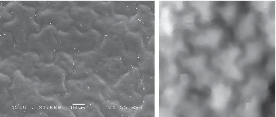
[image:6.595.73.524.125.316.2]individual stoma. The stomata of the English Ivy consist of guard cells surround by cuticle wax (Figure 2).
Fig. 1. To scale images of the adaxial surface of English Ivy (A) SEM image (B) SICM image.
7
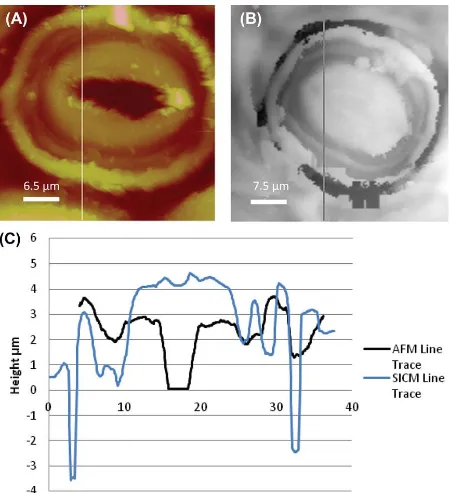
[image:7.595.73.523.121.614.2]image beyond the stomata area (Figure 3), with this area having a similar “hillock” structure as the adaxial surface.
Fig. 2. Images of stomata from the adaxial surface of English Ivy. (A) AFM image, line shows the path of the line trace. (B) SICM image, line shows the path of the line trace. (C) AFM and SICM line traces of the stomata, showing changes in topography.
8
9
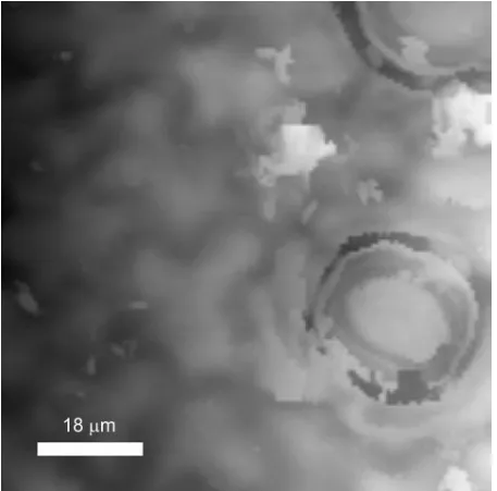
Fig. 3. SICM image of the adaxial surface of the English Ivy, showing stomata and the undulating hillock structure.
The use of SICM to image hydrophobic leave surfaces
[image:9.595.71.528.71.522.2]10
[image:10.595.73.525.153.335.2]formation at a surface (Feng et al., 2008). If this were to occur the surface would poses ‘hidden’ regions to the SICM due to air-electrolyte interfaces. Whist a challenge to SICM imaging this also presents an opportunity to study wetting processes at surface in situ using the SICM to image liquid-air and liquid-solid interfaces as a solution wets a surface.
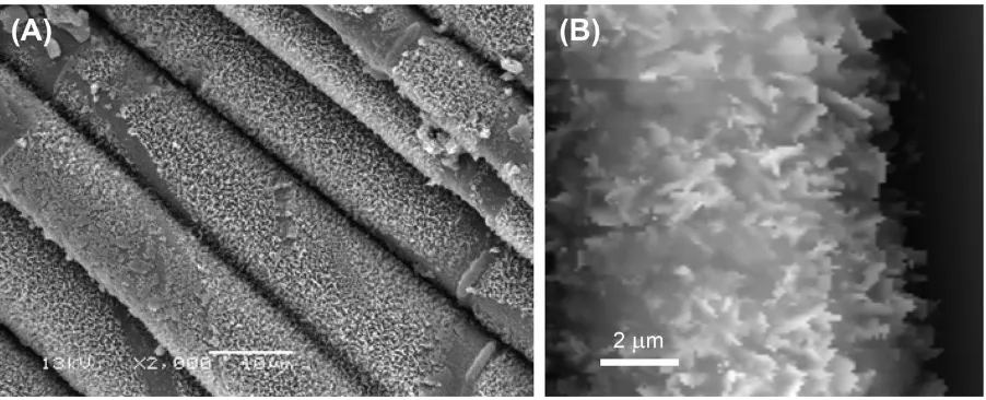
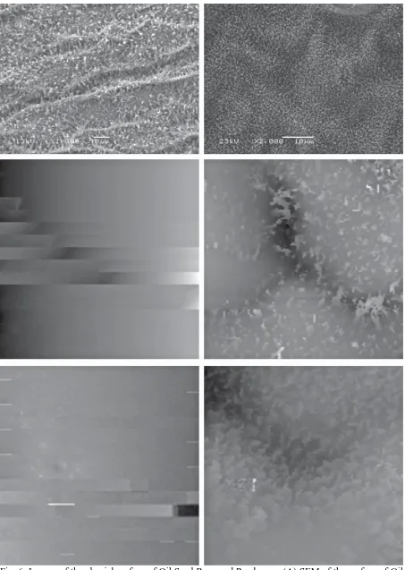
Fig. 4. Images of the abaxial surface of the Strawberry leaf. (A) SEM image showing the convoluted cuticle with filament features. (B) High-resolution SICM image of the filament features.
Fig. 5. Images of the abaxial surface of the Festuca grass (A) SEM images showing the platelet structure. (B) High-resolution SICM image of the platelet features, imaging above and the side of the features.
[image:10.595.73.524.371.554.2]11
the electrolyte solution can be in contact with the entire surface and still allow conductance between the two electrodes.
The leaves of the Oil Seed Rape and Pea plants also posses a hydrophobic surface (Gniwotta et al., 2005). For such surfaces only a small fraction of the droplet is believed to be in actual contact with the surface due to trapped air pockets at the interface separating the drop and the leaf surface (Marmur, 2004). Oil Seed Rapeand Pea have differing structures (Figure 6a and b). The Oil Seed Rape leaf surface is highly convoluted with the EW composed of tubule crystal features with amorphous wax between the features, Pea consists of a high density of crystal platelet feature, like Festuca, although the surface is not as convoluted.
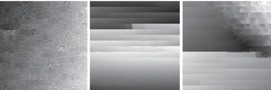
For both leaf surfaces, when the electrolyte solution is applied alone the solution is not in contact with the entire surface of the leaves. This is evidenced by considering the SICM data in (Figure 6c and e). The images show that the SICM probe is not imaging the leaf surface and we propose that it is detecting the liquid/gas interface of micro air pockets trapped at the surface. For Oil Seed Rape (Figure 6c) this interface was smooth showing no protruding leaf structures. In contrast for Pea (Figure 6e), the SICM image shows some disruption of the interface, indicating that the apex of the platelet crystals are submerged in the electrolyte. This is consistent with a Cassie-Baxter wetting regime where only a relatively small fraction of the droplet is in contact with the surface of the leaf with small compartments of gas trapped under the liquid (Marmur, 2003).
12
13
14
Fig. 7. SICM images of Pea leaf and the liquid–gas interface before and after adding small
concentration of tween 20. (A) Interface before adding the surfactant. (B) Image after 5 minutes of adding the surfactant, showing a collapse in the interface. (C) Image after 30 minutes of adding the surfactant, showing another collapse and the leaf surface being exposed.
Conclusions
15 References
Bargel, H., Koch, K., Cerman, Z.& Neinhuis, C. (2006) Structure-function relationships of plant
cuticle and cuticular waxes – a smart material ? Functional Plant Biology. 33, 893-910. Barthlott, W., Neinhuis, C., Cutler, D., Ditsch, F. Meusel, I., Theisen, I. & Wilhelmi, H. (1998)
Classification and terminology of plant epicuticular waxes. Botanical Journal of the Linnean Society. 126, 237-260.
Bhushan, B. & Jung, Y. C. (2006) Micro – and nanoscale characterization of hydrophobic and
hydrophilic leaf surfaces. Nanotechnology. 17, 2758-2772.
Canet, D., Rohr, R., Chamel, A. & Guillain, F. (1996) Atomic force microscopy study of isolated ivy
leaf cuticles observed directly and after embedding in Epon. New Phytologist, 134, 571-577. Del Linz, S., Willman, E., Caldwell, M., Klenerman, D., Fernadez, A. & Moss, G. (2014)
Contact-free scanning and imaging with the scanning ion conductance microscopy. Analytical Chemistry. 86, 2353-2360.
Feng, L., Zhang, Y. A., Xi, J. M., Zhu, Y., Wang, N., Xia, F. & Jiang, L. (2008) Petal effect: A
superhydrophobic state with high adhesive force. Langmuir. 24, 4114-4119.
Gniwotta, F., Vogg, G., Gartmann, V., Carver, T. L. W., Riederer, M. & Jetter, R. (2005) What do
microbes encounter at the plant surface ? Chemical composition of pea leaf cuticular waxes.
Plant Physiology. 139, 510-530.
Hammiche, A., Reading, M., Pollock, H. M., Song, M. & Hourston, D. J. (1996) Localized thermal
analysis using a miniaturized resistive probe. Review of Scientific Instruments. 67, 4268-4274. Holloway, P. J (1969) Effects of superficial wax on leaf wettability. Annals of Applied Biology. 63,
145 – 153
Holloway, P. J (1993) Structure and chemistry of plant cuticle. Pesticide Science. 37, 203-206. Jeffree, C. E. (2006) The fine structure of the plant cuticle. Riederer, M & Muller, C. (eds) Biology of
plant cuticle. Blackwell publishing. Oxford, England.
Jenks, M. A., Joly, R. J., Peters, P. J., Rich, P. J., Axtell, J. D. & Ashworth, E. N. (1994)
Chemically-induced cuticle mutation affecting epidermal conductance to water-vapor and disease
susceptibility in Sorghum Bicolor (L) Moench. Plant Physiology. 105, 1239-1245.
Kerstiens, G. (1996) Signalling across the divide: A widerperspective of cuticular structure-function
relationships. Trends in Plant Science. 1, 125-129.
Koch, K. & Barthlott, W. (2009). Superhydrophobic and superhydrophilic plant surfaces: an
inspiration for bimimetic materials. Philosophical Transactions of the Royal Society a-Mathematical Physical and Engineering Sciences. 367, 1487-1509.
Koch, K., Bhushan, B., Ensikat, H. J, & Barthlott, W. (2009) Self-healing voids in the wax coating on
16
Koch, K. & Ensikat, H. J. (2008) The hydrophobic coatings of plant surfaces: Epicuticular wax
crystals amd their morphologies, crystallinity and molecular self-assembly. Micron. 39, 759-772.
Korchev, Y. E., Bashford, C. L., Milovanovic, M., Vodyanoy, I. & Lab, M. J. (1997) Scanning ion
conductance microscopy of living cells. Biophysical Journal. 73, 653-658.
Lee, K. S., Ivanova, N., Starov, V. M., Hilal, N. & Dutschk, V. (2008) Kinetics of wetting and
spreading by aqueous surfactant solutions. Advances in colloid and interface Science. 144, 54-65.
Liu, X., Yang, X., Zhang, B., Zhang, X. F., Lu, H. J., Zhang, J.N. & Zhang, Y. J. (2011)
High-resolution morphological identification and characterization of living neuroblastoma
SK-N-SH cells by hopping probe ion conductance microscopy. Brain Research. 1386, 35-40. Long , L. M., Patel, H. P., Cory, W. C. & Stapleton, A. E. (2003) The maize epicuticular wax layer
provides UV protection. Functional Plant Biology. 30, 75-81.
Mackerron, D. K. L. (1976) Wind damage to surface of strawberry leaves . Annals of Botany. 40,
351 – 354.
Marmur, A. (2003) Wetting on hydrophobic rough surfaces: To be heterogenous or not to be?
Langmuir. 19,8343-8348.
Marmur, A. (2004) The lotus effect: Superhydrophonicity and metastability. Langmuir. 20, 3517-3519.
Neděla, V., Tihlaříková, E. and Hřib, J. (2014) The low-temperature methode for study of coniferous
tissues in the environmental scanning electron microscope. Microsc. Res. Tech.. Early View Article first published online: 20 SEP 2014, doi: 10.1002/jemt.22439 -
Novak, P et al. (2009) Nanoscale live-cell imaging using hopping probe ion conductance microscopy.
Nature methods. 6, 279 - 281.
Perkins, M. C., Roberts, C. J., Briggs, D., Davies, M. C., Friedmann, A., Hart, C. & Bell., G. (2005)
Macro and microthermal analysis of plant wax/surfactant interactions: plasticizing effects of
two alcohol ethoxtlated surfactants on an isolated cuticular wax and leaf model. Applied Surface Science. 243, 158-165.
Pollard, M., Beisson, F., Li, Y. H. & Ohlrogge, J. B. (2008) Building lipid barriers: biosynthesis of
cutin and suberin. Trends in Plant Science. 13, 236-246.
Rheinlaender, J., Geisse, N. A., Proksch, R. & Schaffer, T. E. (2011) Comparison of scanning Ion
Conductance Microscopy with Atomic Force Microscopy for cell imaging. Langmuir. 27, 697-704.
Richardson, A., Wojciechowski, T., Franke, R., Schreiber, L., Kerstiens, G., Jarvis, M. & Fricke, W.
(2007) Cuticular permeance in relation to wax and cutin development along the growing
17
Riederer, M. & Schreiber, L. (2001) Protecting against water loss: analysis of the barrier properties of
plant cuticles. Journal of Experimental Botany. 52, 2023-2032.
Santer, S. & Chamel, A. (1998) Reassessment of the role of cuticular waxes in the transfer of organic
molecules through plant cuticles. Plant Physiology and Biochemistry. 36, 225-231.
Shevchuk, A. I et al (2006) Imaging proteins in membranes of living cells by high-resolution scanning
ion conductance microscopy. Angewandte chemie-International Edition. 45, 2212-2216. Stabentheiner E, Zankel A, Pölt P (2010) Environmental scanning electron microscopy (ESEM) - a
versatile tool in studying plants. Protoplasma 246, 89–99.
Ying, L. M et al (2005) The scanned nanopipette: A new tool for high resolution bioimaging and