ORIGINAL RESEARCH
ADULT BRAIN
Arterial Spin-Labeling Perfusion MR Imaging Demonstrates
Regional CBF Decrease in Idiopathic Normal
Pressure Hydrocephalus
XJ. Virhammar,XK. Laurell,XA. Ahlgren, andXE.-M. Larsson
ABSTRACT
BACKGROUND AND PURPOSE: Regional cerebral blood flow has previously been studied in patients with idiopathic normal pressure hydrocephalus with imaging methods that require an intravenous contrast agent or expose the patient to ionizing radiation. The purpose of this study was to assess regional CBF in patients with idiopathic normal pressure hydrocephalus compared with healthy controls using the noninvasive quantitative arterial spin-labeling MR imaging technique. A secondary aim was to compare the correlation between symptom severity and CBF.
MATERIALS AND METHODS:Differences in regional cerebral perfusion between patients with idiopathic normal pressure hydrocephalus and healthy controls were investigated with pseudocontinuous arterial spin-labeling perfusion MR imaging. Twenty-one consecutive patients with idiopathic normal pressure hydrocephalus and 21 age- and sex-matched randomly selected healthy controls from the population registry were prospectively included. The controls did not differ from patients with respect to selected vascular risk factors. Twelve different anatomic ROIs were manually drawn on coregistered FLAIR images. The Holm-Bonferroni correction was applied to statistical analyses.
RESULTS:In patients with idiopathic normal pressure hydrocephalus, perfusion was reduced in the periventricular white matter (P⬍.001), lentiform nucleus (P⬍.001), and thalamus (P⬍.001) compared with controls. Cognitive function in patients correlated with CBF in the periventricular white matter (r⫽0.60,P⬍.01), cerebellum (r⫽0.63,P⬍.01), and pons (r⫽0.71,P⬍.001).
CONCLUSIONS: Using pseudocontinuous arterial spin-labeling, we could confirm findings of a reduced perfusion in the periventricular white matter, basal ganglia, and thalamus in patients with idiopathic normal pressure hydrocephalus previously observed with other imaging techniques.
ABBREVIATIONS:ASL⫽arterial spin-labeling; DWMH⫽deep white matter hyperintensities; iNPH⫽idiopathic normal pressure hydrocephalus; MMSE⫽ Mini-Mental State Examination; QRAPMASTER⫽Quantification of Relaxation Times and Proton density by Multiecho acquisition of a saturation recovery with Turbo spin-echo Readout
I
diopathic normal pressure hydrocephalus (iNPH) is a condi-tion with balance and gait disturbances, cognitive dysfunccondi-tion, and urinary incontinence.1The symptoms can be reversed withimplantation of a shunt system, with improvement in 50%– 80% of patients.2-4
Regional CBF has been assessed in patients with iNPH to find explanations for the pathophysiology and in search of better methods to identify shunt responders. Several studies have shown reduced perfusion in the frontal cortex, periventricular WM, basal ganglia, thalamus, and cerebellum in patients with iNPH compared with controls, but the results have been divergent.5-11In a recent study, reduced hippocampal CBF was reported as well.9
These previous studies have used different methods to esti-mate CBF: SPECT, PET, DSC MR imaging, and CT perfu-sion.8,9,12,13These methods require administration of a radioac-tive tracer or a contrast agent, and SPECT, PET, and CT expose the patient to ionizing radiation.
Pseudocontinuous arterial spin-labeling (ASL) is an MR im-aging technique with the advantage of allowing quantitative mea-Received March 16, 2017; accepted after revision June 12.
From the Departments of Neuroscience, Neurology (J.V.) and Surgical Sciences, Radiology (E.-M.L.), Uppsala University, Uppsala, Sweden; Department of Pharma-cology and Clinical Neuroscience (K.L.), Unit of Neurology, O¨ stersund, Umeå Uni-versity, Umeå, Sweden; and Department of Medical Radiation Physics (A.A.), Lund University, Lund, Sweden.
Johan Virhammar was supported by the independent Swedish foundation Erik, Karin och Go¨sta Selanders Stiftelse; Katarina Laurell received research grants from the Region Ja¨mtland Ha¨rjedalen.
Please address correspondence to J. Virhammar, MD, PhD, Department of Neuro-science, Neurology, Uppsala University, Akademiska sjukhuset, ing 85, 751 85 Uppsala, Sweden; e-mail: [email protected]; @johanvirhammar
Indicates article with supplemental on-line tables and appendix. Indicates article with supplemental on-line photo.
surements of CBF without injection of any contrast agent.14The method is completely noninvasive, with a relatively short scan time (⬃5 minutes) and can therefore easily be added to a standard MR imaging protocol. To the best of our knowledge, no previous study has investigated differences in CBF between healthy con-trols and patients with iNPH with an ASL technique.
The purpose of this study was to use ASL to investigate whether regional cerebral perfusion is reduced in patients with iNPH compared with healthy controls and whether the level of clinical symptoms correlates with CBF.
MATERIALS AND METHODS
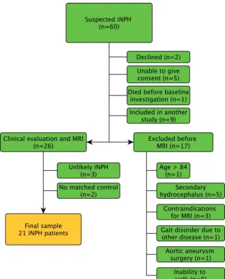
Patients and Healthy ControlsSixty patients were prospectively and consecutively recruited from the waiting list of patients referred to Uppsala University Hospital for evaluation of suspected iNPH. Nine patients were excluded for participating in an ongoing multicenter study of the CSF tap test. Patients with complete inability to walk were ex-cluded because of the intention to study the relationship between CBF and gait and balance function. Other exclusion criteria were secondary or congenital hydrocephalus, contraindications to MR
imaging, older than 84 years of age, se-vere cognitive dysfunction with an in-ability to give informed consent, refusal to undergo evaluation or shunt surgery, and gait problems that could be ex-plained by other known diseases. The in-clusion process is illustrated in the flow chart (Fig 1). Of the 26 patients exam-ined clinically, 17 were classified as hav-ing probable iNPH accordhav-ing to the iNPH guidelines.15 Six patients were classified as having possible iNPH, 1 be-cause of only 1 triad symptom (gait dis-turbance), 1 due to severe white matter changes, 1 because of a small cyst com-municating with 1 of the lateral ventri-cles, and 3 due to a history of head trauma, probably unrelated to the hy-drocephalus. Three patients were con-sidered unlikely to have iNPH, 2 because of symptoms of Parkinson disease and 1 with an aqueductal stenosis. The pa-tients examined in this study have been described in detail in a previous study.16 Controls were randomly recruited from the county of Uppsala using the Swedish population registry and were matched with patients with respect to age (⫾2 years) and sex. Exclusion criteria were any known neurologic disease, stroke, di-abetes mellitus, history of myocardial infarction that required acute treatment or resulted in persistent electrocardiogram changes, dependence on walking aids, or any terminal disease. Antihypertensive medication, aspirin, and common pain medications were allowed. Information was sent to 105 potential elderly controls. Forty-nine did not reply; 31 responded that they did not want to participate or could not participate according to the exclusion criteria. The remaining 25 accepted participation and were examined clinically by a neurologist. MR imaging examination was not possible in 2 of the 25 controls because the MR imaging scanner was upgraded before those 2 controls were examined, and another 2 controls did not match any patient. Thus, a total of 21 matched pairs of patients (15 with probable iNPH and 6 with possible iNPH) and controls were available and included in the statistical analysis. The study was approved by the local ethics committee in Uppsala, Sweden.
Clinical Examination
Neurologists, especially trained physiotherapists, and occupational therapists performed the clinical examinations. Besides a standard neurologic examination, the tests included the modified Rankin Scale, Mini-Mental State Examination (MMSE), incontinence scale,17Romberg test, and time and number of steps required to walk 10 m at a maximum pace. Tests of motor function were performed twice, and the mean result was used in the statistical analyses.
[image:2.594.56.380.45.446.2]Imaging
Details of the imaging method, scanner protocol, and postpro-cessing have been described previously.16The first MR imaging was performed between 8:00AMand 10:00AM. The second MR imaging was performed 60 minutes after the first scan to assess the repeatability of the method. The patients left the scanner after the first investigation. Between the investigations, the patients rested in the supine position in a quiet, isolated room next to the scan-ner. The patients were not allowed to use caffeine on the day of the MR imaging, and all transportation was performed with the pa-tient in bed or in a wheelchair to minimize variations in CBF. Healthy controls were only examined once with MR imaging.
Scanner and Imaging Protocol
Imaging was performed with a 3T MR imaging scanner (Achieva; Philips Healthcare, Best, the Netherlands) with a 32-channel head coil. A 3D T1-weighted gradient-echo and a T2-weighted FLAIR sequence were included in the protocol. Perfusion MR imaging was performed with a background-suppressed pseudocontinuous ASL sequence, with a single-shot echo planar imaging readout, with the following protocol: FOV⫽264⫻264 mm2, in-plane resolution⫽2.75⫻2.75 mm2, section thickness⫽5 mm, num-ber of sections⫽20, TR⫽4100 ms, TE⫽14.5 ms, label dura-tion⫽1650 ms, postlabeling delay⫽1600 ms, and 30 repetitions.
To allow quantitative CBF values, we obtained a fast reference scan.
Synthetic MR imaging data were acquired with the multisection, multi-echo, and multisaturation delay sequence Quantification of Relaxation Times and Proton density by Multiecho acquisition of a saturation recovery with Turbo spin-echo Readout (QRAPMASTER).18 Be-cause of technical problems, synthetic MR imaging data were not acquired in 2 pa-tients and in 1 control.
Perfusion MR Imaging Postprocessing
For perfusion quantification, the compart-ment model proposed by Alsop and Detre was used.19The magnetization difference (perfusion-weighted signal) is given by
⌬M⫽2M0a␣f T1a共1⫺e⫺/T1a兲e⫺w/T1a,
whereM0ais the arterial blood magneti-zation,␣is the labeling efficiency,fis the perfusion,T1ais the T1 of arterial blood,
is the labeling duration, andwis the postlabeling delay. Labeling efficiency was set to 70%,T1awas set to 1664 ms, and M0awas estimated with the signal intensity of CSF (see Alsop and Detre19 for more detail).
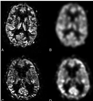
As an anatomic reference, the FLAIR images were coregistered to the perfu-sion maps by using a 12-degree affine transform (elastix; http://elastix.isi.uu. nl/). To minimize the effect of nonideal image coregistration and to reduce the intrasubject variation caused by varying delineation of regions, we spatially smoothed the CBF maps (Gaussian kernel with 4-mm full width at half maximum,Fig 2).
ROIs and Morphologic Evaluations
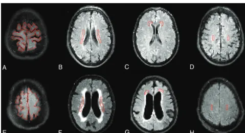
Twelve different ROIs were drawn manually on transverse T2 FLAIR images using the in-house software Eval Gui (developed by Markus Nilsson, Lund University, Lund, Sweden). We covered the following regions: cerebellum, pons, high-convexity cortex, medial frontal cortex, lentiform nucleus, medial temporal lobe, supplementary motor area, thalamus, frontal WM, lateral WM, superior WM, and periventricular WM. The number of sections and voxels for all ROIs is listed inTable 1. Four different ROIs are illus-trated inFig 3, and all ROIs in both patients and controls are illus-trated in the On-line Appendix. All sections of all scans were visually inspected, and care was taken not to include artifacts or large vessels in the ROIs. The ROIs were drawn by the first author (J.V., 4 years of experience) and reviewed by the last author (E.-M.L.,⬎30 years of experience). When ROIs were drawn, the investigator was blinded to the patients’ clinical data. CBF values were calculated for every voxel, and a mean CBF was provided in each ROI.
For descriptive purposes, we assessed the following morphologic
[image:3.594.54.374.46.393.2]MR imaging features: the Evans index,20the callosal angle,21and deep white matter hyperintensities (DWMH) according to the visual grading scale of Fazekas et al.22Disproportionately enlarged sub-arachnoid space hydrocephalus was defined as a combination of en-larged ventricles (Evans index of⬎0.3), narrow sulci at the high con-vexity or parafalcine and dilated Sylvian fissures (Table 2).3The measurements of morphologic features were performed by the last author (E.-M.L.). She was blinded to CBF data.
Volumetric Measurement
Measurements of CSF volumes were performed by using the QRAPMASTER sequence,18 which provides quantification of
longitudinal relaxation time (T1), transverse relaxation time (T2), and proton density. The software SyMRI Brain Studio (Syn-theticMR, Linko¨ping, Sweden) uses combinations of T1, T2, and proton density to estimate voxelwise partial volumes of GM, WM, and CSF, which allow volumetric estimation of these tissues. The volume of both lateral ventricles was calculated semiautomati-cally in both patients and controls after manual delineation on each section that included the lateral ventricles.
Statistical Analysis
[image:4.594.53.533.279.539.2]The mean CBF in each ROI from the 2 scans of the patients was used in the statistical analyses. Each patient was paired with an
Table 1: Cerebral blood flow values in patients and controlsa
ROI Sections Voxelsb CBF, Repeatabilityc
CBF (Mean) (SD) (mL/100 g/min)
PValued
Patients Controls
Cerebellum 2 924 (144) 0.92 20.7 (9.3) 27.2 (7.0) .006
MFC 3 133 (30) 0.90 24.9 (5.6) 27.4 (5.1) .23
Lentiform nucleus 1 115 (15) 0.75 20.4 (2.7) 25.0 (4.0) ⬍.001e
MTL 1 114 (15) 0.86 30.4 (5.6) 31.7 (3.8) .86
HCC 2 1350 (459) 0.72 22.5 (5.5) 21.6 (3.0) .47
Pons 3 68 (17) 0.90 22.0 (7.0) 24.1 (5.3) .19
SMA 2 130 (18) 0.72 25.1 (5.1) 25.2 (3.7) .93
Thalamus 2 116 (19) 0.92 26.8 (10.2) 34.1 (5.6) ⬍.001e
Frontal WM 2 41 (8) 0.91 11.0 (5.0) 10.7 (2.2) .76
Lateral WM 2 136 (33) 0.89 12.2 (3.0) 13.8 (1.7) .014
Superior WM 2 67 (7) 0.88 14.2 (5.0) 12.6 (2.5) .17
Periventricular WM 1 186 (15) 0.88 8.0 (2.5) 13.1 (3.1) ⬍.001e
Note:—HCC indicates high-convexity cortex; MFC, medial frontal cortex; MTL, medial temporal lobe; SMA, supplementary motor area.
a
Data are mean with SD in parentheses unless otherwise indicated.
bMean (SD) voxels in each ROI in patients. c
Repeatability of perfusion measurements between the 2 baseline investigations using the intraclass correlation coefficient.
dUnadjustedPvalues tested with the paired-samplesttest. e
Significant after Holm-Bonferroni correction.
age- and sex-matched control. A paired-samplesttest was used for the difference in CBF between patients and controls. Differ-ences in descriptive data and comorbidity were analyzed with the Wilcoxon signed rank test or the McNemar test. Difference in age was tested with the Mann-WhitneyUtest. Correlations were as-sessed with the Spearman correlation. An intraclass correlation coefficient (2-way mixed, single measures) was used to calculate the repeatability of CBF between the 2 investigations in patients. For the analysis of CBF in patients and controls in the 12 ROIs, the Holm-Bonferroni correction for multiple comparisons was ap-plied to reduce type I errors. Holm-Bonferroni correction works by sorting the NPvalues from the multiple tests and testing the smallestPvalue against␣/N, the second smallest against␣/(N-1), and so on. As soon as a null hypotheses is not rejected, all
subse-quent null hypotheses are also deemed not rejected, resulting in an effective sig-nificance threshold. With␣⫽.05, the effective significance threshold for ROI analysis in patients and controls wasP⬍
.006. Correlations were tested among all 12 ROIs, and the 4 clinical tests as well as the 4 morphologic imaging features. Only correlations with r ⬎ 0.60 were considered strong and included in the results section. All analyses were per-formed with SPSS Statistics for Macin-tosh, Version 22.0 (IBM, Armonk, New York).
RESULTS
Background DataThe radiologic morphologic measure-ments and the results of gait and balance tests, urgency, and MMSE differed markedly between patients and healthy con-trols (Table 2). However, there were no significant differences between patients and controls concerning age, the radiologic finding DWMH, and the use of antihypertensive drugs, acetyl-salicylic acid, antidiabetic drugs, or statins, and there were no differences in the history of stroke, traumatic brain injury, meningitis, and previous or present smoking.
Cerebral Blood Flow
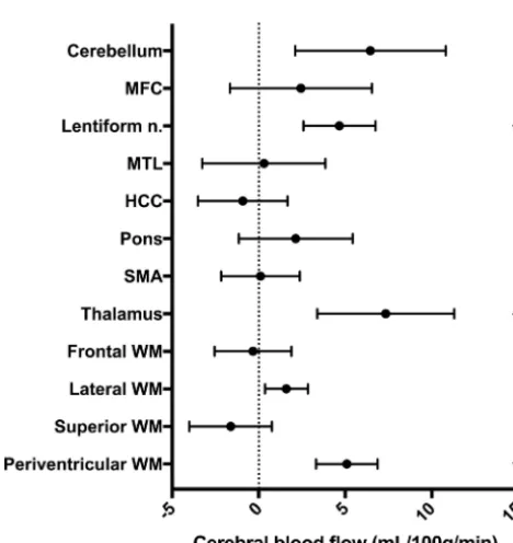
The CBF in all patients and controls is presented inTable 1. In patients, the CBF was significantly lower in the periventricular WM, lentiform nucleus, and thalamus after Holm-Bonferroni correction (Fig 4). In 3 patients with iNPH, the ASL maps con-tained vascular artifacts and apparent hypoperfusion in the brain parenchyma. If these 3 patients were excluded from statistical analyses, CBF was still significantly lower in patients in the periventricular WM, lentiform nucleus, and thalamus. There was no difference in age or symptom severity among the 3 patients with vascular artifacts compared with the other patients.
Morphologic Features and CBF
In controls, there were negative correlations between the Evans index and perfusion in frontal WM (r⫽ ⫺0.75,P⬍.001), lateral WM (r⫽ ⫺0.73,P⬍.001), and periventricular WM (r⫽ ⫺0.66,
P⬍.01). In controls, there were also negative correlations be-tween quantified lateral ventricular volume with Synthetic MR imaging and perfusion in the frontal WM (r⫽ ⫺0.72,P⬍.001), lateral WM (r⫽ ⫺0.67,P⬍.01), periventricular WM (r⫽ ⫺0.65,
P⬍.01), and medial frontal cortex (r⫽ ⫺0.61,P⬍.01). There were no correlations between ventricular volume and CBF in patients.
Clinical Symptoms and CBF
In patients, the MMSE score correlated significantly with CBF in the pons (r⫽0.71,P⬍.001), cerebellum (r⫽0.63,P⬍.01), and periventricular WM (r⫽0.60,P ⬍.01), but not in controls. Neither in patients nor in controls did gait, balance, or urgency incontinence correlate with CBF values. See On-line Tables 1 and 2 for all correlation coefficients in patients.
[image:5.594.55.377.56.201.2]FIG 4. Mean difference with 95% CI between patients with iNPH and healthy controls. MFC indicates the medial frontal cortex; HCC, high-convexity cortex; MTL, medial temporal lobe; SMA, supplementary motor area. Theasteriskindicates a significant difference after Holm-Bonferroni correction.
Table 2: Demographics and background data in patients and controlsa
Patients (n= 21) Controls (n= 21) PValue
Age (median) (range) 74 (65–81) 74 (65–82) NSb
No. of men (%) 11 (52%) 11 (52%) NSc
MMSE 25 (22–27) 30 (29–30) ⬍.001d
Urgency scale 3 (1–4) 1 (1–1) ⬍.001d
Modified Rankin Scale score 2 (2–3) 0 (0–0) ⬍.001d
Romberg test (sec) 18 (4–60) 60 (60–60) ⬍.001d
10-m Walk time (sec) 12 (8–17) 5 (5–6) ⬍.001d
10-m Walk (No. of steps) 22 (16–30) 12 (11–13) ⬍.001d
Evans index score 0.35 (0.34–0.39) 0.28 (0.24–0.30) ⬍.001d
DWMH 1 (1–3) 1 (1–2) NSd
DESH (No.) (%) 14 (67%) 0 (0%) ⬍.001c
Callosal angle 66° (60°–73°) 113° (104°–121°) ⬍.001d
Lateral ventricular volume (mL) 130 (111–136) 31 (24–54) ⬍.001d Note:—DESH indicates disproportionately enlarged subarachnoid space hydrocephalus; NS, not significant.
a
Unless indicated otherwise, data are median, with interquartile range in parentheses.
b
Mann-WhitneyUtest.
c
McNemar test.
d
[image:5.594.52.286.252.500.2]Repeatability
Repeatability for the CBF values obtained in the 2 investigations in patients is presented inTable 1as intraclass correlation coeffi-cients. The intraclass correlation coefficients were in the range of 0.72– 0.92.
Volumetric Measurement
The median lateral ventricular volume in patients was signifi-cantly larger than in controls, 130 mL (interquartile range⫽111– 136 mL) and 31 mL (interquartile range, 24 –54 mL), respectively.
DISCUSSION
In this study, pseudocontinuous ASL, a noninvasive perfusion method without ionizing radiation or contrast agent, was used to compare cerebral perfusion between patients with iNPH and healthy controls. In agreement with previous studies using ana-tomic ROIs in patients with iNPH, CBF was reduced in the periventricular WM, basal ganglia, and thalamus.5,9There was a trend indicating that CBF was also reduced in the cerebellum in patients with iNPH, but the difference was not significant after Holm-Bonferroni correction. In patients, MMSE correlated sig-nificantly with CBF in the pons, cerebellum, and periventricular WM. The repeatability of the CBF measurements was acceptable to high, which strengthens our findings.
In the present study, reduced CBF in patients with iNPH was found in the basal ganglia, thalamus, and periventricular WM, whereas previous studies also reported hypoperfusion in the fron-tal cortex, temporal lobes, and cerebellum.5,9,13The more conser-vative correction for multiple analyses performed in our study could be one explanation for this difference, but another could be differences in the selection of healthy controls as well as the dif-ference in imaging methods.
The mechanism of the reduced regional CBF in iNPH is likely multifactorial. One hypothesis is that transependymal passage of CSF into the parenchyma leads to reversal of interstitial fluid flow, initiating local CSF edema in the periventricular WM. The accu-mulation of interstitial fluid may cause local compression of small vessels and, more important, reduce elimination of vasoactive metabolites. The region mainly affected by hypoperfusion, ac-cording to this hypothesis, should be the periventricular WM, which is in line with our results and has also been reported by others.6,9The reduced CBF in the thalamus and lentiform nucleus may be an indirect result of periventricular WM edema, by affect-ing the penetrataffect-ing arteries that supply the thalamus and lenti-form nucleus.
The supplementary motor area, basal ganglia, thalamus, mes-encephalon, or the white matter tracts connecting these regions have been suggested as the most important anatomic structures behind gait and balance disturbances in patients with iNPH.23,24 There were no correlations between gait function and CBF in the present study. These findings indicate that the etiology of the gait disturbance in iNPH is complex and might be caused by factors other than reduced CBF.
However, there were correlations in the present study between impaired cognitive function measured with MMSE and reduced CBF in the pons, cerebellum, and periventricular WM. Perfusion in the brain stem has not been well-studied in iNPH, but Tullberg
et al25reported a relative CBF increase in the mesencephalon in patients with improved wakefulness after shunt surgery. Distur-bances in cognitive function, especially in attention and executive ability, have been reported after insults to the pons,26and such symptoms are often seen in iNPH as well.27The connection be-tween impaired cognitive function and reduced CBF in the cere-bellum is harder to explain; however, there are reports that the cerebellum is involved in cognitive function.28,29
Hypertension and vascular risk factors30and MR imaging ev-idence of vascular disease such as DWMH are overrepresented in patients with iNPH.31These vascular risk factors are associated with reduced CBF32; therefore, differences in CBF could be over-estimated if controls are a highly selected sample of healthy el-derly. In the present study, the controls were randomly selected from the population, with a limited number of exclusion criteria. There was no significant difference in the burden of DWMH or the use of acetylsalicylic acid or antihypertensive drugs between patients and controls, which strengthens our findings.
In controls, there were correlations between the size of the lateral ventricles and reduced CBF in WM regions. A potential reason may be asymptomatic small-vessel disease with reduction of WM volume and secondary dilation of the lateral ventricles, indicating that even moderately enlarged ventricles may not be considered a normal aging phenomenon.
The pseudocontinuous ASL technique has some advantages compared with other perfusion methods. The repeatability in healthy controls and in patients with Alzheimer disease is high, and CBF values have been validated against PET.33Like PET, perfusion values can be quantified. Therefore, no reference re-gions are needed to calculate relative perfusion values. In SPECT and DSC perfusion, relative values are used, with the risk that pathologic perfusion in the reference regions may affect the rela-tive perfusion values. In a recent study, CT perfusion was evalu-ated in iNPH and may be a promising alternative because it is available in many sites and is not as sensitive to artifacts from the shunt valve after shunt implantation as with MR imaging.8
In the iNPH guidelines from 2005, the SPECT-acetazolamide challenge is the only perfusion method mentioned as a supportive diagnostic technique.15 A SPECT-acetazolamide challenge re-quires a separate imaging investigation, is time-consuming, and exposes the patient to ionizing radiation from the injected tracer. The ASL sequence is rapid and noninvasive; these features make it ideal for research studies in patients with iNPH, but the diagnostic value of the method is uncertain. Although CBF differences were found between patients and controls on a group level, the effect may not be large enough in individual cases to allow the use of ASL as a supportive diagnostic technique in clinical routine.
The strength of this study was that prospectively and consecu-tively included patients were compared with age- and sex-matched controls who were randomly selected from the population.
delays would be preferable in elderly patients with pathologic CBF.
The CBF values in both our patients and controls were low compared with those reported in PET studies,5,35but they were in line with other ASL studies in patients with iNPH.36Also, it has been reported that quantified perfusion values are underesti-mated in elderly individuals with ASL.37Our main results were probably not affected by this limitation because we compared patients with age-matched controls.
White matter perfusion estimation with ASL is challenging, due to a longer arterial transit time and lower blood volume com-pared with gray matter. Thus, the WM perfusion signal had a low signal-to-noise ratio, and the corresponding results should be treated with some caution. On the other hand, 2 recent studies suggest that though pixel-wise analysis is challenging, ROI-based analysis of WM perfusion is feasible in single subjects.38,39This finding increases the credibility of our group-level results based on ROI analysis in 21 patients and 21 controls.
Spatial smoothing was applied to reduce the effects of nonideal coregistration but can potentially introduce partial volume effects on CBF measurements. This might have influenced the results of small ROIs with contamination of data from surrounding tissue. The MMSE cognitive test used in this study may underesti-mate subcortical deficits that are very relevant in iNPH. More sophisticated neuropsychological tests could have revealed more information about the relationship between cognition and CBF in both patients and controls.40
CONCLUSIONS
Pseudocontinuous ASL was used to compare perfusion of pa-tients with iNPH versus healthy controls. In papa-tients, perfusion values were reduced in the periventricular WM, basal ganglia, and thalamus, and there was a correlation between cognitive dysfunc-tion and reduced CBF. Because ASL is a noninvasive quantitative perfusion method without ionizing radiation or contrast agent, it is a suitable perfusion method for research studies in patients with iNPH.
ACKNOWLEDGMENTS
The authors thank Markus Nilsson for the software Eval Gui, Markus Fahlstro¨m for technical assistance, and Emma Jansson for assistance with examinations of healthy controls. The authors also thank our normal pressure hydrocephalus team and the MR im-aging staff at Uppsala University Hospital, especially Britt-Mari Bolinder. Finally, the authors thank Selanders Stiftelse for its support.
Disclosures: Johan Virhammar—RELATED:Grant: Selanders Foundations,Comments: Swedish independent foundation*;UNRELATED:Payment for Lectures Including
Ser-vice on Speakers Bureaus: Medtronic,Comments: I have received 1 educational lecture
honoraria from Medtronic for 1 lecture. Andre´ Ahlgren—UNRELATED:Grant: Swedish Research Council.* *Money paid to the institution.
REFERENCES
1. Adams RD, Fisher CM, Hakim S, et al.Symptomatic occult
hydro-cephalus with “normal” cerebrospinal-fluid pressure: a treatable syndrome.N Engl J Med1965;273:117–26CrossRef Medline
2. Klinge P, Hellstro¨m P, Tans J, et al.One-year outcome in the
Euro-pean multicentre study on iNPH. Acta Neurol Scand2012;126:
145–53CrossRef Medline
3. Hashimoto M, Ishikawa M, Mori E, et al; Study of INPH on
neuro-logical improvement (SINPHONI).Diagnosis of idiopathic normal
pressure hydrocephalus is supported by MRI-based scheme: a pro-spective cohort study.Cerebrospinal Fluid Res2010;7:18CrossRef Medline
4. Sundstro¨m N, Malm J, Laurell K, et al.Incidence and outcome of
surgery for adult hydrocephalus patients in Sweden.Br J Neurosurg
2017;31:21–27CrossRef Medline
5. Owler BK, Momjian S, Czosnyka Z, et al.Normal pressure
hydro-cephalus and cerebral blood flow: a PET study of baseline values.
J Cereb Blood Flow Metab2004;24:17–23CrossRef Medline
6. Momjian S, Owler BK, Czosnyka Z, et al.Pattern of white matter
regional cerebral blood flow and autoregulation in normal pres-sure hydrocephalus.Brain2004;127:965–72CrossRef Medline
7. Sasaki H, Ishii K, Kono AK, et al.Cerebral perfusion pattern of
id-iopathic normal pressure hydrocephalus studied by SPECT and statistical brain mapping.Ann Nucl Med2007;21:39 – 45CrossRef Medline
8. Ziegelitz D, Arvidsson J, Hellstro¨m P, et al.Pre-and postoperative
cerebral blood flow changes in patients with idiopathic normal pressure hydrocephalus measured by computed tomography (CT) perfusion. J Cereb Blood Flow Metab 2016;36:1755– 66 CrossRef Medline
9. Ziegelitz D, Starck G, Kristiansen D, et al.Cerebral perfusion
mea-sured by dynamic susceptibility contrast MRI is reduced in patients with idiopathic normal pressure hydrocephalus.J Magn Reson
Im-aging2014;39:1533– 42CrossRef Medline
10. Corkill RG, Garnett MR, Blamire AM, et al.Multi-modal MRI in
normal pressure hydrocephalus identifies pre-operative haemody-namic and diffusion coefficient changes in normal appearing white matter correlating with surgical outcome.Clin Neurol Neurosurg
2003;105:193–202CrossRef Medline
11. Owler BK, Pena A, Momjian S, et al.Changes in cerebral blood flow
during cerebrospinal fluid pressure manipulation in patients with normal pressure hydrocephalus: a methodological study.J Cereb
Blood Flow Metab2004;24:579 – 87CrossRef Medline
12. Owler BK, Pickard JD.Normal pressure hydrocephalus and cerebral
blood flow: a review.Acta Neurol Scand2001;104:325– 42CrossRef Medline
13. Larsson A, Bergh AC, Bilting M, et al.Regional cerebral blood flow in
normal pressure hydrocephalus: diagnostic and prognostic as-pects.Eur J Nucl Med1994;21:118 –23Medline
14. Wu WC, Ferna´ndez-Seara M, Detre JA, et al.A theoretical and
ex-perimental investigation of the tagging efficiency of pseudocon-tinuous arterial spin labeling.Magn Reson Med2007;58:1020 –27
CrossRef Medline
15. Relkin N, Marmarou A, Klinge P, et al.Diagnosing idiopathic
nor-mal-pressure hydrocephalus.Neurosurgery2005;57:S4 –16;
discus-sion ii-vMedline
16. Virhammar J, Laurell K, Ahlgren A, et al.Idiopathic normal pressure
hydrocephalus: cerebral perfusion measured with pCASL before and repeatedly after CSF removal.J Cereb Blood Flow Metab2014;34:
1771–78CrossRef Medline
17. Hellstro¨m P, Klinge P, Tans J, et al.A new scale for assessment of
severity and outcome in iNPH.Acta Neurol Scand2012;126:229 –37
CrossRef Medline
18. Warntjes JB, Leinhard OD, West J, et al.Rapid magnetic resonance
quantification on the brain: optimization for clinical usage.Magn
Reson Med2008;60:320 –29CrossRef Medline
19. Alsop DC, Detre JA.Reduced transit-time sensitivity in noninvasive
magnetic resonance imaging of human cerebral blood flow.J Cereb
Blood Flow Metab1996;16:1236 – 49CrossRef Medline
20. Evans W.An encephalographic ratio for estimating ventricular
en-largement and cerebral atrophy.Arch Neurol Psychiatry1942;47:
931–37CrossRef
measured on MRI as a predictor of outcome in idiopathic normal-pressure hydrocephalus.J Neurosurg2014;120:178 – 84CrossRef Medline
22. Fazekas F, Chawluk JB, Alavi A, et al.MR signal abnormalities at 1.5
T in Alzheimer’s dementia and normal aging.AJR Am J Roentgenol
1987;149:351–56CrossRef Medline
23. Lundin F. Idiopathic Normal Pressure Hydrocephalus, Aspects on
Pathophysiology, Clinical Characteristics and Evaluation Methods [PhD thesis]. Linko¨ping: Linko¨ping University; 2012
24. Hellstrom P.The Neuropsychology of Idiopathic Normal Pressure
Hy-drocephalus[PhD thesis]. Gothenburg: University of Gothenburg;
2011
25. Tullberg M, Hellstro¨m P, Piechnik SK, et al.Impaired
wakeful-ness is associated with reduced anterior cingulate CBF in pa-tients with normal pressure hydrocephalus.Acta Neurol Scand
2004;110:322–30CrossRef Medline
26. Garrard P, Bradshaw D, Ja¨ger HR, et al.Cognitive dysfunction after
isolated brain stem insult: an underdiagnosed cause of long term morbidity.J Neurol Neurosurg Psychiatry2002;73:191–94CrossRef Medline
27. Ogino A, Kazui H, Miyoshi N, et al. Cognitive impairment in
patients with idiopathic normal pressure hydrocephalus.Dement
Geriatr Cogn Disord2006;21:113–19CrossRef Medline
28. E KH, Chen SH, Ho MH, et al.A meta-analysis of cerebellar
contri-butions to higher cognition from PET and fMRI studies.Hum Brain
Mapp2014;35:593– 615CrossRef Medline
29. Moore DM, D’Mello AM, McGrath LM, et al.The developmental
relationship between specific cognitive domains and grey matter in the cerebellum.Dev Cogn Neurosci2017;24:1–11CrossRef Medline
30. Malm J, Graff-Radford NR, Ishikawa M, et al.Influence of
comor-bidities in idiopathic normal pressure hydrocephalus: research and clinical care—a report of the ISHCSF task force on comorbidities in INPH.Fluids Barriers CNS2013;10:22CrossRef Medline
31. Krauss JK, Regel JP, Vach W, et al.White matter lesions in patients
with idiopathic normal pressure hydrocephalus and in an age-matched control group: a comparative study.Neurosurgery1997;40:
491–95; discussion 495–96Medline
32. Bastos-Leite AJ, Kuijer JP, Rombouts SA, et al.Cerebral blood flow
by using pulsed arterial spin-labeling in elderly subjects with white matter hyperintensities.AJNR Am J Neuroradiol2008;29:1296 –301
CrossRef Medline
33. Xu G, Rowley HA, Wu G, et al.Reliability and precision of
pseudo-continuous arterial spin labeling perfusion MRI on 3.0 T and com-parison with 15O-water PET in elderly subjects at risk for Alzhei-mer’s disease.NMR Biomed2010;23:286 –93CrossRef Medline
34. Zaharchuk G, Bammer R, Straka M, et al.Arterial spin-label imaging
in patients with normal bolus perfusion-weighted MR imaging findings: pilot identification of the borderzone sign. Radiology
2009;252:797– 807CrossRef Medline
35. Klinge PM, Berding G, Brinker T, et al.A positron emission
tomog-raphy study of cerebrovascular reserve before and after shunt sur-gery in patients with idiopathic chronic hydrocephalus.J Neurosurg
1999;91:605– 09CrossRef Medline
36. Ivkovic M, Reiss-Zimmermann M, Katzen H, et al.MRI assessment
of the effects of acetazolamide and external lumbar drainage in id-iopathic normal pressure hydrocephalus.Fluids Barriers CNS2015; 12:9CrossRef Medline
37. Ambarki K, Wåhlin A, Zarrinkoob L, et al.Accuracy of parenchymal
cerebral blood flow measurements using pseudocontinuous arte-rial spin-labeling in healthy volunteers.AJNR Am J Neuroradiol
2015;36:1816 –21CrossRef Medline
38. Wu WC, Lin SC, Wang DJ, et al.Measurement of cerebral white
matter perfusion using pseudocontinuous arterial spin labeling 3T magnetic resonance imaging: an experimental and theoretical in-vestigation of feasibility.PLoS One2013;8:e82679CrossRef Medline
39. Skurdal MJ, Bjornerud A, van Osch MJ, et al.Voxel-wise perfusion
assessment in cerebral white matter with PCASL at 3T: is it possible and how long does it take?PLoS One2015;10:e0135596CrossRef Medline
40. Hellstro¨m P, Edsbagge M, Archer T, et al.The neuropsychology of
patients with clinically diagnosed idiopathic normal pressure hy-drocephalus. Neurosurgery 2007;61:1219 –26; discussion 1227–28