Image Processing Techniques for Brain Tumor
Detection: A Review
Vinay Prasad Tamta1
1School of Engineering & Technology, HNBGU Srinagar Garhwal Uttarakhand, India
Abstract
A tumor is an abnormal growth of tissues. That can be distinguished from the surrounding fabric for its
construction. The image of the magnetic resonance generates a better contrast indicating the regular and irregular
tissues. The tumor is an uncontrolled development of tissues in any region of the body. A tumor can cause the
leading cause of death and is responsible for around 16% of all deaths worldwide. Detection and segmentation of
brain tumors is a very difficult task in MRI. In-depth knowledge and experience in radiology are mandatory for the
precise detection of tumors in medical images. Automation of tumor detection is necessary because there may be a
deficit of experienced radiologists at a time of great need. The tumor is an uncontrolled growth of tissues in any
region of the body. Detection of tumor in early stages facilitates treatment. Here, we will discuss an abbreviated
review of the different segmentation methods used for the detection of magnetic resonance tumors (MRI) of the
brain.
Index Terms - Brain Tumor, Image Segmentation, Magnetic Resonance Imaging.
1.
INTRODUCTION
The basic reasons of tumor in the brain are abnormal cells in the brain. There are two cases of cancer: malignant
and benign tumors. Malignant tumor occurs due to abnormal cell increases with the positivity of attacking or
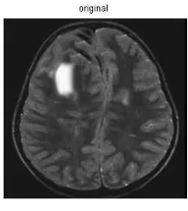
spreading to the other body part. Figure 1 shows a tumor in the human brain. The doctor administers the treatment
for shots instead of the treatment for the tumor. This event is why tumor detection is important for that treatment.
Medical imaging techniques play a significant role in the diagnosis and early detection of tumors. MRI, CT, digital
mammography and other imaging processes allow an efficient mean of detecting types of diseases from magnetic
resonance imaging. If we can identify the location of tumors, it can be very helpful for radiologists in analyzing
the tumor and in turn performing surgery to remove that tumor. Magnetic resonance use radiofrequency and
magnetic field without ionized radiation. Malignant tumor leads to abnormal cell growth with the possibility of
attacking or spreading to other regions of the body parts. Normally, the brain tumor affects the cerebrospinal fluid
and can cause the shots. With the help of a brain MRI, it is possible to locate tumors and provide useful
information to radiologist, which enables them with an easy diagnosis and effective surgical approach for its
Figure 1. MRI of Human Brain. (The Presence of a Tumor Towards The left side)
1.1
MAGNETIC RESONANCE IMAGING
MRI is normally helpful in the medical field for detecting and displaying details in the internal structure of a body.
It is commonly helpful to find out differences in body tissues that have different properties than the surrounding
tissues [1]. Therefore, it is a technique that has become a very effective, particularly for brand change and cancer
imaging. The CT is helpful for ionizing radiation, while the magnetic resonance uses a secure magnetic area.
These are as align to the nuclear magnetization. A product signal can be processed later to get more data about the
physique [1]
2.
LITERATURE REVIEW
An MRI uses the magnetism and radio waves to produce two dimensional images of internal structure of body.
Because of its detailed information nature it is a favorite prescription from neurosurgeons. Once we have the
scanned image of brain, image processing plays an important role in identifying the tumor, its size and its position.
Image processing software can highlight the areas of probably tumor which reduces half of the analysis effort of
medical expert.
I will now talk about the image processing techniques which can help in identifying the tumor. For brain tumor
identification we require to separate an MRI brain image in two regions (one region with brain tumor cells and
other containing the normal brain cells). We can combine FCM, seed region growth and similarity coefficient
algorithm for measuring tissues with tumor.
Alternatively SVM based on the core of the radial base function (RBF) and classification of brain tumors based on
the principal component analysis (PCA), produces a relatively very high accurate results. A method was proposed
by Sharma et al. [2] which use the primitive plot characteristics with the artificial neural network (ANN) for
Mark Schmidt et al. presented a procedure that quantitatively evaluates the performance of four different parts of
alignment-based (AB) characteristics that encode spatial anatomical information for use in the classification of
supervised pixels [3]. This is the first study to liken various types of alignment-based (AB) feature. It’s a way to
add various types of AB functions and analyze the combination of AB characteristics with the characteristics of
the plot in a learning framework.
Mohd Fauzi Bin et al. [4] performed two methods for brain tumors (i) wavelet-based feature extraction method
(ii)Support Vector Machine (SVM). The extraction of the features was performed using the Daubechies wavelet
(db4) and the approximation coefficients of the MRI brain images were used as vectors of characteristics for
classification. Their accuracy was 65%. Therefore, the SVM method led to limited precision.
Dzung L. Pham et al. [5] proposed a method which is based on a threshold. In this method a threshold needs to be
identified. Based on this threshold image is partitioned into two classed. One class has pixel values greater than the
threshold and other have pixel values less than the threshold. Similarly, multiple thresholds were taken and this
approach is called as
Andac Hamamci et al. [6] proposed a method of targeted tumor segmentation based on T1 magnetic resonance
cellular automata with contrast, which standardizes the volume of interest (VOI) and seed selection.
3.
TUMOR SEGMENTATION TECHNIQUES
In tumor segmentation, different ways can be used for image segmentation. Such as thresholds, region growth,
classifier, grouping, AN networks, atlas-driven, established-level methods.
A.
Thresholding
This is an older method for image segmentation. This method is very simple and easy to apply in all types of
images. The segmentation is prepared by grouping all the pixels with intensity between two thresholds of this type
in a class. Generally we know threshold have two types one is local and another is global. The global threshold is
best when the intensity is homogeneous in all the images and the local threshold is best when the image contains
more than one region with different objects [7].
B.
Region Growing
Because of simplicity and good performance, the growing segmentation of the region is one of the most popular
techniques. This technique allows you to group pixels or regions that have similar properties based on predefined
criteria [8]. Regional growth techniques benefit from the crucial fact that neighboring pixels have similar gray
scale values. The image is loaded and the homogeneity criterion is selected. The difference in intensity and the
difference is selected experimentally and the user can adjust the values of the vector parameters. In this technique,
we use two different approaches first is manually and second is automated. In the first approach, we choose the
seed point and the selection of seed point are based upon the user criteria and iteration. Now we analyzed the
region criteria and compare them with neighbors’ pixel values. The pixel values are compared based upon the
homogeneity criteria and the process will be continuing until all the pixels belong to some regions. In the second
approach, we set a threshold values and compare to other pixel values.
C.
Classifier
The classifier is also known as supervised method. In this method we use pattern recognition techniques [9]. It
divides the feature space, which is derived from the images using data values, with known labels. A simple
classifier is the classifier of the closest neighbor, where every image element is classified in the same class known
as the training data set with the nearest intensity. The nearest k-neighbor classifier is a generalization of this
approach. The nearest k-neighbor classifier is considered a non-parametric classifier because it does not make a
basic assumption on the statistical structure.
D.
Mean Shift
Mean Shift is also known as mode-seeking algorithm. In this algorithm we use a non-parametric feature space
analysis technique. According to this technique the user used density function to locate the maxima. Applications
of this algorithm are cluster analysis and image processing in computer vision. In this technique first we use a
spherical window. In this spherical window, we specify the radio r in the data sets. Then we compute the average
points within the window. This process is repeated in each point to compute its peak value. After this, the spherical
window moves towards forward means and repeated until its convergence. So now we can clearly say that with
each iteration the spherical window approaches a dense part of the data set up to maxima of peak values [10].
E.
Clustering
Clustering means to collect the objects which are alike object together. So the different objects belonging to other
groups. In the clustering techniques we use two approaches first is exclusive and second is superimposed. In the
first approach, the collective database contains only one group like k-mean clustering algorithm. In the second
approach, the collective database contains two or more groups like fuzzy c-mean clustering [11].
F.
Watershed Segmentation
This is a classic algorithm and also known as basin algorithm. The main idea of this algorithm comes from
segmentation of topological areas, where an area with maximum watershed has to be identified [12]. Areas where
the water is filled in called basins. The pixel values of an image considered as a local topography. This method is
based on a distance function which is used to identify various regions in an image. A gray scale image is used in
this method and a watershed transformation is applied on this image, until a local maxima or highest peak is
identifies the region of dam need to be prepared. Similar approach is applied to identify the tumor in brain image.
This method generally produces images with high noise and sometimes results in over-segmentation [13]. Results
from this method highly depend on the gradient level of images. Images with low contrast result in gray scale
images with small area of gradient. This will ultimately result in mixed regions.
G.
Based on Morphology
The morphological processing of the image segmentation is a compilation of non-linear operations. That is related
to the morphology of the characteristics of a picture [14]. This technique is based on exclusively relative order of
pixel value. The technique is also useful for grayscale pictures. In this technique we explore the pictures with a
tiny form or model. Such models are also called a structuring element. These elements are placed in all possible
positions of the picture. These elements, than, compared with their corresponding neighbor’s pixel values. We use
different operations to check whether these elements fit in the neighbor or hit the neighborhood. Two basic
operations used are Dilation and Erosion. In the dilation operation we add pixels (dilate) the image while in
erosion we remove pixels from the boundaries of an object which results in shrinking of images. The
morphological operations are used in the extraction of the limits, in the filling of the region, in the extraction of the
connected components, in the thinning / thickening, in the skeletonization.
4.
CONCLUSION
Image processing plays an important role in today's world. Image processing applications are helpful in different
fields like biomedical, remote sensing, and electronics, etc. In this article different methods of segmentation were
explored. The knowledge of the previous discussions can conclude a detection of tumors in the medial images
using the image segmentation is effective and being used vitally. In the future, additional methods for
segmentation can be introduced which may extend fuzzy logic for image segmentation and wavelets. In current
model only 2 D images were discussed, however using 3 D modeling additional information about nature of
tumors can be gathered.
REFERENCES
[1] A. Mustaqeem, A. Javed and T. Fatima, “An Efficient Brain Tumor Detection Algorithm Using Thresholding
Based Segmentation,” International Journal of Image, Graphics and Signal Processing, Volume 4, No.10, 2012,
34-39.
[2] N. Sharma, A. Ray, S. Sharma, K. Shukla, S. Pradhan and L. Aggarwal, “Segmentation and classification of
medical images using texture-primitive features: Application of BAM-type artificial neural network,” Journal of
[3] M. Schmidt, I. Levner, R. Greiner, A. Murtha and A. Bistritz, “Segmenting Brain Tumors using Alignment
Based Features,” International Conference on Machine Learning and Applications, 2005.
[4] M. F. B. Othman and N. B. Abdullah, “MRI Brain Classification using Support Vector Machine,” IEEE
Transaction, 2011.
[5] D. L. Pham, C. Xu and J. L. Prince “Current Methods in Medical Image Segmentation”, Annual Review of
Biomedical Imaging, Volume 2, 2000, 315-337.
[6] A. Hamamci, N. Kucuk, K. Karaman, K. Engin and G. Unal “Tumor-cut: Segmentation of Brain Tumors on
Contrast Enhanced MR Images for Radiosurgery Applications”, IEEE Transactions on Medical Imaging, Volume
31, No. 3, 2012, 790 –804
[7] H. D. Cheng, Y. H. Chen and X. H. Jiang, “Thresholding Using Two Dimensional Histogram and Fuzzy Entropy Principle,” IEEE Transaction on Image Processing, Volume 9, 2000, 732-735.
[8] S. Kamdi and R. K. Krishna, “Image segmentation and region growing algorithm”, International Journal of
Computer Technology and Electronics Engineering, Volume 2, No. 1, 2012, 103–107.
[9] V. C. Chijindu, H. C. Inyiama and G. Uzedhe, “Medical Image Segmentation Methodologies - A Classified
Overview,” African Journal of Computing & ICT, Volume 5, No. 5, 2012, 100-108.
[10] M. A. Carreira-Perpinán, “A review of mean-shift algorithms for clustering”, arXiv, 2015
[11] B. R. Jipkate and V. V. Gohokar, “ A Comparative Analysis of Fuzzy C-means Clustering and K-means Clustering Algorithms”, International Journal of Computational Engineering Research, Volume 2, No. 3, 2012,
737-739.
[12] J. Roerdink and A. Meijster, “The Watershed Transform: Definitions, Algorithms and Parallelization
Strategies,” Fundam. Informaticae, Volume 41, No. 1–2, 2000, pp. 187–228.
[13] W. Bieniecki, “Oversegmentation avoidance in watershed-based algorithms for color images”, Mod. Probl.
Radio Eng. Telecommun. Comput. Sci. 2004. Proc. Int. Conf., 2004, pp. 169–172.
[14] J. W. Lee, P. Y. Wen, S. Hurwitz, P. Black, S. Kesari, J. Drappatz, A. J. Golby, W. M. Wells III, Simon K. Warfield, R.
Kikinis, and E. B. Bromfield, “Morphological Characteristics of Brain Tumors Causing Seizures”, Arch Neural, Volume 67,
No. 10, 2010, pp. 336-342.