R E S E A R C H
Open Access
Boolean network-based model of the Bcl-2
family mediated MOMP regulation
Tomas Tokar, Zdenko Turcan and Jozef Ulicny
**Correspondence: [email protected] Department of Biophysics, University of PJ Safarik, Jesenna 5, 040 01, Kosice, Slovakia
Abstract
Background: Mitochondrial outer membrane permeabilization (MOMP) is one of the
most important points in the majority of apoptotic signaling cascades and it is controlled by a network of interactions between the members of the Bcl-2 family. Methods: To understand the role of individual members of this family within the MOMP regulation, we have constructed a Boolean network-based model of interactions between the Bcl-2 proteins.
Results: Computational simulations have revealed the existence of trapping states which, independently from the incoming stimuli, block the occurrence of MOMP. Our results emphasize the role of the antiapoptotic protein Mcl-1 in the majority of these configurations. We demonstrate here the importance of the Bid and Bim for activation of effectors Bax and Bak, and the irreversibility of this activation. The model further points to the antiapoptotic protein Bcl-w as a key factor preventing Bax activation. Conclusions: In spite of relative simplicity, the Boolean network-based model provides useful insight into main functioning logic of the Bcl-2 switch, consistent with experimental findings.
Keywords: Boolean network, Bcl-2 family, Apoptosis, Mitochondrial outer membrane permeabilisation
Introduction
Apoptosis is a process of programmed cellular death, distinct from necrosis [1,2], which can be well distinguished by its morphology [3]. It is an important homeostatic mechanism, and its defects may cause a variety of serious diseases, including neu-rodegenerative disorders [4], autoimmune diseases [5], or even cancer [6-8]. Signals leading to an apoptosis initiation can originate from an extracellular environment or from a cell’s internal space [8,9]. Apoptotic signals further proceed through an apop-totic signaling and regulatory network, that contains several control points [8,9]. One, highly important of such points is formed by a family of Bcl-2 (B-cell lymphoma 2) proteins [10,11]. An interplay between the Bcl-2 family’s members controls one of the most crucial apoptotic events - the mitochondrial outer membrane permeabilization (MOMP) [12,13].
MOMP allows the release of apoptotic key players - Smac/DIABLO and a cytochrome c, from the mitochondrial, intermembrane space to the cytosol [12,13]. In the pres-ence of ATP, released cytochrome c binds to a cytosolic protein Apaf-1, causing Apaf-1
oligomerization and the recruitment of an inactive pro-caspase-9, leading to the for-mation of a multi-protein complex known as an apoptosome [14-16]. Within the apop-tosome, pro-caspase-9 subsequently undergoes processing and activation [14-16]. The active caspase-9 proteolytically activates caspase-3 [17]. Smac/DIABLO, once released to the cytosol, inhibits XIAP (X-linked inhibitor of apoptosis) - the most prominent suppres-sor of caspases -3 and -9 [18]. Caspase-3 and other effector caspases (caspases -6 and -7) are the primary executioners of apoptosis [8,19]. Activation of these makes the point of no-return, after which the irreversible phase of apoptosis is executed [20]. Although other, mitochondria-independent apoptotic signaling pathways also exist [21], the mitochon-drial (also known as intrinsic) pathway is the major one [22].
The MOMP is carried in “all or nothing” manner, where no intermediate MOMP states are possible. A control mechanism of such an event can be modeled by, in terms of complex systems science, a bistable switch.
This interesting property has made the Bcl-2 family an attractive subject of mathemati-cal modeling and computer simulations. There are several works regarding modeling and a simulation of the Bcl-2 family and the control of MOMP, revealing and examining a vari-ety of non-linear system behaviors such as robustness, stimulus-response ultrasensitivity [23] and bistability [24-26]. Besides these, the Bcl-2 family was involved in several other, more general models of apoptosis signaling [27-29].
All the above-mentioned models are continuous, dynamically simulating the chemical reaction kinetics of the studied system. These models reduce their complexity through aggregating proteins with similar function into functional groups. The most prominent group member is taken as the representative of the given group. Although the above-mentioned models of Bcl-2 regulatory network are of various levels of detail, they all adopt such simplification. This is done usually by grouping the Bcl-2 family’s mem-bers into three or four groups according to their structural and functional classification. Such division provides reasonable trade-offs between the model’s simplicity and plau-sibility. However, when grouped together, certain important functional specifics are ignored.
The critical factor which limits the development of more detailed, quantitative models of the Bcl-2 family is the availability of quantitative data, that is still a systems biol-ogy bottleneck [30]. However, the works of Chen et al [31] and Dai et al [32] provided affinity measurements of most Bcl-2 protein–protein bindings – major type of Bcl-2 intra-familiar interactions. Furthermore, Dussmann and colleagues [33] measured single-cell dynamics of MOMP commitment and supported his measurements by Bcl-2 family model similar to those mentioned above. Recently, Lindner et al [34] translated the western-blot quantifications of several Bcl-2 proteins and clinical findings into currently the most detailed model of great predictive power.
Although, BN does not model continuous time dynamics of the studied system, it may reveal properties of state transition dynamics [37]. For the first time BN model involving members of the Bcl-2 family appeared in work of Calzolari et al [38]. Mai and Liu [35] and few months later Schlatter et al [30], published the most recent BN-based models of apop-tosis, both containing simplified mechanism of Bcl-2 family MOMP control. However, as far as we know, no comprehensive modeling work involving the whole Bcl-2 family has been published yet.
Modeling and simulations
Model and its biological relevance
Bcl-2 family’s members are functionally classified as either antiapoptotic, or proapop-totic. Structurally, Bcl-2 proteins can be categorized according to the number of Bcl-2 homology domains (BH) in theirα-helical regions [8,39]. Antiapoptotic members (Mcl-1, A(Mcl-1, Bcl-xL, Bcl-2, Bcl-w and Bcl-B) are characterized by the presence of four BH domains (BH1-4) [40,41]. Their role is to prevent MOMP by inhibition of proapoptotic family members [40,41]. Proapoptotic members can be divided to BH3-only proteins and multidomain proteins - effectors [8]. BH3-only proteins can be further subdivided based upon their role in apoptotic signaling. BH3-only subgroup members, termed sen-sitizers (Noxa, Bad, Puma, Hrk, Bmf and Bik), can only bind to antiapoptotic Bcl-2 proteins, forming inactive dimers [39]. Members of another BH3 subgroup, termed acti-vators (Bim and Bid), can act in the same way [39], but in addition, actiacti-vators can directly activate effectors [40,42]. Effectors, once activated, undergo oligomerization and form pores in mitochondrial outer membrane (MOM), leading eventually to MOMP. [13,43]. Therefore, effectors are the primary target of inhibition by their antiapoptotic relatives [42].
Altogether, interactions between Bcl-2 family members can be classified into only three types: i) Binding and mutual inhibition between antiapoptotic and BH3-only pro-teins. ii) Binding and mutual inhibition between antiapoptotic proteins and effectors. iii) Activation of effectors by BH3-only proteins. However, the situation ceases be so sim-ple when we focus on the interaction between individual molecules. E.g., the BH3-only sensitizer Noxa can bind to and inhibit only two antiapoptotic proteins (see Table 1) [8,31,39], but the other BH3-only sensitizer, Puma is able to inhibit five of six major antiapoptotic proteins [8,31,39]. On the other hand, while it seems that the antiapop-totic protein Bcl-B is not bound or inhibited by any of the BH3-only proteins [44], the other antiapoptotic protein Bcl-xL is bound by seven of them [8,31]. There is also a strong asymmetry in the level of inhibition of effectors by antiapoptotic proteins. While Bak is inhibited only by three antiapoptotic proteins, Bax is inhibited by all six of them [8,39].
The knowledge about interactions between Bcl-2 family members was encoded in the Boolean network-based model we present here. The model contains 14 nodes, represent-ing the Bcl-2 family’s members. Each member of the Bcl-2 family is represented by one of the nodes. The only exception was Bad & Bmf and Hrk & Bik pairs, coupled together due to their identical intra-familiar interaction profiles (see Table 1).
Table 1 Binding and inhibition between individual members of the Bcl-2 family
Bcl-2 Full name of the protein Binds to and inhibits Ref.
protein
Antiapoptotic Members:
Mcl-1 Myeloid cell leukemia sequence-1 Noxa, Bim, Puma, Bax, Bak [8,31]
Bcl-2 B-cell lymphoma 2 Bad, Bim, Puma, Bmf, Bax [8,31]
A1 Bcl-2 related protein Noxa, Bim, Puma, tBid, Hrk, Bik, Bax, Bak [8,31]
Bcl-xL Bcl-2-like Bad, Bim, Puma, tBid, Hrk, Bmf, Bik, Bak, Bax [8,31]
Bcl-w Bcl-2-like-2 Bad, Bim, Puma, tBid, Hrk, Bmf, Bik, Bax [8,31]
Bcl-B Bcl-2-like-10 Bax [44]
BH3-only
Members:
Noxa Phorbol-12-myristate-13-acetate-induced Mcl-1, A1 [8,31,39]
protein 1
Bad Bcl-2 antagonist of cell death Bcl-xL, Bcl-w, Bcl-2 [8,31,39]
Bim Bcl-2like-11 Bcl-xL, Bcl-w, Bcl-2, Mcl-1, A1 [8,31,39]
Puma Bcl-2-binding component-3 Bcl-xL, Bcl-w, Bcl-2, Mcl-1, A1 [8,31,39]
tBid truncated BH3-interacting Bcl-xL, Bcl-w, A1 [31,39]
domain death agonist
Hrk Harakiri Bcl-xL, Bcl-w, A1 [31]
Bmf Bcl-2-modifying factor Bcl-xL, Bcl-w, Bcl-2 [31,39]
Bik Bcl-2-interacting killer Bcl-xL, Bcl-w, A1 [31]
Effectors:
Bak Bcl-2-antagonist/killer-1 Bcl-xL, Mcl-1, A1 [8,39]
Bax Bcl-2-associated X protein Bcl-xL, Bcl-w, Bcl-2, Bcl-B, Mcl-1, A1 [8,39]
Transition rules
Each of the nodes can be either active or inactive. Each of the nodes is affected by received inputs from one or several other upstream nodes. The state of the nodeiin the next time stepsi(t+1)is defined by the following transition rule:
si(t+1)= ⎧ ⎪ ⎨ ⎪ ⎩
1, i>0,
si(t), i=0,
0, i<0,
(1)
i= ei+
j
rijsj(t) (2)
Here,rijspecifies the relation of thej-th node toi-th node, and it may have three
pos-sible values:rij = 1 ifj-th node activatesi-th node,rij = −1 ifj-th node inhibitsi-th
node andrij =0, if nodesjandiare not connected (the relationships between nodes are
depicted in the Figure 1). The value ofeidefines the expression of the protein represented
by thei-th node (see following section).
Since Bcl-2 family members inhibit each other by mutual binding and formation of inac-tive dimers, our model treats the inhibitory relationships between two nodes as bipartite (ifrij= −1, thenrji= −1).
No
xa
Bad/Bmf Bim Puma tBid Hr
k/Bik
Mcl-1 A1 Bcl-Xl Bcl-2 Bcl-w Bcl-B Bak Bax
Input Noxa
Bad/Bmf Bim Puma tBid Hrk/Bik Mcl-1 A1 Bcl-Xl Bcl-2 Bcl-w Bcl-B Bak Bax
Node
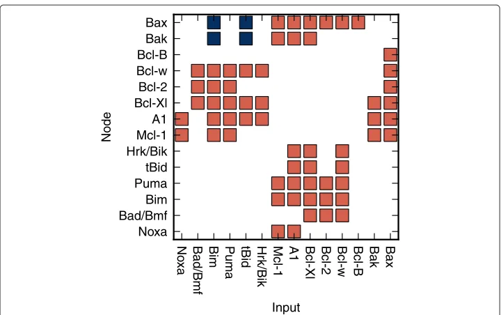
Figure 1 Relationships between the nodes of the model.The red squares represent the negative relationships of the inputs toward the related nodes. Negative relationship corresponds to mutual binding and inhibition between two members of the Bcl-2 family (see Table 1). The blue squares represent the positive relationships of the inputs toward the related nodes. Positive relation corresponds to activation of the effectors Bax/Bak by certain BH3-only proteins - activators (tBid, Bim) [40,41].
Influence of the external conditions
The transition function of thei-th node is dependent on the value ofei. The value of
binary vectorE(E= {e1,e2,. . .e16}) represents here, what we termed the “expression” of the Bcl-2 family proteins. The valueei =1, corresponds to the cellular conditions
allow-ing the synthesis and, if required, the post-translational/post-transcriptional activation (e. g. activation of Bid requires proteolytic cleavage by Caspase-8 [45]) of thei-th protein. Alternatively, the valueei = 0, corresponds to the conditions preventing the synthesis
and/or post-translations activation of thei-th protein.
Since the model contains 14 nodes, the vector of expressions can have 214 = 16384 possible values. However, since other than BH3-only mediated activation of Bax/Bak is irrelevant to our work (the subject of our study is the Bcl-2 family mediated regulation of MOMP), we exclude here the expression vectors whereeBax = 1 oreBak = 1, reducing
thus the number of possible values to 212 = 4096. The value of the vectorE remains constant during each simulation.
Terminal states
The simulation is terminated at the time t once the state of the model S (S =
{s1,s2,. . .s14}) satisfies the following condition:
S(t)=S(t−n), n=1, 2,. . . (3)
The stateS(t), satisfying the condition (3) is the model’s terminal stateS(tend). If the S(tend) involves the states of both effectors,sBax(tend) = 0 and sBak(tend) = 0, then
theS(tend)is denoted as the “survival” state. If theS(tend)involves the states of one of the
effectors, eithersBax(tend)=1, orsBak(tend)=1, then theS(tend)is simply denoted as the
“pro-MOMP” state.
Results
We have identified 1046 of the “survival” states, in which the model preserve the effectors Bak and Bax inactive
The very first step was to find the terminal states in which the model is allowed to persist without the activation of effectors (Bak and Bax) - survival states.
Therefore, for each of the 4096 expression vectors we performed 4096 simulations, each simulation starting from one of the 4096 of the initial states (4096 = 212, that is the number of possible initial states, including bothsBax(t0)=0 andsBak(t0)=0).
We have identified 1046 unique survival states. The 388 of these states are logical steady-states, remaining 678 of the survival states are oscillating. Hereafter, we assume that these 1046 states represent basal, cellular conditions. To lead the cell out from such survival state it requires the change of an expression vector which would initiate the state transition. In the next step, we have investigated the transitions from the survival states to other terminal states. To analyze these transitions, for each of the 1046 survival states, we performed 4096 simulations. In each simulation we used one of the 4096 expression vectors and one of the survival states to define the initial conditions. Thus we “exposed” individual survival states to all the expression vectors and simulated the effect of changes of cellular conditions on actual state of the Bcl-2 proteins family.
Around 70% of the 4.2 million (1046×4096) simulations led to survival, the remaining 30% of the simulations led to pro-MOMP terminal states, i.e. the states where at least one of the effectors was found active.
We found 200 of the trapping states
During the analysis of the transitions between the survival states and the pro-MOMP states, we have revealed an interesting finding. We have discovered the existence of 200 survival states from which the model cannot achieve the pro-MOMP states, regardless of the vector of expressions. Moreover, the model, once found in such a “trapping” state, can only be transitioned to another trapping state. The trapping of the Bcl-2 regulatory mech-anism in one of these states would cause fatal malfunctioning of the MOMP regulation. The cell arrested in one of such states becomes resistant to external apoptotic stimuli - a condition which is one of the hallmarks of cancer cells [46]. Therefore, we denoted these states as the “tumor” states.
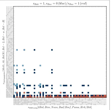
Figure 2 Model’s survival states.The figure depicts the survival states – Bax, Bak representing nodes are inactive. The squares are depicting the terminal states of the model within the “configuration space”, where the configurations of states of nodes representing the antiapoptotic and BH3-only proteins are arranged along y- and x-axis, respectively. The black squares represent the “tumor” states, while the green squares represent the functionally “semioptimal” (light green) and “optimal” (dark green) survival states. While the first mentioned allow only the activation of Bak (transitionT4in Figure 3), but not Bax, “optimal” states allow activation of both effectors.
As we found, the cell can be “liberated” from a trapping state by pharmacological inhibition of activity of antiapoptotic proteins (e.g. by competitive inhibition to prevent neutralization of proapoptotic proteins). Especially effective would be the inhibition of the Mcl-1, since the tumor states are overly abundant among the survival states involving the activity of Mcl-1.
The existence of trapping tumor states indicates that relationships in Bcl-2 family allow the establishment of the molecular populations of the Bcl-2 proteins which could be very insensitive to apoptotic signaling. The apparent relationship between the activity of the Mcl-1 and tumor states suggests that inhibition of Mcl-1 can be of special therapeutic relevance of targeting tumor cells.
There are two functionally distinct subsets of survival states. Those which allow model to activate the Bak, but not Bax and the states allowing activation of both effectors
where the only active effector is Bak. From the 792 survival states of the second group, the model can be turned to states with a single effector (Bak) activity, as well as to the pro-MOMP states, where both effectors are active. The first group we denote as “semioptimal” (the light green squares in Figure 2), the second one we denote as “optimal” (the dark green squares in Figure 2).
Similarly, we distinguish several functionally distinct subsets among the pro-MOMP states. Firstly, every pro-MOMP state may be classified according to the activity of effec-tors. We have found 108 of the pro-MOMP states in which Bak is active, but Bax remains inactive (the blue squares in Figure 3). Besides these, we also have found 132 of the pro-MOMP states, in which both effectors are active (the red squares in Figure 3). However, we haven’t found any such terminal state, where the Bax was active, while the Bak not, indicating that such state is unaccessible by the model, regardless of expressions or initial conditions.
Secondly, the first of the mentioned groups – Bak-active only, can further be divided in two functionally distinct subgroups: states which allow additional activation of Bax (the light blue squares in Figure 3), and those which don’t (the dark blue squares in Figure 3).
It is very interesting that, while the first subgroup is accessible only from the “optimal” survival states, the second one, can be accessed from both, “optimal” and “semioptimal” survival states.
Survival to MOMP transition is irreversible
We have performed another series of simulations in which each of the pro-MOMP states was used as the initial state of the model and each of the 4096 expression vectors were iteratively applied to the model. We have found that it is impossible to turn the model from any of the pro-MOMP states back to the survival one, regardless of the expression vectors.
Irreversibility of the transition to pro-MOMP states originates in activity of effectors itself. Mutual inhibitory relationships between effectors and corresponding antiapoptotic proteins compensate the influence of the expression vectors on a given antiapoptotic pro-tein. According to the rule described in eq. 1, in such a case inactive antiapoptotic protein remains inactive and inhibition of effectors remains insufficient to suppress their activity, regardless of the expression vector.
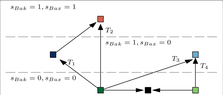
The transitions from the survival to MOMP are caused by expression changes of four distinct types
When taking previous results together, we may distinguish six distinct groups of model’s terminal states. Three of these groups associate survival states, characterized by no effec-tors activity. Two groups associate pro-MOMP states, involving the activity of Bak, but not Bax. The sixth group associates the states involving the activity of both effectors. Assuming that the single effector activation is sufficient for the MOMP occurrence, four types of survival-to-MOMP transitions can be distinguished (T1–T4, see Figure 4).
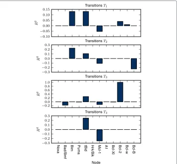
We have analyzed the influence of the expression of the given protein on the initiation of these transitions by means of multiple correlation coefficient -R2(for more details see Appendix).
Figure 5 shows that regardless of the transition type, the necessary condition for the transition to MOMP is the expression of at least one of the activators (Bim, tBid). On the other hand it seems that the expression of the Mcl-1 is the most significant factor
Figure 5 Multiple determination coefficients.R2of the protein expressions calculated across the sets of unique expressions vectors causing the survival-to-MOMP transitions of given type (for more details see Appendix).
that prevents the MOMP. The statistical importance of the expression of activators and the absence of Mcl-1 is common for bothT1andT2transitions. Nevertheless, the tran-sitions of typeT2- the activation of both effectors, additionally requires the absence of Bcl-B expression. This finding is not unexpected as the Bcl-B is the only inhibitor of Bax that is not suppressed by any of the BH3-only proteins [44]. Furthermore, the model pre-dicts that after theT1transition, the subsequent downregulation of Bcl-B can cause the additional activation of Bax (the arrow pointing from the dark blue square to the red one, Figure 4).
Transitions of the typeT3are similar to theT1andT2. The only difference is that tran-sitions of the typeT3 occurs only in presence of the Bcl-2 expression. In other words, expression of the antiapoptotic protein Bcl-2 prevents the transitions of typeT3andT4– transitions from optimal survival states to “dark blue” and “red” pro-MOMP states. If model is located in one of the “light blue” states, subsequent down-regulation of Bcl-2 would allow its relocation to “dark blue” states (not depicted in Figure 4), but not to “red” states.
We have found (data not shown) that the trapping of the model within the “trap-ping” state occurs as the antiapoptotic expression disproportionately dominates over the expression of BH3-only proteins. This points to the necessity of the balance between the presence and synthesis of the both pro- and antiapoptotic Bcl-2 proteins within the cell.
Discussion
We have analyzed the Bcl-2 family interaction network using the Boolean network-based computational model. Bcl-2 family members have been represented by nodes, with binary encoded activity. The active (ON state) of the given node represents the biologically active form of the represented protein. Nodes are mutually interacting according to the given transition rules and the pre-defined relationship matrix, represent-ing the currently known interactions among the Bcl-2 proteins. In addition, the model operates under the influence of the vector of expressions that represent the biological conditions. Change of vector of expressions is the primary driving force of the Bcl-2 regulatory mechanism. The expression vector allows introduction of synthesis and the post-translational/-transcriptional activation of zymogens, if relevant for given protein.
Computational simulations of the model show that the majority of the expression vec-tors lead the Bcl-2 family into set of states avoiding Bax and/or Bak activation. Our results suggest, that once the antiapoptotic proteins significantly outnumber the BH3-only pro-teins’ activity, the Bcl-2 family regulation may be seriously disrupted. Our model predicts, that once this happens, even the subsequent activation of proapoptotic BH3-only pro-teins cannot recover the proper MOMP. We have noted that the defects of MOMP regulation are often associated with presence of Mcl-1 activity. However, the existence of such “tumor facilitating” trap shows the importance of the balance between pro- and antiapoptotic proteins maintained by their continuous expression.
Depending on the current state of the Bcl-2 family, certain configurations of the incoming signals cause the activation of effectors Bak, and/or Bax. Statistically, the most important signals are the truncation of Bid to tBid, the activation of Bim and the downregulation of Mcl-1.
The ability of tBid to initiate apoptosis through MOMP has been well documented by numerous works [45,47]. Similarly, extensive experimental support exists, proving that activation of Bim [48-50] and downregulation of Mcl-1 [51,52] lead to apoptosis in cells.
It seems that the BH3-only mediated activation of Bak requires less specific conditions, compared to the activation of Bax. This finding is well supported by several experimental works, suggesting that MOMP initiated by BH3-only proteins occurs mainly through the activation of Bak, not Bax [53-56]. According to the model, the activation of Bax is asso-ciated with the downregulation of Bcl-w. Finally, our results confirm the irreversibility of the effectors activation, which has been previously experimentally shown [57-59].
In spite of the simplicity of the Boolean-based approach the model provides remarkable predictive and explanatory power. Moreover, the proposed model can be reutilized for further analyses of robustness and stability of Bcl-2 family regulation of apoptosis.
Appendix: A calculation of multiple determination coefficients
Let’s have set ofnunique expression vectors –E= {e1,e2,. . .e12}that cause the studied transition. For each couple of nodesi,jwe can calculate the phi coefficient:
φij=
n11n00−n10n01
√
n1•n0•n•0n•1
, (4)
wheren00,n01,n10,n11, are counts of the following combinations of valuesei,ejacross
the set of expression vectors:
ei=1 ei=0 total
ej=1 n11 n10 n1• ej=0 n01 n00 n0•
total n•1 n•0 n
The phi coefficient is a measure of association for two binary variables, similar to Pearson correlation coefficient [60].
The matrix of phi coefficients -Rφ, is then used as to calculate the coefficient of multiple determination –R2:
Ri2=cTiRφ−,1ici, (5)
where the ci is the vector of values φij, j = 1, 2. . .12,i = j.ci is actually the vector
of correlations between the independent variables and the target variable –ei.cTi is the
transpose ofc. TheRφ,iis the matrixRφ, reduced by removing thei-th line andi-th
col-umn.Rφ,i is actually the matrix of correlations between the independent variables and R−φ,1i is the inverse of the matrixRφ,i.
Finally, theR2i was multiplied by−1 if the count ofei=0 appearances was greater than
the count ofei = 1, across the set of expressions – the expression of thei-th node was
mostly absent among the expression vectors causing the given transition.
In case the values ofi−thprotein expression had no variability – eitherei = 0, or ei=1 among all the expression vectors, theR2i was arbitrary set either to−1, or 1,
respec-tively. The correlations of any of the other expression with theeiwere then excluded from
any other calculations. Such situation occurs in case of expression of Bcl-2 among the transitions of the typeT3(see Figure 4).
Competing interests
The authors declare that they have no competing interests.
Authors’ contributions
TT proposed the Boolean networks-based model of Bcl-2 family, implemented the model within the Python environment and performed most of the simulations. Furthermore, TT processed and analyzed the obtained data, prepared all the illustrations and major part of the manuscript. ZT participated on the implementation of the model and its simulations. Furthermore, ZT contributed to this study by critical revision of the manuscript. JU substantively contributed to this work, by extensive revision of the manuscript. Furthermore, JU gave the final approval of the version to be published. In addition, all the authors have contributed to this work, by numerous valuable ideas and proposals. All authors read and approved the final manuscript.
Acknowledgements
This work was funded by Slovak Research and Development Agency, grant no. APVV-0242-11, from the project SEPO-II, grant no. ITMS 26220120039, FP7 EU project CELIM 316310 and from the Scientific Grant Agency of the Ministry of Education of the Slovak Republic, grant no. VEGA-1/4019/07. Authors strongly appreciate this support.
References
1. Ulukava E, Acilan C, Yilmaz Y:Apoptosis: why and how does it occur in biology?Cell Biochem Funct2011,
29:468–80.
2. Wyllie AH:“Where, o death, is thy sting?” A brief review of apoptosis biology.Mol Neurobiol2010,42:4–9. 3. Elmore S:Apoptosis: a review of programmed cell death.Toxicol Pathol2007,35:495–516.
4. Mattson MP:Neuronal life-and-death signaling, apoptosis, and neurodegenerative disorders.Antioxid Redox Signal2006,8:1997–2006.
5. Nagata S:Apoptosis and autoimmune diseases.Ann N Y Acad Sci2010,1209:10–16.
6. Burz C, Berindan-Neagoe I, Balacescu O, Irimie A:Apoptosis in cancer: Key molecular signaling pathways and therapy targets.Acta Oncol2009,48:811–821.
7. Fulda S:Tumor resistance to apoptosis.Int J Cancer2009,124:511–515.
8. Strasser A, Cory S, Adams JM:Deciphering the rules of programmed cell death to improve therapy of cancer and other diseases.EMBO J2011,30:3667–3683.
9. Strasser A, O’Connor L, Dixit VM:Apoptosis signaling.Annu Rev Biochem2000,69:217–245. 10. Danial NN, Korsmeyer SJ:Cell death: critical control points.Cell2004,116:205–219.
11. Chipuk JE, Green DR:How do BCL-2 proteins induce mitochondrial outer membrane permeabilization?
Trends Cell Biol2008,18:157–164.
12. Tait SWG, Green DR:Mitochondria and cell death: outer membrane permeabilization and beyond.Nat Rev Mol Cell Biol2010,11:621–632.
13. Landes T, Martinou JC:Mitochondrial outer membrane permeabilization during apoptosis: the role of mitochondrial fission.Biochim Biophys Acta2011,1813:540–545.
14. Mace PD, Riedl SJ:Molecular cell death platforms and assemblies.Curr Opin Cell Biol2010,22:828–836. 15. Perez-Paya E, Orzaez M, Mondragon L, Wolan D, Wells JA, Messequer A, Vincent MJ:Molecules that modulate
Apaf-1 activity.Med Res Rev2011,31:649–675.
16. Kulikov AV, Shilov ES, Mufazalov IA, Gogvadze V, Nedospasov SA, Zhivotinsky B:Cytochrome c: the Achilles’ heel in apoptosis.Cell Mol Life Sci2012,69:1787–1797.
17. Wurstle ML, Laussmann MA, Rehm M:The central role of initiator caspase-9 in apoptosis signal transduction and the regulation of its activation and activity on the apoptosome.Exp Cell Res2012,318:1213–1220. 18. Martinez-Ruiz G, Maldonado V, Caballos-Cancino G, Grajeda JP, Melendez-Zajgla J:Role of Smac/DIABLO in cancer
progression.J Exp Clin Cancer Res2008,27:48.
19. Olsson M, Zhivotinsky B:Capases and cancer.Cell Death Differ2011,18:1441–1449.
20. Green DR, Amarate-Mendes GB:The point of no return: mitochondria, caspases, and the commitment to cell death.Results Probl Cell Differ1999,24:45–61.
21. Wallach D, Kang TB, Kovalenko A:The extrinsic cell death pathway and the elan mortel.Cell Death Differ2008,
15:1533–1541.
22. Estaquier J, Vallette F, Vayssiere JL, Mignotte B:The mitochondrial pathways of apoptosis.Adv Exp Med Biol2012,
942:157–183.
23. Chen C, Cui J, Zhang W, Shen P:Robustness analysis identifies the plausible model of the Bcl-2 apoptotic switch.FEBS Lett2007,581:5143–5150.
24. Cui J, Chen C, Lu H, Sun T, Shen P:Two independent positive feedbacks and bistability in the Bcl-2 apoptotic switch.PLoS ONE2008,3:1469.
25. Sun T, Lin X, Wei Y, Xu Y, Shen P:Evaluating bistability of Bax activation switch.FEBS Lett2010,584:954–960. 26. Tokar T, Ulicny J:Computational study of Bcl-2 apoptotic switch.Physica A2012,391:6212–6225.
27. Bagci EZ, Vodovotz Y, Billiar TR, Ermentrout GB, Bahar I:Bistability in Apoptosis: Roles of Bax, Bcl-2, and Mitochondrial Permeability Transition Pores.Biophys J2006,90:1546–1559.
28. Abeck JG, Burke JM, Aldridge BB, Zhang M, Lauffenburger DA, Sorger PK:Quantitative analysis of pathways controlling extrinsic apoptosis in single cells.Mole Cell2008,30:11–25.
29. Harrington H, Lo KL, Ghosh S, Tung K:Construction and analysis of a modular model of caspase activation in apoptosis.Theor Biol Med Model2008,90:1546–1559.
30. Schlatter R, Schmich K, Avalos Vizcarra I, Scheurich P, Sauter T, Borner C, Ederer M, Merfort I, Sawodny O:ON/OFF and beyond–a boolean model of apoptosis.PLoS Comput Biol2009,5:e1000595.
31. Chen L, Willis SN, Wei A, Smith BJ, Fletcher JI, Hinds MG, Colman PM, Day CL, Adams JM, Huang DCS:Differential targeting of prosurvival Bcl-2 proteins by their BH3-Only ligands allows complementary apoptotic function.Molecular Cell2005,17:393–403.
32. Dai H, Meng XW, Lee SH, Schneider PA, Kaufmann SH:Context-dependent Bcl-2/Bak interactions regulate lymphoid cell apoptosis.J Biol Chem2009,284:18311–18322.
33. Düssmann H, Rehm M, Concannon CG, Anguissola S, Würstle M, Kacmar S, Völler P, Huber HJ, Prehn JHM:
Single-cell quantification of Bax activation and mathematical modelling suggest pore formation on minimal mitochondrial Bax accumulation.Cell Death Diff2009,17:278–290.
34. Lindner AU, Concannon CG, Boukes GJ, Cannon MD, Llambi F, Ryan D, Boland K, Kehoe J, McNamara DA, Murray F:
Systems analysis of BCL2 protein family interactions establishes a model to predict responses to chemotherapy.Cancer Res2013,73:519–528.
35. Mai Z, Liu H:Boolean network-based analysis of the apoptosis network: Irreversible apoptosis and stable surviving.J Theor Biol2009,259:760–769.
36. Kauffman SA:Metabolic stability and epigenesis in randomly constructed genetic nets.J Theor Biol1969,
22:437–467.
37. Helikar T, Kochi N, Konvalina J, Rogers JA:Boolean modeling of biochemical networks.Open Bioinform J2011,
5:16–25.
39. Elkholi R, Floros KV, Chipuk JE:The role of BH3-Only proteins in tumor cell development, signaling, and treatment.Gen Cancer2011,2:523–537.
40. Chipuk JE, Moldoveanu T, Llambi F, Parsons MJ, gREEN DR:The Bcl-2 Family Reunion.Mol Cell2010,37:299–310. 41. Placzek WJ, Wei J, Kitada S, Zhai D, Reed JC, Pellecchia M:A survey of the anti-apoptotic Bcl-2 subfamily
expression in cancer types provides a platform to predict the efficacy of Bcl-2 antagonists in cancer therapy.Cell Death Dis2010,6:e40.
42. Westhpal D, Dewson G, Czabotar PE, Kluck RM:Molecular biology of Bax and Bak activation and action.
Biochimica Biophysica Acta2011,4:521–531.
43. Dejean LM, Ryu SY, Martinez-Caballero S, Teijido O, Peixoto PM, Kinnally KW:MAC and Bcl-2 family proteins conspire in a deadly plot.Biochimica Biophysica Acta2010,1797:1231–1238.
44. Rautureau GJP, Day CL, Hinds MG:The structure of Boo/Diva reveals a divergent Bcl-2 protein.Proteins2010,
78:2181–2186.
45. Kantari C, Walczak H:Caspase-8 and bid: caught in the act between death receptors and mitochondria.
Biochimica Biophysica Acta2011,4:558–563.
46. Hanahan D, Weinberg RA:The hallmarks of cancer.Cell2000,100:57–70.
47. Rehm M, Huber HJ, Hellwig CT, Anguissola S, Dussmann H, Prehn JHM:Dynamics of outer mitochondrial membrane permeabilization during apoptosis.Cell Death Diff2009,16:613–623.
48. O’Connor L, Strasser A, O’Reilly LA, Hausmann G, Adams JM, Cory S, Huang DCS:Bim: a novel member of the Bcl-2 family that promotes apoptosis.EMBO J1998,17:384–395.
49. Mendez G, Policarpi C, Cenciarelli C, Tanzarella C, Antoccia A:Role of Bim in apoptosis induced in H460 lung tumor cells by the spindle poison Combretastatin-A4.Apoptosis2011,16:940–949.
50. Faber AC, Ebi H, Costa C, Engelman JA:Apoptosis in targeted therapy responses: the role of BIM.Adv Pharmacol
2012,65:519.
51. Shore GC, Warr MR:Unique biology of Mcl-1: therapeutic opportunities in cancer.Curr Mol Med2008,8:138–147. 52. Polier G, Ding J, Konkimalla BV, Eick D, Ribeiro N, Köhler R, Giaisi M, Efferth T, Desaubry L, Krammer PH:Wogonin and
related natural flavones are inhibitors of CDK9 that induce apoptosis in cancer cells by transcriptional suppression of Mcl-1.Cell Death Dis2011,2:e182.
53. Carton PF, Juin P, Oliver L, Martin S, Meflah K, Vallette FM:Nonredundant role of Bax and Bak in bid-mediated Apoptosis.Mol Cell Biol2003,23:4701–4712.
54. Lindenboim L, Kringel S, Braun T, Borner C, Stein R:Bak but not Bax is essential for Bcl-xS-induced apoptosis.
Cell Death Diff2005,12:713–723.
55. Zhang W, Wang X, Chen T:Resveratrol induces apoptosis via a Bak-mediated intrinsic pathway in human lung adenocarcinoma cells.Cell Signal2012,24:1037–1046.
56. Zhou C, Pan W, Wang XP, Chen TS:Artesunate induces apoptosis via a Bak-mediated caspase-independent intrinsic pathway in human lung adenocarcinoma cells.J Cell Phys2012,227:3778–3786.
57. Madesh M, Antonsson B, Srinivasula SM, Alnemri ES, Hajnoczky G:Rapid kinetics of tBid-induced Cytochrome c and Smac/DIABLO release and Mitochondrial depolarization.J Biol Chem2002,277:5651–5659.
58. Rehm M, Dussmann H, Prehn JHM:Real-time single cell analysis of Smac/DIABLO release during apoptosis.
J Cell Biol2003,162:1031–1042.
59. Hellwig CT, Kohler BF, Lehtivarjo AK, Dussmann H, Courtney MJ, Prehn JHM, Rehm M:Real time analysis of tumor necrosis factor-related Apoptosis-inducing Ligand/Cycloheximide-induced Caspase activities during Apoptosis initiation.J Biol Chem2008,283:21676–21685.
60. Carroll J:The nature of the data, or how to choose a correlation coefficient.Psychometrika1961,26:347–372.
doi:10.1186/1742-4682-10-40
Cite this article as:Tokaret al.:Boolean network-based model of the Bcl-2 family mediated MOMP regulation. Theoretical Biology and Medical Modelling201310:40.
Submit your next manuscript to BioMed Central and take full advantage of:
• Convenient online submission
• Thorough peer review
• No space constraints or color figure charges
• Immediate publication on acceptance
• Inclusion in PubMed, CAS, Scopus and Google Scholar
• Research which is freely available for redistribution