Cover Page
The handle
http://hdl.handle.net/1887/20616
holds various files of this Leiden University
dissertation.
Author
: Lichtenbelt, Bart Jan
Title
:
PK-PD modelling of the interaction of propofol and midazolam : implementation
and future perspectives
PK-PD modelling of the interaction of
Propofol and Midazolam
Implementation and future perspectives
PK-PD modelling of the Interaction of Propofol and Midazolam
Implementation and Future Perspectives
Proefschrift
ter verkrijging van
de graad van Doctor aan de Universiteit Leiden, op gezag van Rector Magnificus prof.mr. C.J.J.M. Stolker
volgens besluit van het College voor Promoties ter verdediging op donderdag 14 maart 2013
klokke 15.00 uur
door
Bart Jan Lichtenbelt
Promotiecommissie:
Promotores: Prof. Dr. A. Dahan
Prof. Dr. M.M.R.F. Struys (UMCG)
Co-Promotor: Dr. J. Vuyk
Overige Leden: Prof. Dr. L. Aarts Prof. Dr. J. Bovill
Prof. Dr. A. Absalom (UMCG)
Prof. Dr. T.W.L. Scheeren (UMCG)
CONTENT
1. Introduction
2. Strategies to Optimize Propofol-Opioid Anesthesia
Clinical Pharmacokinetics 2004; 43(9): 577-593
3. Propofol reduces the ditribution and clearance of midazolam
Anesthesia and Analgesia 2010 june 1; 110(6): 1597-606
4. Low Bispectral Index values in awake volunteers receiving a combination of Propofol
and Midazolam
Anesthesiology 2004 jan; 100 (1); 179-81
5. Propofol Reduces the Distribution and Clearance of Midazolam
Anesthesia and Analgesia 2009 may; 108(5): 1522-30
6. Response surface modeling of the propofol-midazolam interaction to define the optimal concentration combination that assures unconsciousness and hemodynamic stability.
Submitted
7. Optimizing Intravenous drug administration by applying pharmacokinetic / pharmacodynamic concepts
Br J Anaesth. 2011 Jul;107(1):38-47.
8. Summary and Conclusions
9. Samenvatting en Conclusies
10. Curriculum Vitae
Introduction
The Basic Equation; Pharmacokinetics, Pharmacodynamics and Training.
Basic research in anesthesia has given anaesthesiologist a new window of opportunity to further improve management of patients. The introduction of Target Controlled Infusion (TCI) has proven to be one of the key elements with which can (visually) help the anaesthesiologist understand the action-reaction chain of events when inducing and maintaining anesthesia. With increasing possibilities for the anaesthesiologist to incorporate pharmacokinetic principles in the operating theatre, one must seek new possibilities to incorporate all of this information into a practical tool. 1
Even though basic research is not in the spotlights, it is essential for every day practice 2. Knowledge of pharmacokinetic parameters of different drugs will give the anaesthesiologist the advantage of knowing how and when to differ in strategy. In current practice, intravenous drugs are commonly administered using standard dosing guidelines, an approach which ignores inter- and intra-individual variability in the dose-response relation. It has been proven that incorporating pharmacokinetic-dynamic information as an additional input to guide clinical anaesthesia can result in better patient care.3
This research will partially open the so-called “black box”. Understanding interactions between different drugs is the fundament for patient safety.
In this thesis we have studied hypnotic-hypnotic interaction. Medication given to patients to decreases preoperative stress interacts with induction of anesthesia, and although this is taken into consideration when inducing anesthesia, the interaction has never been studied. Knowledge of this interaction helps to redefine the induction dose needed for a safe and efficient induction with the least hemodynamic changes.
TCI can further improve safety for a wide range of patients. Incorporating Bispectral indexing (BIS) completes this equation. Not only a comprehensive understanding of the pharmacokinetic, but also the dynamic principles, of intravenous medication is needed to understand the hemodynamic alterations during induction and maintenance of anesthesia. BIS has developed into a basic tool in the operating theatre, which can visually help guide the anaesthesiologist in dosing the necessary medication needed for the operation, although a low BIS value itself is not a full guarantee for a deep anaesthesia 4
In chapter 1 we describe the optimization of opioid drug combinations. For every combination an optimal dose has been described and a TCI model has been developed. In this review we also give a comprehensive overview of the use of BIS monitoring.
In chapter 2 we have studied the influence of midazolam on the pharmacokinetics of propofol. Volunteers were studied in a randomized crossover manner during two separate sessions with a minimum of two weeks between the two sessions. During the first session they were given a bolus of propofol of 1 mg.kg-1, and an infusion of 2 mg.kg-1, for 59 minutes. In the second session we have given midazolam with a target controlled infusion to reach a steady state and the same dosing scheme of propofol. Blood samples were drawn to measure midazolam and propofol levels. Additional hemodynamic measurements were recorded using the LiDCO® non invasive hemodynamic monitor and stored for later use. With these data we constructed a model for the pharmacokinetic influence of midazolam on propofol, with the use of additional parameters.
In Chapter 3 we describe three case reports from our study. Volunteers were deeply sedated, resulting in low BIS values but were able to answer simple questions. Hypnotic – hypnotic interaction without the use of muscle relaxants can cause this phenomenon.
In Chapter 4 we have studied the influence of propofol on the pharmacokinetics of midazolam. Volunteers were studied in a randomized crossover manner during two separate sessions with a minimum of two weeks between the two sessions In one session they were given midazolam in a bolus and a continuous infusion. In the second sessions they received an additional infusion with propofol guided by TCI. Blood samples were drawn to measure midazolam and propofol levels. Additional hemodynamic measurements were recorded using the LiDCO® non-invasive monitor and stored for later use. With these data we constructed a model for the pharmacokinetic influence of propofol on midazolam with the use of additional parameters.
References
1. Sahinovic MM, Absalom AR, Struys MM: Administration and monitoring of intravenous anesthetics. Curr Opin Anaesthesiol 2010; 23: 734-40 2. Gin T: Clinical pharmacology on display. Anesth Analg 2010; 111: 256-8
3. Struys MM, De Smet T, Mortier EP: Simulated drug administration: an emerging tool for teaching clinical pharmacology during anesthesiology training. Clin Pharmacol Ther 2008; 84: 170-4
4. Vuyk J, Lichtenbelt BJ, Vieveen J, Dahan A, Engbers FHM, Burm AGL: Low
Strategies to Optimise Propofol-Opioid Anaesthesia
Bart Jan Lichtenbelt MD, Martijn Mertens MD.PhD., Jaap Vuyk MD.PhD
Introduction
Anaesthesia facilitates a wide variety of surgical procedures. Patients generally receive a combination of anaesthetic and analgesic agents to induce and maintain an adequate depth of anaesthesia and analgesia. In addition to anaesthesia and analgesia, muscle relaxation is provided using muscle relaxants, facilitating the surgical procedure. Next to the positive effects of anaesthetic agents in maintaining unconsciousness, analgesia and muscle relaxation, these agents potentially compromise the autonomic stability of the patient. Thorough knowledge of the pharmacokinetics and pharmacodynamics of these agents enables the anaesthesiologist to administer a combination that offers the most stable anaesthetic with the shortest possible induction and recovery times and optimal operating conditions with the least incidence of adverse effects.
In contrast to the past practice of administering anaesthesia on the basis of knowledge of the needs of the population, modern anaesthesia focuses on the individual needs of the patient. To focus the administration of intravenous anaesthetics on the individual needs of the patient, the anaesthesiologist has three strategic tools.
The first and most important tool is the pharmacological knowledge that has been gathered over the past 20-30 years. From this body of knowledge, the anaesthesiologist may take data that allows him or her to adjust the administration of the various anaesthetic agents to the specific need of the individual patient. In this way, each individual patient may experience rapid induction, stable maintenance and rapid recovery from anaesthesia without serious adverse effects.
The second tool to optimize intravenous anaesthesia is the application of state-of-the-art intravenous drug administration techniques. Until recently, intravenous anaesthetic agents were administered either as a bolus doses or by manually controlled infusion pumps, but now target-controlled infusion is the state of the art and is increasingly gaining interest from the clinical anaesthesiologist. Target controlled infusion offers significant advantages over conventional administration methods for intravenous agents and thereby allows for further optimization and individualization of intravenous anaesthesia.
monitoring (BIS). The bispectral index, a mathematical derivative of the electroencephalogram (EEG), closely correlates with the state of the unconsciousness and the concentration of various anaesthetic agents. As such, it may be used to guide the administration of intravenous agents and may thus lead to a more controlled anaesthesia that again is better tailored to the individual needs of the patient.
1. Pharmacokinetic-Pharmacodynamic Knowledge
In everyday clinical practice, anaesthesiologists are faced with dose-effect relationships of both opioids and intravenous anaesthetic agents that exhibit a wide interindividual variability. This interindividual dose-effect variability of anaesthetic agents is caused by both pharmacokinetic and pharmacodynamic differences between patients. The pharmacokinetic variability is in the order op 70%. With a propofol infusion rate of 10 mg/kg/h, blood propofol concentrations may vary between patients between 3 and 5 mg/L. Differences in cardiac output, hepatic perfusion, protein binding and enzyme activity are responsible for these interindividual pharmacokinetic differences.(1-6)
The pharmacodynamic variability is much larger, in the order of 300-400%. During induction of anaesthesia with a target-controlled infusion of propofol, some patients already lose consciousness at a target of 1mg/L, whereas others need 4-5 mg/L to experience the same effect. Factors that are responsible for this huge pharmacodynamic interindividual variability still remain obscure, but genetic differences in receptor pharmacology may play an important role. (4)
Next to the pharmacokinetic and pharmacodynamic variability of single agents, the administration of two or more agents together gives rise to pharmacokinetic and pharmacodynamic interactions. Anaesthesiologists combine anaesthetic agents on a daily basis because the provision of anaesthesia on the basis of a single agent is associated with significant adverse effects compromising hemodynamic and/ or respiratory function, affecting operating conditions, and/or postponing postoperative recovery. Because of the small therapeutic window, a detailed characterization of anaesthetic agents and their interactions is required to allow a proper selection of the various intravenous agents and their combinations, and to obtain an optimal therapeutic pharmacological effect in the absence of significant adverse effects.
1.1 Pharmacology of propofol
Propofol, a lipophilic agent, has a fast onset and short duration of action due to a rapid penetration through the blood-brain barrier and distribution to and from the CNS followed by redistribution to inactive tissue depots such as muscle and fat. (9) Propofol pharmacokinetics are best described on the basis of a three compartment model (table I). The short effect-site equilibration half-life and the small central compartment are responsible for its time peak effect of only two minutes. The larger volumes of distribution, combined with a clearance that equals hepatic perfusion, are associated with a context sensitive half-time that only increases from about 20 to about 30 minutes with infusion durations increasing from 2 to 8 hours. Consequently, propofol is very well suited for continuous infusion techniques. Its high clearance and redistribution, even after prolonged infusion, allow for a rapid return to consciousness even after many hours of anaesthesia. Propofol as a single agent for anaesthesia, without opioid pre-treatment, causes loss of consciousness in 50% of the patients (EC50) at a blood concentration of 3.4 mg/L. Propofol may be used as a
monoanaesthetic agent during surgery. Then blood concentrations in excess of 10-12 mg/L are required to suppress responses evoked by surgical stimulation. (10-12)
Propofol dosage schemes should be adjusted for age and sex. Schnider et al. (13) described the relation ship between dose, age and blood concentrations for loss of consciousness in healthy non –premedicated volunteers. In this study, the EC50 for loss of consciousness was
2.4, 1.8 and 1.3 mg/L in volunteers aged 25, 50 and 75 years, respectively(13). Children require a higher induction dose as result of a larger central compartment, (14) whereas elderly patients require a lower induction dose as a result of smaller central compartment and a reduced clearance. (15,16) As well as the relatively larger central compartment in children, the clearance is increased to a lesser extent. The application of target-controlled infusions of propofol in children using adult pharmacokinetic parameter sets will therefore cause a divergence of the blood concentration from the desired target concentration. Elderly female patients need a higher dosage of propofol compared with males because of a higher clearance rate. (17)
Table I. Pharmacokinetic and pharmacodynamic parameters of propofol and the opioids. Various pharmacokinetic parameter sets are available
in the literature for all of these agents, but population pharmacokinetic data are available only for propofol, remifentanil and alfentanil. These
population pharmacokinetic parameter sets may therefore be best applicable in a population that varies greatly in age, weight and gender.
Parameter and unit Propofol(20)a Fentanyl(21) Remifentanil(22) Alfentanil(23) Sufentanil(24)
V1 (L)
V2 (L)
V3 (L)
CL1 (L/min)
CL2 (L/min)
CL3 (L/min)
t½,keO (min) EC50 (µg/L)
4.27 24.0 238 0.68 1.60 0.836 2.40 3400b 8.9 50.3 295.5 0.63 4.83 2.23 4.70 1.1c 4.98 9.01 6.54 2.46 1.69 0.065 0.90 4.7c 8.9 13.8 12.1 0.36 0.93 0.15 1.10 90c 14.3 63.1 261.6 0.92 1.55 0.33 5.87 0.14c
a Model estimation for patient 40 years, 180 cm and 80 kg.
b For loss of consciousness
c Optimal EC50 in the presence of propofol
Cl1 = elimination clearance; Cl2 = rapid distribution clearance; Cl3 = slow distribution clearance; EC50 = 50% effective concentration for loss of
consciousness (propofol) or adequate analgesia (opioids); t½½½½keO = effect site equilibration half-time; V1 = volume of central compartment;
Propofol inhibits CYP 2A1 (phenacetin O-de-ethylation), CYP2C9 (tolbutamide 4’-hydroxylation), CYP2D6 (dextromethorphan O-demethylathion) and CYP3A4 (testosterone 6β-hydroxylation) activities with 50% inhibitory concentrations (IC50) of 40, 49, 213 and 32
µmol/L, respectively. (25)
Propofol induces a marked loss of sympathetic tone in healthy volunteers. Cardiac and sympathetic baroslopes are significantly reduced with propofol, especially in response to hypotension, suggesting that propofol induced hypotension may be mediated by an inhibition of the sympathetic nervous system and impairment of baroreflex regulatory mechanisms. (26)
Loss of vascular tone in arteries, as a result of a reduced Ca2+ influx, may also contribute to the hypotension following induction with propofol. (27) Reduction of cardiac muscle contraction is a result of reduced free systolic Ca2+ concentration in myocardial cells (28) resulting in a negative inotropic state of the cardiac muscle by propofol. Especially in elderly patients, this may contribute to propofol induced hypotension, giving rise to the need for adjusted induction schemes for propofol in the elderly. Propofol, even at low doses, depresses the ventilatory response to acute hypoxic incidents. The depression of the acute hypoxic response results from an exclusive effect within the central chemoreflex loop at the central chemoreceptor.
(29,30)
1.2. Pharmacology of Opioids
The pharmacology of the four most commonly used opioids, fentanyl, alfentanil, remifentanil and sufentanil, has been studied extensively. The opioids differ in their pharmacokinetics but, by acting at similar receptor sites, exhibit comparable pharmacodynamics. Table I gives an overview of representative pharmacokinetic parameters of the four opioids. The effect site equilibration half time (t½,keO) is fastest for alfentanil and remifentanil. (Table I) The context
sensitive half time of the four opioids gives an indication of the suitability of these agents to be given by prolonged infusion.
Remifentanil has the most rapid pharmacokinetics of the four opioids. It has the shortest time to peak effect as a result of its small central compartment and short t½,keO. As a result of its
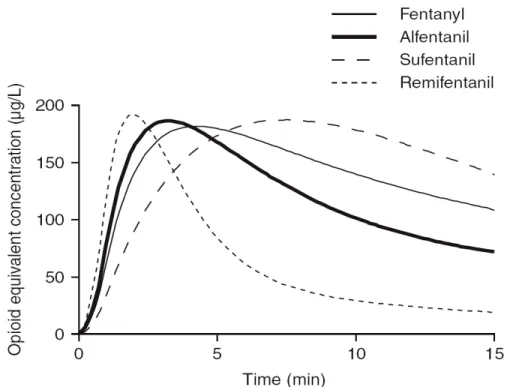
high rate of clearance of tissue esterases, remifentanil the shortest context-sensitive half-time of only a few minutes even after continuous infusion for many hours or days. The measured context-sensitive half-time of remifentanil after a 3 hour infusion was 3 minutes, with an offset of respiratory depressant effect of about 5 minutes, whereas the measured context-sensitive half-time of alfentanil was 47 minutes with an offset of about 54 minutes. (31) Increasing the infusion duration hardly increases the time to a 50% reduction in the blood remifentanil concentration after termination of the infusion. This is caused by the fact that remifentanil reaches steady state very rapidly and thus becomes context-insensitive. Alfentanil an sufentanil become context-insensitive after a few hours of infusion (figure 1), whereas in the clinical situation fentanyl does not reach this state. Consequently, remifentanil is generally administered by continuous infusion.
Figure 1: Context-sensitive Half-times (CSHT; the time required after termination of an infusion for the blood concentration to drop by 50%) for the opioids fentanyl, alfentanil, sufentanil and remifentanil.
Age and lean body mass significantly influence opioid distribution and clearance. With increasing age from 20 to 80 years, t½keO increases by approximately 50%; effect site
equilibration is thus considerable slower in the elderly. (35) Lean body mass is also a significant covariate in the distribution of remifentanil. In both young and elderly obese patients, remifentanil dosage should be based on lean body mass rather than total body mass. (22,36)
1.3 Pharmacokinetic interactions between Propofol and Opioids
The first suggestion of pharmacokinetic interactions between propofol and the various opioids go back to 1993 when Schütller and Ihmsen (16), revealed, on the basis of a mixed
effects modelling population pharmacokinetic analysis, that fentanyl and alfentanil both decreased the volume of the central compartment and the clearance of propofol. More recently, Pavlin et al. (38) showed that in the presence of alfentanil at plasma concentrations of 40 µg/L, with patients still breathing spontaneously, blood propofol concentrations were increased by 20%. Furthermore, Matot et al. (39) showed that the first pass pulmonary uptake reduced from 60-40% after pretreatment with fentanyl. A reduced first-pass uptake of propofol may indeed increase the initial blood propofol concentration after bolus dose administration.
Conversely, both Gepts et al. (40) and Pavlin et al.(38) reported increased alfentanil concentrations in the presence of propofol. This may be the result of inhibition by propofol of the oxidative metabolism of alfentanil by CYP, which so far has only been described in vitro.
(41,42)
Also, sufentanil metabolism appears to be inhibited in the presence of propofol. Other sedative agents that interfere with the metabolism of opioids are midazolam and dexmedetomidine, which have been shown to inhibit the metabolism of alfentanil and eltanolone (pregnanolone). (41,43)
Recently, two pure pharmacokinetic interaction studies have shed more light on interactions between propofol and opioids. In the presence of a constant blood propofol concentration of 1.5 mg/L, the pharmacokinetics of alfentanil were significantly altered.(44) Propofol increased mean plasma alfentanil concentrations by approximately 15%. Propofol decreased the elimination clearance (Cl1) of alfentanil by 15%, rapid distribution clearance (CL2) by 68%,
slow distribution clearance (CL3) by 51% and lag-time by 62%. Mean arterial pressure and
systemic vascular resistance were significantly lower in the presence of propofol, suggesting that the hemodynamic changes induced by propofol may be the cause of the pharmacokinetic interaction. This pharmacokinetic interaction was furthermore expressed by the prolonged context-sensitive half-time of alfentanil during combined infusion with propofol. Propofol increased the context sensitive half time of alfentanil by 10-15% on average for durations of infusion from 6-240 minutes, at which time alfentanil reaches a steady state and decay becomes context insensitive.
bradycardia blood propofol concentrations tended to be elevated. The authors conclude that propofol has a flow limited clearance; all processes that influence liver blood flow might influence blood propofol concentration. Tachycardia induced by perioperative stress or fever, or bradycardia induced by β-adrenoreceptor agonists or co administered opioids, may, through changes in cardiac output, significantly affect dose-concentration relationship for propofol, thereby affecting its dose-effect relationship.(45)
1.4 Pharmacodynamic interactions between propofol and opioids.
1.4.1 Terminology
Bovill(46) reviewed the methodology of the study of drug interactions in anaesthesia and described four methods of interaction analysis: fractional analysis, isobolographic analysis, the method of Plummer and Short and the parallel line assay. The response surface modelling technique described recently by Minto et al.(47) is the latest branch of the pharmacodynamic modelling tree. Each of these modelling techniques uses more or less the same terminology. In general, four classes of drug interactions can be defined as follows.(48,49)
Zero interaction is said to occur when the effect of the combination of two drugs is exactly the sum of the individual agents. This is more often referred as an additive interaction. This occurs when two agents do not really interact but simply provide their action next to one another without influence. Inhalational anaesthetic agents generally exhibit an additive interaction.
When the effect of the combination is greater than expected, as based on the concentration-effect relationships of the individual agents, the interaction is said to be synergistic. Supra-additivity or potentiation are often used as synonyms for synergism. One then needs relatively less of the combination obtain a certain effect compared to when the agents are given alone.
An infra-additive interaction is said to occur when the effect of the combination is less than the sum of the effects of the individual agents. One needs relatively more of the combination to obtain a certain effect to when the agents are give alone.
Lastly, antagonism is the situation where the effect of the combination is less than that of one of the constituents. For example, the combined effect of alfentanil and nalaxone is less than that of alfentanil alone.
1.4.2 Interactions is practice
Combinations of propofol (0.1-1 mg/L) and fentanyl (40µg/L) have enhanced the sedative and analgesic properties. Although propofol has no analgesic properties, it can be used as a monoanaesthetic agent at blood concentrations exceeding 10-12 mg/L in the absence of opioids. Furthermore, propofol offsets the emetic effects of alfentanil (EC50 0.5 mg/L),
whereas alfentanil induced pruritis persists.(38) With these concentrations, ventilation is only moderately affected. Resting minute ventilation decreases by approximately 25%, in the presence of a somewhat smaller reduction in CO2 production of approximately 15%, resulting
Both fentanyl and alfentanil have been shown to decrease propofol requirements for induction of anaesthesia in a synergistic manner.(10,50) A fentanyl concentration of 3 µg/L and a plasma alfentanil concentration of 122 µg/L both reduce the blood propofol EC50 for loss of
consciousness by 40%. Although alfentanil reduces propofol requirements, the reduced dosage requirements of propofol do not assure a more haemodynamically stable induction of anaesthesia in American Society Anaesthesiology (ASA) status classification 1-2 patients, because alfentanil potentiates the haemodynamically depressant effects of propofol to a similar degree as it potentiates the its sedative effects. The interaction between fentanyl and propofol is also a source of hemodynamic changes. Billard et al.(51) have shown that the mean decrease in systolic pressure after induction with propofol alone was 28 mm Hg, but 53 mmHg in the presence of fentanyl 2µg/kg. Hemodynamic changes post-intubation were not different with increasing doses of propofol.(51)
Intraoperative, propofol is also potentiated by opioids.(8,12) Propofol concentrations required to blunt motor responses to skin incision in 50% of the patients (EC50,INC) diminished greatly
with plasma fentanyl concentrations increasing from 0 to 3μg/L.(10) Higher plasma fentanyl concentrations, did not further reduce the EC50,INC of propofol, demonstrating a ceiling effect
for propofol dosage reduction by fentanyl. Intraoperatively, with a 5-fold increase in the propofol concentration from 2-10 mg/L, alfentanil requirements were reduced by over 10-fold in female patients undergoing gynaecological surgery.(8,12) For both alfentanil and fentanyl, the magnitude of the interaction with propofol increases with the strength of the stimulus (the concavity of the isobole for loss of eyelash reflex or loss of consciousness < skin incision < intra-abdominal surgery). Lastly, alfentanil has been shown to affect the propofol concentrations at which patients awake postoperatively. In the presence of still significant alfentanil concentrations of 150µg/L, the blood propofol concentration had to decrease to 0.5-1 mg/L before patients regained consciousness, whereas with plasma concentrations of alfentanil below 50 µg/L patients awoke at blood propofol concentrations of 2-3 mg/L.(8) For remifentanil and propofol, the interaction for intraoperative endpoints and awakening run parallel to those between alfentanil and propofol. In general, one may conclude that propofol concentrations at which patients regain consciousness are affected by the degree of painful stimulation postoperatively and the opioid concentration. The extend of reduction in propofol EC50 for intraoperative anaesthetic stability is similar for alfentanil and remifentanil, with a
potency ratio of alfentanil to remifentanil of 35:1.(8,12)
patients.(8) This optimal propofol-alfentanil concentration combination has been determined to be a blood propofol concentration of 3.5 mg/L in the presence of 85 µg/L alfentanil. After termination of a 5-hour target controlled infusion with these concentrations, 50% of the patients will regain consciousness after 16 minutes. With higher propofol concentrations the postoperative surplus of propofol will postpone recovery, whereas in the presence of lower propofol concentrations the higher intraoperative alfentanil concentrations will delay recovery. With the use of pharmacokinetic-pharmacodynamic computer simulation, this optimal propofol concentration is affected by both the choice of the opioid as well as the infusion duration. The steeper the decay in the opioid concentration relative to the decay in the propofol concentration, the more the optimal propofol-opioid concentration shifts to a lower propofol and a higher opioid concentration. As a consequence, the optimal propofol concentration is much lower when it is combined with remifentanil compared when it is combined with fentanyl, sufentanil or alfentanil. For example, the optimal propofol concentration (EC95 for no response to surgical stimuli) when combined with fentanyl is in the
order of 5 mg/L, whereas the optimal propofol concentration when combined with remifentanil is 2.5 mg/L.(8) The exact optima of these propofol-opioid concentrations are defined on the basis of steepness of the concentration decay of propofol relative to those of the opioids, as well on the position of the interaction curves associated with a 50 or 95% probability of no response to a surgical stimulus, relative to the position of the interaction curve associated with a 50% probability of return of consciousness postoperatively. Consequently, the optimal propofol concentration decreases in the presence of various opioids in the order of fentanyl > alfentanil > sufentanil >> remifentanil (with the order of alfentanil and sufentanil changing after approximately 180 minutes (see figure 1)). The duration of infusion is the second factor influencing the decay of the two agents and thereby the optimal propofol-opioid concentrations. However, with increasing duration of infusion the optimal effect-site concentrations change only marginally.
concentrations recovery, even after prolonged infusion, is still rapid. To avoid a delayed return to consciousness, these data suggest that intraoperative responses may be best counteracted by additional propofol in combination with fentanyl, alfentanil or sufentanil and by additional remifentanil during propofol remifentanil anaesthesia.
Furthermore, when spontaneous breathing is desired, lower (than optimal) effect-site opioid concentrations (e.g. effect-site alfentanil concentrations, < 50 µg/L) in the presence of corresponding higher (than optimal) effect-site propofol concentrations should be given. In contrast, in the cardiovascular compromised patient, haemodynamic function may become less depressed in the presence of higher (than optimal) effect-site opioid and correspondingly lower (than optimal) effect-site propofol concentrations. In spontaneously breathing patients and cardiovascular compromised patients, suboptimal (with respect to speed of recovery) propofol-opioid concentrations thus are indicated intraoperatively at the expense of a prolonged recovery.
From the optimal propofol-opioid concentrations, optimal propofol and opioid infusion schemes have been derived that assure adequate anesthesia and the most rapid return of consciousness after termination of the infusion when propofol is combined with one of the opioids fentanyl, alfentanil, sufentanil or remifentanil (table II). These infusion schemes should be used as guidelines and adjustments must be made to the meet the individual needs in anticipation of factors such as age, sex, and stimulus intensity related to the type of surgery.
1.5 Can We Benefit From Drug Interactions?
For various clinical endpoints one may now evaluate, on the basis of existing pharmacokinetic-dynamic interactions data, if it is possible to benefit clinically from the interactions between propofol and the various opioids.
1. Is it possible to increase the speed of induction on the basis of propofol-opioid interactions? Two factors govern speed of induction with a single agent. These are the speed of administration and time to peak effect. Time to peak effect is determined by the initial distribution of a drug (V1, K12, and K13 with a three compartment model) and the equilibration
rate between blood and effect site (ke0). It is possible to improve speed of induction using
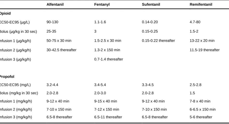
Table II. Infusion schemes of propofol and opioids required to maintain effect site concentrations of these agents, when given in combination, with ±15% of
the effect-site concentrations that are associated with a 50% and 95% probability of no response to surgical stimuli (EC50 and EC95) and the most rapid return of consciousness after termination of the infusions. These optimal infusion schemes have been derived from data in female patients undergoing lower abdominal surgery. They should be uses as guidelines and be adjusted to the individual needs of the patients. (vuyk et al. (8))
Alfentanil Fentanyl Sufentanil Remifentanil
Opioid
EC50-EC95 (µg/L) 90-130 1.1-1.6 0.14-0.20 4.7-80
Bolus (µg/kg in 30 sec) 25-35 3 0.15-0.25 1.5-2
Infusion 1 (µg/kg/h) 50-75 x 30 min 1.5-2.5 x 30 min 0.15-0.22 thereafter 13-22 x 20 min
Infusion 2 (µg/kg/h) 30-42.5 thereafter 1.3-2 x 150 min 11.5-19 thereafter
Infusion 3 (µg/kg/h) 0.7-1.4 thereafter
Propofol
EC50-EC95 (mg/L) 3.2-4.4 3.4-5.4 3.3-4.5 2.5-2.8
Bolus (mg/kg in 30 sec) 2.0-2.8 2.0-3.0 2.0-2.8 1.5
Infusion 1 (mg/kg/h) 9-12 x 40 min 9-15 x 40 min 9-12 x 40 min 7-8 x 40 min
Infusion 2 (mg/kg/h) 7-10 x 150 min 7-12 x 150 min 7-10 x 150 min 6-6.5 x 150 min
lower effect-site propofol concentrations are needed for loss of consciousness and these are reached more rapidly. Because the time to peak effect differs for propofol and the various opioids, the timing of the opioid bolus relative to that of propofol is critical in this respect. Times to peak effect for propofol, remifentanil, alfentanil, fentanyl and sufentanil are 2, 1.2, 2.3, 4.3 and 7.5 minutes, respectively (figure 2). To benefit most from the ability opioids to reduce anesthetic requirements, sufentanil should be given well in advance of propofol, more so than remifentanil or alfentanil. 2. Is it possible to increase the hemodynamic stability of the induction or maintenance of anaesthesia on the basis of the current knowledge of propofol-opioid interactions? Opioids reduce the anaesthetic dose requirements for induction of anaesthesia. In theory, this may lead to an improved hemodynamic profile of the induction of anaesthesia. However in ASA 1-2 patients this dose reduction does not leas to a more stable induction of anaesthesia. (12) In elderly patients or patients with cardiovascular instability, high opioid/low propofol anaesthesia may be associated with increased hemodynamic stability during induction of anaesthesia. However, not data are yet available to support this supposition.
3. Is it possible to decrease the time to awakening postoperatively on the basis of propofol-opioid interactions? With the use of optimal propofol-opioid concentrations, it is clearly possible to optimize intravenous, anaesthetic drug delivery. The propofol and opioid infusion regimens described in table II can be used as guidelines and will allow adequate anesthesia associated with a rapid recovery after termination of the propofol and opioid infusions. (8) In general, propofol-remifentanil anesthesia is associated with the most rapid return of consciousness after any infusion duration compared with fentanyl, alfentanil or sufentanil. Another benefit of remifentanil is that even at suboptimal high concentrations, return of consciousness is only marginally postponed.
4. What are the optimal propofol-opioid concentrations for anesthesia that allow spontaneous respiration? So far, no clinical relevant data regarding propofol-opioid interactions for spontaneous respiration have been described. Bouillon et al. (52) described for a single agent, alfentanil, the clinical profile in this respect. The EC50 for adequate ventilation during normocapnia is 60 µg/L. With higher
plasma alfentanil concentrations, the arterial pressure of CO2 has to increase considerable to
maintain adequate ventilation. Similarly, for propofol is has been shown that with increasing concentrations the responses to both hypercapnia and hypoxia are diminished.(30) This means that in the presence of propofol hypoxia will be deeper and hypercapnia more severe before a ventilatory response will be evoked by these stimulants. Because no interaction data exist, and nor are data available regarding the effect of nociception on propofol-opioid respiratory depression, optimal propofol-opioid concentrations that assure adequate anesthesia and adequate respiration cannot yet be defined.
2. State-of-the-art Administration Techniques
Target-controlled infusion as used in modern anaesthetic practice refers to the use of an infusion pump with an integrated pharmacokinetic dataset. With this technique, the user does not set an infusion rate but rather sets the desired blood concentration, i.e. the so-called target-concentration. The computer then uses the incorporated pharmacokinetic dataset to calculate the infusion rate required to reach and maintain the desired blood concentration. Next, the computer triggers the infusion pump to actually administer the infusion rate calculated. The pump will initially at a high infusion rate, thus giving a loading dose. In addition, the pump will repeatedly calculate the running rate required to maintain a constant blood concentration. After the initial loading dose, the calculated maintenance infusion rate decreases logarithmically to maintain a constant blood concentration. The logarithmic decrease in infusion rate is the result of the gradual saturation of the various pharmacokinetic compartments. When a lower target is set, the computer will stop the infusion of the drug until, as a result of clearance and redistribution, the desired concentration is reached.
The development of computer-controlled infusion systems date back to 1983 when Schüttler et al. (53) described the use of a computer to perform the ‘bolus elimination and transfer’ infusion scheme with a system called CATIA (computer assisted total intravenous anesthesia). Many other systems followed, including that of Alvis et al. (54) who compared target-controlled infusion-controlled anesthesia with that from a manual administration scheme. This has led to the introduction of the clinically available target-controlled infusion pump registered for the administration of propofol, the Diprifusor®. The Diprifusor® is provided with prefilled propofol syringes containing either 10 or 20 mg/mL of propofol. The prefilled syringes are equipped with a passive magnetic device that serves as a recognition tag for the target-controlled infusion device to indentify the drug and the solution of the drug in the syringe. Two important features of the Diprifusor® are the display of the predicted effect-site concentration and the prediction of the time to reach a lower blood concentration. With this last feature, anaesthesiologist now is capable of predicting the time to recovery in patient irrespective of the infusion duration.
five datasets tested varied between 20% and 100%, stressing the importance of installing a proper pharmacokinetic parameter set.
Similarly, Mertens reported on the predictive performance of remifentanil target-controlled infusion using the Minto parameter set. In general, measured remifentanil concentrations were on average 18% lower than predicted by the target-controlled infusion device. In an offline analysis, Mertens and colleagues reported on the improved predictive performance with the Egan remifentanil pharmacokinetic parameter dataset.(57) Although the parameter set of Egan and colleagues(58) performed best in in the analysis of Mertens et al., a population pharmacokinetic parameter set like that of Minto(35) may prove to beneficial in a more heterogeneous group of patients.
In conclusion, target-controlled infusion devices have been shown to be capable of predicting the actual measured concentrations quite closely, although proper selection of a matching pharmacokinetic parameter set remains important. The Diprifusor® has been shown to accurately predict the measured concentration in a wide variety of patients.
In general, the target-controlled infusion mode of administration of drugs provides a number of practical advantages to the user compared with conventional infusion;
• Improved control and predictability of pharmacodynamic effect achieved; • Therapeutic concentration achieved rapidly and maintained constant;
• Control over onset time by slow upward titration of target if desired in the elderly; • Proportional changes in blood concentration rapidly achieved;
• Improved titratability;
• Avoidance of peak blood concentrations and possible risk of toxicity; • No need for calculating of infusion rates;
• Automatic adjustment for differences in body weight, lean body mass, age or sex if complex model available;
• Displayed effect-site concentration facilitates titration of the blood concentrations; • Estimation of the time required to reach a lower plasma concentration;
• Target concentration regained automatically after syringe change; • A more logical and modern approach.
However, may of these advantages have not been proven in outcome studies. Lastly, target-controlled infusion systems can either target the blood concentration or the effect compartment concentration. The only clinically available system, the Diprifusor®, targets and controls the blood concentration.
3. Bispectral Index Monitoring
In 1875, Richard Caton (59) described the EEG as a way of determining cerebral activity on the cortical surface of the skull of animals. Then, in 1937, Gibbs and colleagues(60) discovered that the EEG activity was affected by the administration of anesthetic agents. Because the raw EE is hardly interpretable online, this quest for a clinically useful parameter derived from the EEG has great importance.
In this search, time domains, frequency domain and higher order statistical analysis techniques have been evaluated for their usefulness in the analysis of a depth of anesthesia parameter. Time domain-derived parameters are, for example, the change in total power or median frequency in time, the occurrence of activity in time in certain EEG frequency bands or the frequency of occurrence of burst-suppression. The effect of various anesthetic agents on time domain-derived EEG parameters have been described and claimed to be clinically useful.(61,62)
However, apart from various publications in this field, time domain EEG parameters have never been exploited on a large scale in clinical practice.
The most often used frequency domain analytical method for EEG data is the Fast Fourier Transformation (FFT). During FFT, the EEG signal is sliced into small time period of a few seconds, called epochs. The FFT analysis then results in the projection of the power spectrum versus the EEG frequency in, e.g. the 0-30 Hz range, during each epoch. The FFT in its turn gives rise to the derivation of clinically useful parameters. Two of the most studied FFT derived EEG parameters are the spectral edge and the median frequency. The spectral edge (SE95) is the FFT-derived frequency below which 95% of the power spectrum in the
FFT spectrum is found; the median frequency (SE50) is defined as the frequency below which
50% of the power in the FFT spectrum is found. Both SE95 and SE50 decrease with
increasing depth of anaesthesia and increasing blood and CNS concentrations of anaesthetic agents.
Opioid Concentrations correlates very well with the FFT derived parameters. (63,64) With increasing opioid concentration, the EEG changes from a low amplitude high frequency signal to a high amplitude low frequency signal. This results in the FFT as an increase in power at lower frequencies (0-5Hz) with a reduction of power at higher frequencies (10-30Hz) and results in a decrease of the SE95 and SE 50.
domain-derived parameters have never been used on a broader scale in clinical practice. Consequently, the search went on an resulted in the application of higher order statistical analysis of the EEG in recent years, which in the end has resulted in the introduction of the BIS monitor.
Bispectral analysis focuses on the correlation between the phases of the various wave components of which the raw EEG is built. It is a computation of the burst suppression ratio (BSR) and QUAZI, two time domain-derived parameters, the β-ratio, a frequency domain parameter defining the power in the 30-47Hz band relative to the 11-20Hz band, and lastly the SyncFastSlow parameter determined from the bispectrum peaks in the 0.5-47Hz band relative to the 40-47Hz frequency band.(66) An important feature in the calculation of the bispectral index is that the weight of any of these four subparameters in the final calculation (BSR, QUAZI, β-ratio and SyncFastSlow) changes with the level sedation. The β-ratio weighs heavier in the final computation at levels of light sedation, the SyncFastSlow parameter dominates at excitation and surgical levels of anaesthesia and the BSR and QUAZI are more important in the calculation at the most deep levels of EEG depression. The specific weight of the parameters of the BIS at various clinical states has been determined, during the development of the BIS by Aspect Medical Systems, on the basis of a dataset gathered from a group of patients that received various anaesthetics while EEG and behavioral data were collected. In practice, the BIS is determined as a running average over 15-30 seconds of EEG signal collection and visualized as a dimensionless nonlinear parameter between 0 and 100, with 0 equalling no electrical activity and 100 defining the awake state (figure 4). The BIS reflects the awake state at values exceeding 95, a state of sedation at BIS values 65-85, an arousal state depression suited for general anaesthesia at BIS values of 40-65 and burst suppression patterns become evident al BIS levels below 40.(67)
The most promising application of the BIS may be as a monitor of awake-sedation-unconsciousness levels. In the absence of CNS monitoring, anaesthetic agents are often administered on the basis of the prescribed administration regimens (12-10-8 mg/kg/h step down propofol infusion scheme) that may be adjusted to the response of the individual patient. The prescribed regimens do not take into account the pharmacokinetic of ± 70% or the pharmacokinetic variability of ± 300-400% between patients. This huge interindividual pharmacokinetic-dynamic variability, next to the sometime poor predictability of the surrogate measures of sedation and anaesthesia (e.g. hemodynamic parameters, movement responses to nociception), is the cause of frequent overdosage or underdosage of individual patients during sedation and general anaesthesia. Monitoring of the BIS allows for almost instant focusing, out of the huge inter- and intraindividual pharmacokinetic-pharmacodynamic variability, on the specific needs of the individual patient at any time.
Lastly, BIS monitoring has been incorporated in closed loop systems with a target-controlled infusion device for anaesthesia drug administration with BIS value as the control parameter. In these systems, the target-controlled infusion system thus determines the infusion rate on the basis of the difference between the measured and desired BIS value. Using this system provided safe and reliable anaesthesia, although an initial overshoot in BIS value occurred during induction of anaesthesia (70,71) as well as some oscillation around the set BIS. (72)
The use of BIS has some limitations. Some agents like nitrous oxide and ketamine, induce their effects by mechanisms that the BIS monitor is unable to track. Adding ketamine or nitrous oxide deepens the anaesthetic level but increases the BIS. In the presence of these agents, the BIS monitor should not be used. Electrocautery will make the BIS disappear or increase; pacemakers have also been described to increase the BIS. Electromyographic activity has been claimed to increase the BIS, but later versions like the XP may be less susceptible to this. Lastly, hypothermia decreases the BIS by 1.12 units per ºC decline in body temperature.
although monitoring of auditory evoked potentials has proven to be be of value for research purposes, at this moment its clinical value remains unclear.
As with the other two strategic tools, the implementation of EEG monitoring by means of the bispectral index, or perhaps in the future through monitoring the auditory evoked potentials, further enhances the ability of the anaesthesiologist to rapidly obtain information on the specific needs of the individual patient
4. Conclusion
References
1. Kuipers JA, Boer F, de Roode A, et al. Modeling population pharmacokinetics of lidocaine: should cardiac output be included as a patient factor? Anesthesiology 2001;94:566-73.
2. Kuipers JA, Boer F, Olofsen E, et al. Recirculatory and compartmental pharmacokinetic modeling of alfentanil in pigs: the influence of cardiac output. Anesthesiology
1999;90:1146-57.
3. Kuipers JA, Boer F, Olofsen E, et al. Recirculatory pharmacokinetics and pharmacodynamics of rocuronium in patients: the influence of cardiac output. Anesthesiology 2001;94:47-55.
4. Kharasch ED, Jubert C, Senn T, et al. Intraindividual variability in male hepatic CYP3A4 activity assessed by alfentanil and midazolam clearance. J Clin Pharmacol
1999;39:664-9.
5. Labroo RB, Paine MF, Thummel KE, Kharasch ED. Fentanyl metabolism by human hepatic and intestinal cytochrome P450 3A4: implications for interindividual variability in disposition, efficacy, and drug interactions. Drug Metab Dispos 1997;25:1072-80. 6. van den Nieuwenhuyzen MC, Engbers FH, Burm AG, et al. Target-controlled infusion of
alfentanil for postoperative analgesia: contribution of plasma protein binding to intra-patient and inter- intra-patient variability. Br J Anaesth 1999;82:580-5.
7. Mertens MJ, Vuyk J, Olofsen E, et al. Propofol alters the pharmacokinetics of alfentanil in healthy male volunteers. Anesthesiology 2001;94:949-57.
8. Vuyk J, Mertens MJ, Olofsen E, et al. Propofol anesthesia and rational opioid selection: determination of optimal EC50-EC95 propofol-opioid concentrations that assure
adequate anesthesia and a rapid return of consciousness. Anesthesiology 1997;87:1549-62.
9. Kanto J, Gepts E. Pharmacokinetic implications for the clinical use of propofol. Clin Pharmacokinet 1989;17:308-26.
10. Smith C, McEwan AI, Jhaveri R, et al. The interaction of fentanyl on the Cp50 of propofol for loss of consciousness and skin incision. Anesthesiology 1994;81:820-8. 11. Stanski DR, Shafer SL. Quantifying anesthetic drug interaction. Implications for drug
dosing. Anesthesiology 1995;83:1-5.
12. Vuyk J, Lim T, Engbers FH, et al. The pharmacodynamic interaction of propofol and alfentanil during lower abdominal surgery in women. Anesthesiology 1995;83:8-22. 13. Schnider TW, Minto CF, Shafer SL, et al. The influence of age on propofol
pharmacodynamics. Anesthesiology 1999;90:1502-16.
14. Saint-Maurice C, Cockshott ID, Douglas EJ, et al. Pharmacokinetics of propofol in young children after a single dose. Br J Anaesth 1989;63:667-70.
16. Schuttler J, Ihmsen H. Population pharmacokinetics of propofol: a multicenter study. Anesthesiology 2000;92:727-38.
17. Vuyk J, Oostwouder C.J., Vletter A, et al. Gender differences in the pharmacokinetics of propofol in elderly patients during and after continuous infusion. Br J Anaesth 2001;86:183-8.
18. Oda Y, Hamaoka N, Hiroi T, et al. Involvement of human liver cytochrome P4502B6 in the metabolism of propofol. Br J Clin Pharmacol 2001;51:281-5.
19. Guitton J, Buronfosse T, Desage M, et al. Possible involvement of multiple human cytochrome P450 isoforms in the liver metabolism of propofol. Br J Anaesth 1998;80:788-95.
20. Schnider TW, Minto CF, Gambus PL, et al. The influence of method of administration and covariates on the pharmacokinetics of propofol in adult volunteers. Anesthesiology 1998;88:1170-82.
21. Scott JC, Stanski DR. Decreased fentanyl and alfentanil dose requirements with age. A simultaneous pharmacokinetic and pharmacodynamic evaluation. J Pharmacol Exp Ther 1987;240:159-66.
22. Minto CF, Schnider TW, Shafer SL. Pharmacokinetics and pharmacodynamics of remifentanil. II. Model application. Anesthesiology 1997;86:24-33.
23. Maitre PO, Vozeh S, Heykants J, et al. Population pharmacokinetics of alfentanil: the average dose- plasma concentration relationship and interindividual variability in patients. Anesthesiology 1987;66:3-12.
24. Gepts E, Shafer SL, Camu F, et al. Linearity of pharmacokinetics and model estimation of sufentanil. Anesthesiology 1995;83:1194-204.
25. McKillop D, Wild MJ, Butters CJ, Simcock C. Effects of propofol on human hepatic microsomal cytochrome P450 activities. Xenobiotica 1998;28:845-53.
26. Ebert TJ, Muzi M, Berens R, et al. Sympathetic responses to induction of anesthesia in humans with propofol or etomidate. Anesthesiology 1992;76:725-33.
27. Sztark F, Ichas F, Mazat JP, Dabadie P. Propofol and cellular calcium homeostasis. Anesthesiology 1995;83:1386.
28. Li YC, Ridefelt P, Wiklund L, Bjerneroth G. Propofol induces a lowering of free cytosolic calcium in myocardial cells. Acta Anaesthesiol Scand 1997;41:633-8.
29. Nieuwenhuijs D, Sarton E, Teppema L, Dahan A. Propofol for monitored anesthesia care: implications on hypoxic control of cardiorespiratory responses. Anesthesiology 2000;92:46-54.
30. Nieuwenhuijs D, Sarton E, Teppema LJ, et al. Respiratory sites of action of propofol: absence of depression of peripheral chemoreflex loop by low-dose propofol.
Anesthesiology 2001;95:889-95.
32. Westmoreland CL, Hoke JF, Sebel PS, et al. Pharmacokinetics of remifentanil
(GI87084B) and its major metabolite (GI90291) in patients undergoing elective inpatient surgery. Anesthesiology 1993;79:893-903.
33. Dershwitz M, Hoke JF, Rosow CE, et al. Pharmacokinetics and pharmacodynamics of remifentanil in volunteer subjects with severe liver disease. Anesthesiology
1996;84:812-20.
34. Hoke JF, Shlugman D, Dershwitz M, et al. Pharmacokinetics and pharmacodynamics of remifentanil in persons with renal failure compared with healthy volunteers.
Anesthesiology 1997;87:533-41.
35. Minto CF, Schnider TW, Egan TD, et al. Influence of age and gender on the pharmacokinetics and pharmacodynamics of remifentanil. I. Model development. Anesthesiology 1997;86:10-23.
36. Egan TD, Huizinga B, Gupta SK, et al. Remifentanil pharmacokinetics in obese versus lean patients. Anesthesiology 1998;89:562-73.
37. Egan TD. Remifentanil pharmacokinetics and pharmacodynamics. A preliminary appraisal. Clin Pharmacokinet 1995;29:80-94.
38. Pavlin DJ, Coda B, Shen DD, et al. Effects of combining propofol and alfentanil on ventilation, analgesia, sedation, and emesis in human volunteers. Anesthesiology 1996;84:23-37.
39. Matot I, Neely CF, Katz RY, Neufeld GR. Pulmonary uptake of propofol in cats. Effect of fentanyl and halothane. Anesthesiology 1993;78:1157-65.
40. Gepts E, Jonckheer K, Maes V, et al. Disposition kinetics of propofol during alfentanil anaesthesia. Anaesthesia 1988;43 Suppl:8-13.
41. Janicki PK, James MF, Erskine WA. Propofol inhibits enzymatic degradation of alfentanil and sufentanil by isolated liver microsomes in vitro. Br J Anaesth 1992;68:311-2.
42. Baker MT, Chadam MV, Ronnenberg WC, Jr. Inhibitory effects of propofol on
cytochrome P450 activities in rat hepatic microsomes. Anesth Analg 1993;76:817-21. 43. Kharasch ED, Hill HF, Eddy AC. Influence of dexmedetomidine and clonidine on human
liver microsomal alfentanil metabolism. Anesthesiology 1991;75:520-4.
44. Vuyk J. Pharmacokinetic and pharmacodynamic interactions between opioids and propofol. J Clin Anesth 1997;9:23S-6S.
45. Mertens MJ, Olofsen E, Burm AG, et al. Mixed-effects modeling of the influence of alfentanil on propofol pharmacokinetics. Anesthesiology 2004;100:795-805. 46. Bovill JG. Adverse drug interactions in anesthesia. J Clin Anesth 1997;9:3S-13S. 47. Minto CF, Schnider TW, Short TG, et al. Response surface model for anesthetic drug
interactions. Anesthesiology 2000;92:1603-16.
49. Berenbaum MC. Concepts for describing the interaction of two agents. Radiat Res 1991;126:264-8.
50. Vuyk J, Engbers FH, Burm AGL, et al. Pharmacodynamic interaction between propofol and alfentanil when given for induction of anesthesia. Anesthesiology 1996;84:288-99. 51. Billard V, Moulla F, Bourgain JL, et al. Hemodynamic response to induction and
intubation. Propofol/fentanyl interaction. Anesthesiology 1994;81:1384-93.
52. Bouillon T, Schmidt C, Garstka G, et al. Pharmacokinetic-pharmacodynamic modeling of the respiratory depressant effect of alfentanil. Anesthesiology 1999;91:144-55. 53. Schuttler J, Schwilden H, Stoekel H. Pharmacokinetics as applied to total intravenous
anaesthesia. Practical implications. Anaesthesia 1983;38 Suppl:53-6.
54. Alvis JM, Reves JG, Govier AV, et al. Computer-assisted continuous infusions of fentanyl during cardiac anesthesia: comparison with a manual method. Anesthesiology 1985;63:41-9.
55. Shafer SL, Siegel LC, Cooke JE, Scott JC. Testing computer-controlled infusion pumps by simulation. Anesthesiology 1988;68:261-6.
56. Vuyk J, Engbers FH, Burm AG, et al. Performance of computer-controlled infusion of propofol: an evaluation of five pharmacokinetic parameter sets. Anesth Analg
1995;81:1275-82.
57. Mertens MJ, Engbers FHM, Burm AGL, Vuyk J. Predictive performance of computer-controlled infusion of remifentanil during propofol/remifentanil anaesthesia. British Journal of Anaesthesia 2003;90:132-41.
58. Egan TD, Minto CF, Hermann DJ, et al. Remifentanil versus alfentanil: comparative pharmacokinetics and pharmacodynamics in healthy adult male volunteers.
Anesthesiology 1996;84:821-33.
59. Caton R. The electrical currents of the brain. British Medical Journal 1875;2:278. 60. Gibbs F, Gibbs E, Lennox W. Effect on the electroencephalogram of certain drugs
which influence nervous activity. Archives of Internal Medicine 1937;60:154-66. 61. Breimer LT, Burm AG, Danhof M, et al. Pharmacokinetic-pharmacodynamic modelling
of the interaction between flumazenil and midazolam in volunteers by aperiodic EEG analysis. Clin Pharmacokinet 1991;20:497-508.
62. Breimer LT, Hennis PJ, Burm AG, et al. Quantification of the EEG effect of midazolam by aperiodic analysis in volunteers. Pharmacokinetic/pharmacodynamic modelling. Clin Pharmacokinet 1990;18:245-53.
63. Scott JC, Ponganis KV, Stanski DR. EEG quantitation of narcotic effect: the comparative pharmacodynamics of fentanyl and alfentanil. Anesthesiology 1985;62:234-41.
65. Kuizenga K, Kalkman CJ, Hennis PJ. Quantitative electroencephalographic analysis of the biphasic concentration-effect relationship of propofol in surgical patients during extradural analgesia. Br J Anaesth 1998;80:725-32.
66. Rampil IJ. A primer for EEG signal processing in anesthesia. Anesthesiology 1998;89:980-1002.
67. Johansen JW, Sebel PS. Development and clinical application of
electroencephalographic bispectrum monitoring. Anesthesiology 2000;93:1336-44. 68. Glass PS, Bloom M, Kearse L, et al. Bispectral analysis measures sedation and
memory effects of propofol, midazolam, isoflurane, and alfentanil in healthy volunteers. Anesthesiology 1997;86:836-47.
69. Lysakowski C, Dumont L, Pellegrini M, et al. Effects of fentanyl, alfentanil, remifentanil and sufentanil on loss of consciousness and bispectral index during propofol induction of anaesthesia. Br J Anaesth 2001;86:523-7.
70. Struys MM, De Smet T, Versichelen LF, et al. Comparison of closed-loop controlled administration of propofol using Bispectral Index as the controlled variable versus "standard practice" controlled administration. Anesthesiology 2001;95:6-17.
71. Mortier E, Struys M, De Smet T, et al. Closed-loop controlled administration of propofol using bispectral analysis. Anaesthesia 1998;53:749-54.
72. Absalom AR, Sutcliffe N, Kenny GN. Closed-loop control of anesthesia using Bispectral index: performance assessment in patients undergoing major orthopedic surgery under combined general and regional anesthesia. Anesthesiology 2002;96:67-73.
73. Bonhomme V, Plourde G, Meuret P, et al. Auditory steady-state response and bispectral index for assessing level of consciousness during propofol sedation and hypnosis. Anesth Analg 2000;91:1398-403.
74. Thornton C, Barrowcliffe MP, Konieczko KM, et al. The auditory evoked response as an indicator of awareness. Br J Anaesth 1989;63:113-5.
Propofol Reduces the Distribution and Clearance of Midazolam
Bart Jan Lichtenbelt, MD.,* Erik Olofsen, M.Sc.,# Albert Dahan, MD., PhD#, Jack W. Van Kleef MD., PhD#, Michel M.R.F. Struys MD., PhD*, Jaap Vuyk, MD., PhD#
*
From the department of Anaesthesiology, University Medical Centre Groningen, Universityof Groningen, Groningen, and # Department of anaesthesiology, Leiden University Medical Centre (LUMC), Leiden, the Netherlands.
I
NTRODUCTIONPreviously, we studied the influence of midazolam on the pharmacokinetics of propofol (1). The most important finding of that study was that midazolam increased blood propofol concentrations by 25% through a reduction in the metabolic, rapid, and slow distribution clearances of propofol. In addition, a reduction in mean arterial blood pressure was associated with propofol pharmacokinetic alterations that increased the blood propofol concentrations even further. In that study, the plasma midazolam concentration, as controlled by target-controlled infusion (TCI), was increased when administered in the presence of propofol, indicative of a possible influence of propofol on the pharmacokinetics of midazolam.
In clinical practice, midazolam is used for preoperative anxiolysis, to assure sedation during regional anesthesia and during ventilation in the intensive care unit and during prolonged procedures to induce and maintain surgical hypnosis perioperatively. In these settings, midazolam is occasionally combined with other sedatives and/or opioids to obtain the desired effect (hypnosis) and limit the side effects (hemodynamic or respiratory depression) (2-5). Various combinations of hypnotic drugs and/or opioids have been shown to exhibit both pharmacokinetic and pharmacodynamic interactions (6), often increasing the effect of the combination. (7-9)
Researchers studying the effect the propofol-midazolam interaction predominantly evaluated the pharmacodynamic interaction. (3,4,10,11) Only 1 study described the pharmacokinetic interaction between propofol and midazolam and reported that propofol affected the clearance of midazolam through a possible competitive inhibition of hepatic CYP 3A4.(8) However in that study, clearance was determined on the basis of the influence of just a 1-hour infusion of propofol on the pharmacokinetics of midazolam that was given as just a single bolus dose. Because propofol and midazolam are at times combined for prolonged periods of time, e.g., for intensive care unit sedation (10), we evaluated this interaction during prolonged infusion.
MATERIALS AND METHODS
Volunteers and Study Protocol
After obtaining approval of the Medical Ethics Committee of the Leiden University Medical Centre and written informed consent, 8 healthy male volunteers, were studied. All volunteers were within 30% of ideal body weight, had no history of renal or hepatic disease standards and were not taking medication within 6 months before or during the investigation. All volunteers denied smoking or consumption of more than 20 g of alcohol per day. Before the investigation, a blood sample was taken for screening of renal or hepatic disease in accordance with Leiden University Medical Centre standards.
Volunteers were studied in a randomized cross-over manner during two sessions. During the first session volunteers received a midazolam bolus dose of 0.035 to 0.05 mg.kg-1 in 1 min followed by an infusion of 0.035 to 0.05 mg.kg-1.h-1 for 59 min (session A, control). During the
second study session (session B) the volunteers received the same midazolam infusion scheme as during session A, but now in the presence of a TCI of propofol for 7 hours at a constant propofol target concentration (CT) of 0.6 or 1.0 µg.ml-1 using the Diprifusor®. The
target controlled infusion of propofol was started 15 min before to the start of the midazolam administration to ensure a semi steady state concentration of propofol at the beginning of the midazolam infusion.
The 2 sessions were separated by a period of at least 2 weeks. Both the CT (0.6 or 1.0
µg.mL-1) and the order of the 2 sessions were randomized, such that in half of the volunteers
the control sessions preceded the other session and half of the volunteers received a CT of
0.6 µg.mL-1 and the other half 1.0 µg.mL-1. Volunteers fasted from midnight on the night before the study until the last blood sample had been collected. During the administration of propofol, they breathed 30% oxygen in air. When indicated, ventilation was assisted using a face mask to maintain the end-tidal CO2 partial pressure at <50 mm Hg. After termination of
Materials
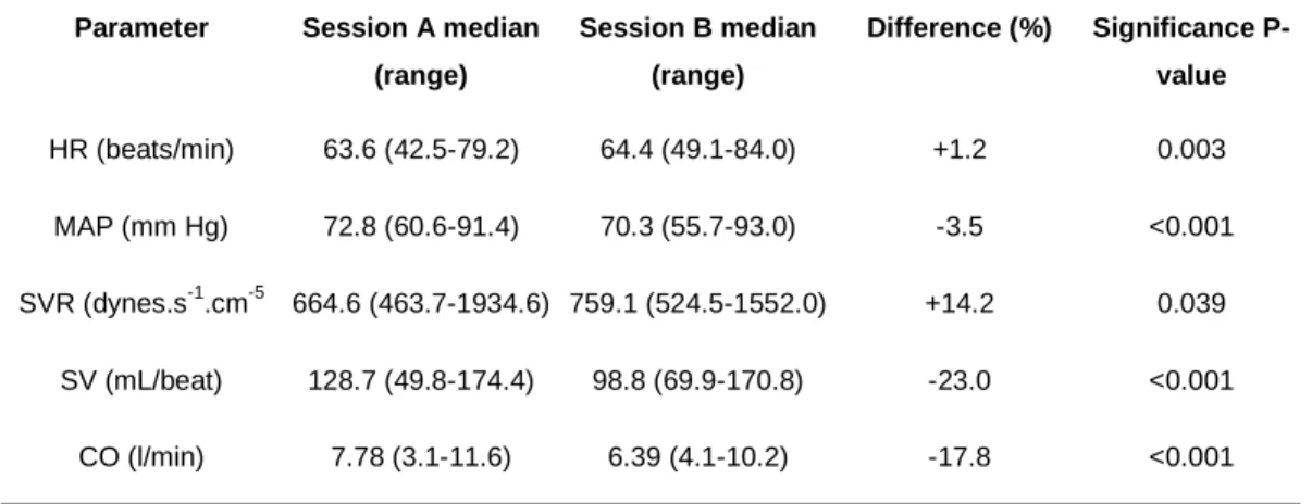
The studies were performed in an operating room. An IV cannula was inserted into a large forearm vein for the infusion of propofol and midazolam and an arterial cannula was inserted into a radial artery for collection of hemodynamic data and blood samples. The electrocardiogram, respiratory rate, peripheral oxygen saturation, the bispectral index and intra-arterial blood pressure were monitored continuously throughout the study. Furthermore, the cardiac output was determined using the pulsecontour methodology on the basis of the intra-arterial blood pressure curve with the LiDCOplus monitor (LiDCOgroup plc, London). The LiDCO monitor was calibrated before each experiment. For this purpose, a lithium sensor was connected to the arterial cannula. After 0.2mmol lithium was injected IV, the LiDCO monitor was calibrated on the basis of the non-invasive online-determined arterial lithium concentration-time curve and the cardiac output calculated. The LiDCO has been found reliable for cardiac output monitoring when compared with traditional thermodilution cardiac output monitoring for up to 8 hours after calibration (LiDCO versus thermodilution; r = 0.86).(12) Blood samples were drawn from the arterial cannula, after calibration of the LiDCO.
Heart rate; cardiac output; cardiac index; systemic vascular resistance, the systolic, mean and diastolic arterial blood pressure were all recorded online and saved for further analysis. All volunteers received an infusion of saline of 2 ml.kg-1.h-1 during each session.
Blood Samples and Assays
During session A, a blank blood sample (10 mL) was obtained. This sample was used for calibration purposes. Additional arterial blood samples (5 mL) for the determination of the plasma midazolam concentration, were taken 1, 3, 5, 10, 20, 30, 45 and 60 min after the start of the midazolam infusion, and 1, 2, 3, 5, 10, 20, 30, 45, 60, 90, 120, 180, 240, 300 and 360 min after termination of the midazolam infusion. Blood samples were taken into heparinised syringes for determination of the plasma midazolam concentration. These samples were centrifuged to obtain plasma which was subsequently stored at -20 °C until analysis. The concentration of midazolam in plasma was determined by reversed-phase high-performance liquid chromatography-UV detection at 216 nm (HPLC).(13) The intra- and interassay coefficients of variation of this method were 2.2% and 2.0 % respectively, for midazolam in plasma in the concentration range of 9.7-1120 ng.mL-1. Midazolam assays were conducted within 12 weeks.