A subset of replication-dependent histone mRNAs are
expressed as polyadenylated RNAs in terminally
differentiated tissues
Shawn M. Lyons
1, Clark H. Cunningham
1, Joshua D. Welch
2, Beezly Groh
3, Andrew
Y. Guo
1, Bruce Wei
1, Michael L. Whitfield
4, Yue Xiong
2,5and William F. Marzluff
1,2,5,*1Department of Biology, University of North Carolina, Chapel Hill, NC 27599, USA,2Department of Computer
Science, University of North Carolina, Chapel Hill, NC 27599,3Department of Biochemistry and Biophysics, University of North Carolina, Chapel Hill, NC 27599, USA,4Department of Genetics, Dartmouth Geisel School of Medicine, Hanover, NH 03755, USA and5Integrative Program for Biological and Genome Sciences, University of North Carolina, Chapel Hill, NC 27599, USA
Received December 18, 2015; Revised June 27, 2016; Accepted June 30, 2016
ABSTRACT
Histone proteins are synthesized in large amounts during S-phase to package the newly replicated DNA, and are among the most stable proteins in the cell. The replication-dependent (RD)-histone mRNAs ex-pressed during S-phase end in a conserved stem-loop rather than a polyA tail. In addition, there are replication-independent (RI)-histone genes that en-code histone variants as polyadenylated mRNAs. Most variants have specific functions in chromatin, but H3.3 also serves as a replacement histone for damaged histones in long-lived terminally differen-tiated cells. There are no reported replacement hi-stone genes for hihi-stones H2A, H2B or H4. We re-port that a subset of RD-histone genes are expressed in terminally differentiated tissues as polyadenylated mRNAs, likely serving as replacement histone genes in long-lived non-dividing cells. Expression of two genes, HIST2H2AA3 and HIST1H2BC, is conserved in mammals. They are expressed as polyadenylated mRNAs in fibroblasts differentiatedin vitro, but not in serum starved fibroblasts, suggesting that their expression is part of the terminal differentiation pro-gram. There are two histone H4 genes and an H3 gene that encode mRNAs that are polyadenylated and ex-pressed at 5- to 10-fold lower levels than the mRNAs from H2A and H2B genes, which may be replacement genes for the H3.1 and H4 proteins.
INTRODUCTION
The bulk of the histone proteins are synthesized coordi-nately with DNA during S-phase and are very stable after incorporation into chromatin. These proteins are encoded by replication-dependent (RD)-histone mRNAs which are the only known cellular eukaryotic mRNAs that do not end in a polyA tail but end instead in a conserved stem-loop. The protein that binds the stem-loop, SLBP, is required for processing of the RD-histone pre-mRNAs and also func-tions during the entire histone mRNA life cycle including transport from the nucleus, translation and mRNA degra-dation (1). Canonical RD-histone processing occurs by en-donucleolytic cleavage following SLBP binding to the stem-loop (2). This cleavage is directed by the U7 snRNP (3–5) that interacts with a second histone specific sequence called the histone downstream element (HDE) (6). These histone mRNAs are expressed at high levels during S-phase increas-ing 35-fold as cells enter S-phase and decreasincreas-ing rapidly at the end of S-phase (7). SLBP levels are also cell cycle regu-lated and the increased levels of SLBP as cells enter S-phase allows increased processing of histone mRNA. The degra-dation of SLBP at the end of S-phase prevents further ac-cumulation of histone mRNAs or protein (8,9).
Most studies of histone mRNAs have focused on their expression and regulation in growing cells. An unresolved question is how histone proteins are synthesized after cells have terminally differentiated and no longer re-enter S-phase. This is a particularly pertinent question for long-lived cells. In dividing cultured cells, histones are stable with
<5% turnover of labelled histones over 8 generations (10). The half-life of histone protein in chicken brain tissue has been estimated at 19 days (11). In a recent proteomics study, histones were identified as among the most stable proteins
*To whom correspondence should be addressed. Tel: +1 919 962 2140; Fax: +1 919 962 1273; Email: [email protected]
Present address: Shawn M. Lyons, Department of Medicine, Harvard Medical School, Division of Rheumatology, Immunology and Allergy, Brigham and Women’s Hospital, Boston, MA 02115, USA.
C
The Author(s) 2016. Published by Oxford University Press on behalf of Nucleic Acids Research.
Nucleic Acids Research, 2016, Vol. 44, No. 19 9191
in mammalian cells (12), with histone H3.1 and H4 signif-icantly more stable than histones H2A and H2B. Histone turnover likely occurs as a result of damage to existing hi-stone proteins. However, many mammalian cells have life spans substantially longer than even the slowly turning over histone proteins (13). How, then, do non-dividing cells such as these maintain proper levels of histone proteins that are critical for genome stability and the regulation of gene ex-pression?
One possibility is that there are distinct genes that are ex-pressed constitutively and encoded by polyadenylated mR-NAs. The histone H3F3 genes found in all multicellular organisms encode the variant histone H3.3, which is syn-thesized constitutively, and plays an important role in gene regulation. It also serves as a replacement histone H3 vari-ant. In mice, histone H3.3 becomes the predominant his-tone H3 protein in non-dividing cells over the lifespan of the mouse (14). Similar to H3.3, histone H1Ois encoded by
a polyadenylated RNA and the amount of H1Oprotein
in-creases after terminal differentiation (15). There are several variants of histone H2A (e.g. H2A.Z, macroH2A), which are expressed from polyadenylated mRNAs, but these have specific functions, and likely do not serve as ‘replacement’ variants. For example, the histone H2A.X protein is ex-pressed from a single gene and is involved primarily in DNA repair (16). In S-phase cells, the H2A.X mRNA ends in a stem-loop and is cell cycle regulated. Outside of S-phase the same gene expresses a longer polyadenylated mRNA (17,18). There are no reports of variant histone H2B genes or histone H4 genes that express only polyadenylated mR-NAs (e.g. like H1Oor H3.3).
Some polyadenylated core histone mRNAs are produced in small amounts in cultured cells as a result of knockdown of a number of factors (NELF (19), ARS2 (20), chromatin modifiers (21), P-TEFb (21), Y-RNAs (22) and SLBP (23)). These treatments likely result in a perturbation of canon-ical histone pre-mRNA processing after the stem-loop. In addition, Kari et al. have shown that there is production of some polyadenylated and spliced histone H2B mRNAs when human fibroblasts are differentiated into adipocytesin vitro(24). In some of these studies, the relative proportion of polyadenylated mRNAs and properly processed mR-NAs were determined and the amount of polyA+ RNA was very small (<5%) (19,22). In other studies, only in-creases in polyadenylated histone mRNAs over the very small amounts of polyadenylated histone mRNAs present in control cells was described (21).
Here, we show that in terminally differentiated tissues, a subset of histone genes in the two RD-histone mRNA clusters remain active and encode polyadenylated mRNAs. These include one histone H1 gene, H1C, previously shown to be expressed in many adult mouse tissues (25), as well as genes for the four core histone proteins. All these mRNAs are polyadenylated and, in most cases, the polyA signal is 3of the stem-loop and extends the 3UTR of the histone mRNA. One or more histone H2B mRNAs (depending on the species) from the HIST1 cluster are formed by splic-ing around the histone stem-loop, resultsplic-ing in a polyadeny-lated histone mRNA lacking the stem-loop. These mRNAs likely encode the ‘replacement’ H2A, H2B and H4 proteins in non-dividing cells. Surprisingly, terminally differentiated
cells also express a number of genes thought to be required only for RD-histone gene expression, including most of the genes required for processing histone mRNA.
MATERIALS AND METHODS
RNA extraction from mouse tissue
Post-natal day 1 (P1 mice) and dissected mouse liver and brain were quick-frozen with liquid nitrogen and stored 80◦C until use. Sections of tissue (100–200 mg) were weighed and placed in a ceramic mortar filled will liquid nitrogen and ground by hand. The resulting tissue powder was transferred to a 15-ml tube and 1 ml of Trizol (Invitro-gen) was added per 50 mg of tissue, and processed as recom-mended by the manufacturer. The RNA pellet was air dried and resuspended in 300 l of 0.3 M sodium acetate (pH 5.6), extracted with phenol/chloroform and ethanol precip-itated.
Preparation of protein lysate from mouse tissues
The tissue powder was prepared as described above and was resuspended in 1 ml of NP-40 lysis buffer (150 mM NaCl, 50 mM Tris [pH 8.0], 0.5% NP-40) per 100 mg of tissue, ro-tated at 4◦C for 20 min, and the cell lysates clarified by cen-trifugation at 16 000xg.The supernatant was transferred to new tubes and the protein concentration was determined by Bradford Assay.
S1 Nuclease protection assays
The S1 nuclease assays were performed as previously de-scribed (26), using either total cell RNA or RNA fraction-ated on oligo(dT) cellulose into polyA+ and polyA−
frac-tions. Each gene was cloned into pUC19 and 5g of each plasmid digested at restriction site in the ORF resulting in the generation of a 5 overhang that can be labelled by incorporation of␣-[32P]-dCTP (3000 Ci/mmol) using the
Klenow fragment of DNA pol I. After labelling the plasmid was then digested with a 2nd restriction site downstream of the 3end. Description of restriction site usage and probe size is found in Supplementary Table S1. After hybridiza-tion and digeshybridiza-tion with S1 nuclease as described previously, the protected fragments were analysed by electrophoresis on a 6% polyacrylamide gel containing 8M urea followed by autoradiography.
3T3-L1 differentiation
quantification of pre-adipocyte and adipocyte marker gene expression (28).
Northern blotting for U7 snRNA
U7 snRNA was detected by Northern blotting as previously described using 5 end labelled antisense oligonucleotides and 25 g of total cell RNA (29). As an internal control the gel was stained prior to blotting and the intensity of 5S rRNA staining determined.
Biotinylated RNA pulldown
Cell lysates were diluted to 1 mg/ml protein in NP-40 lysis buffer (150 mM NaCl, 50 mM Tris [pH 8.0], 0.5% NP-40) and final concentration of EDTA was brought to 20 mM. For each experiment, 100l lysate was incubated with 10l of 10M biotinylated 30 nt stem-loop RNA (Dharmacon) for 1 h as previously described (30). The biotinylated RNA was recovered by binding to streptavidin-agarose beads fol-lowed by centrifugation. The unbound protein was saved for Western blot analysis. Beads were washed 4 times with 1 ml of NP-40 lysis buffer. Bound proteins were recovered in 25l of SDS loading buffer and resolved on an 8% SDS-PAGE gel for Western blot analysis.
Analysis of RNA-seq data
Reads from Illumina Human BodyMap Project were aligned to hg19 using MapSplice2 with default settings. UCSC gene annotations were downloaded and used to cre-ate transcriptome annotations for RSEM. We manually scanned the histone gene cluster to identify any genes ex-pressed in the tissues, and confirmed that there were reads that included the stem-loop and downstream sequences, as well as junction spanning reads that defined the splice sites for all 10 histone genes in each of the human samples. We computed gene expression levels for UCSC genes using RSEM with the settings –estimate-rspd and –paired-end. RSEM aligns reads to a reference transcriptome in a way designed to find all possible genes that each read could have originated from and then uses a Bayesian network model to estimate the abundance of all genes simultaneously. This strategy is ideal for dealing with genes that may have many multimapped reads, as is the case with histone genes. For duplicated genes, RSEM computes a separate expression level for each annotated locus. The expression levels that RSEM reports are in units of FPKM (fragments per kilo-base per million reads). Data from the cow and mouse tis-sues were provided by Jason Merkin and Chris Burge (31).
RESULTS
Histone genes express polyadenylated mRNAs in human tis-sues
There are two major histone gene clusters in mammals (32). The large HIST1 cluster contains more than 50 genes, in-cluding all of the RD-H1 proteins and the H2A.1, H2B.1 and H2B.2, H3.1 and H4 isoforms of core histone genes. The smaller cluster, HIST2, contains about 10 genes for core histones, including a central pair of duplicated histone
H2A and H3 genes that encode the H2A.2 and H3.2 pro-tein variants, and depending on the species, at least one H4 gene and 2–3 other H2A and H2B genes. The structure and gene order in both clusters is conserved in mammals, al-though there are examples of small differences due to cre-ation of pseudogenes and differing extents of the duplica-tion of the central gene pair in HIST2 in some species. In addition, there are three histone genes in the small HIST3 cluster, which are not expressed in somatic cells and a lone H4 gene, HIST4H4, expressed coordinately with the genes in HIST1 and HIST2, all of which have been conserved in mammals. All of these histone genes encode mRNAs that end in the stem-loop. Here, we analysed expression of his-tone mRNAs in human tissues and in tissues of two other mammals, mouse and cow, with sequencing data provided by Jason Merkin and Chris Burge (31).
The non-polyadenylated histone mRNAs are typically not present in the RNAs sequenced in high throughput se-quencing projects as part of the analysis of gene expres-sion. However, Illumina provided human sequences derived from total cell RNA (ribominus) from 15 adult tissues. The mouse and cow sequences were all polyA+ mRNA from terminally differentiated tissues. We also analysed expres-sion of histone genes in human breast tumour tissue (both ribominus and polyA+) and normal adjacent breast tissue (poly A+) from the TCGA project at UNC.
The histone mRNA expression pattern was similar in all 15 human tissues in the Illumina data set. There was essentially no histone mRNA expressed in tissues from most histone genes, as expected, since most of the tissues consisted largely of non-dividing cells. The ‘replication-independent’ histone genes, H3.3, H2A.Z, the polyadeny-lated form of histone H2A.X, histone H1O, and macroH2A,
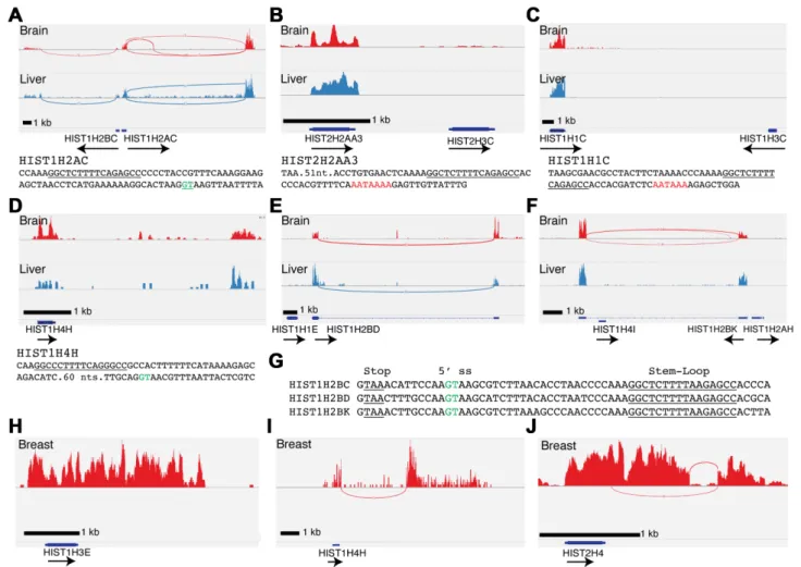
were expressed in all the tissues (Supplementary Fig-ure S1). We were surprised there was expression of the same subset of RD-histone genes in all 15 terminally differentiated human tissues as polyadenylated mRNAs (Table 1). The ten expressed histone mRNAs were the HIST1H2AC, HIST1H2BC (Figure 1A), HIST2H2AA3 (Figure 1B), HIST1H1C (Figure 1 C), HIST1H4H (Fig-ure1D), HIST1H2BD (Figure1E) and HIST1H2BK (Fig-ure 1F), HIST1H3E (Figure 1H), HIST2H4 (Figure 1J) and HIST1H2BE (data not shown). Of these, HIST1H2BC, HIST1H2AC, HIST1H2BD, HIST1H2BK, HIST1H4H and HIST2H4 were expressed as spliced and polyadeny-lated mRNAs (Table 1). The HIST1H1C, HIST1H3E, HIST1H2BE gene and the duplicated HIST2H2AA3 genes were expressed as mRNAs that were polyadenylated, but not spliced. Screenshots of the expression of these genes are shown in Figure1, together with examples of genes adja-cent to HIST2H2AA3 (Figure1B), HIST1H2BD (Figure
Nucleic Acids Research, 2016, Vol. 44, No. 19 9193
Figure 1. A subset of replication-dependent histone mRNAs are expressed in terminally differentiated tissues. (A–F) Sashimi plots RNA-Seq (ribominus) of total RNA from human tissues in the Illumina BodyMap project showing expression of the indicated histone genes in brain (Red) and liver (Blue) for (A) HIST1H2AC and HIST1H2BC; (B) HIST2H3C (not expressed) and HIST2H2AA3; (C) HIST1H3C (not expressed) and Hist1H1C; (D) HIST1H4H; (E) HIST1H1E (not expressed) and Hist1H2BD; (F) Hist1H4I (not expressed), Hist1H2BK and Hist1H2AH (not expressed). Note that there is no expression of adjacent histone genes. Sequences below sashimi plots indicate important sequence motifs; stem-loops(underlined), 5splice sites (green) and polyadeny-lation signals (red). (G) Sequence comparison of HIST1H2B genes whose mRNAs are spliced and polyadenylated. Conserved 5splice site (green) and stem-loop (underlined) are indicated. Note that the location of the 5splice site results in removal of the stem-loop from the polyadenylated mRNA. (H–J) Sashimi plots of normal human breast tissue from a representative TCGA sample for (H) HIST1H3E, (I) HIST1H4H and (J) HIST2H4.
Table 1. Polyadenylated Human histone mRNAs and their mouse orthologs
Human Gene Is the mRNA spliced?
Does the mature mRNA retain its
stem-loop? FPKM Mouse Ortholog
Is the mouse ortholog PolyA+?
Hist1H1C No Yes 14.9 ±3.6 Hist1H1c Yes
Hist1H2AC Yes Yes 21.1 ±5.5 Hist1H2ac No
Hist2H2AA3 No Yes 9.2 ±3.2 Hist2H2aa1 Yes
Hist1H2BC Yes No 1.0 ±0.5 Hist1H2bc Yes
Hist1H2BD Yes No 8.6 ±2.7 Pseudogene N.A.
Hist1H2BK Yes No 29.5 ±4.7 Hist1H2bk No
Hist2H2BE No Yes 10.9 ±3.7 Hist2H2be N.D.
Hist1H3E No Yes 1.1 ±0.3 Hist1H3e N.D.
Hist1H4H Yes Yes 1.3 ±0.2 Hist1H4h N.D.
Hist2H4 Yes Yes 2.9 ±0.9 Hist2H4 Yes
The HIST1H2AC, HIST1H4H and HIST2H4 genes were expressed as polyadenylated mRNAs that were spliced af-ter the normal 3 end of the histone mRNA. Note the green highlighted sequence indicating the 5 splice donor. This processing reaction results in mature mRNA that contains both a stem-loop and a polyA tail, similar to the structure of the polyadenylated histone mRNAs that were not spliced. The HIST1H2AC gene shares a promoter with the adjacent HIST1H2BC, which is also expressed as a spliced and polyadenylated mRNA (Figure 1A), al-beit at lower levels than HIST1H2AC. Two other H2B genes, HIST1H2BD and HIST1H2BK, are also expressed as spliced and polyadenylated mRNAs (Figure1E–F). The HIST1H2BK gene was adjacent to the HIST1H2AH gene and the HIST1H2BD was adjacent to the HIST1H1E gene, neither of which was expressed (Figure1E–F). In contrast to HIST1H2AC, the spliced and polyadenylated H2B genes produced mature mRNAs that lacked the stem-loop, be-cause the 5 splice donor site is located between the stop codon and stem-loop (Figure 1G, green highlighted se-quence). Note that the H2B mRNAs will not be targets for nonsense-mediated decay (NMD). Efficient NMD requires that the stop codon be>55 nts from the exon-junction and that the efficiency of NMD increases only as the distance between the stop codon and multiple exon junctions in-crease (35,36). The distance between the stop codon and 5 splice site is >55 nts for HIST1H4H (Figure1D) and Hist2H4 and this may contribute to the low levels of ex-pression of these genes among the spliced histone mRNAs (Table1, c.f. HIST1H4H and HIST1H2AC, HIST1H2BC, HIST1H2BK, HIST1H2BD).
To further confirm the expression of these genes in nor-mal human tissue, we analysed sequencing data from the TCGA project at UNC-Chapel Hill, where adjacent normal breast tissue as well as breast tumour tissue was sequenced, and where the sequencing was done to a deeper depth. The data from normal tissue confirmed the results from the data obtained from Illumina, and similar levels of expression of the polyadenylated histone mRNAs were found in each sample (Table1). In this data set, we identified a single hi-stone H3 gene, HIST1H3E, which was expressed at a simi-lar level as the HIST1H4H gene (Figure1H), each with an FPKM of about 1, compared to the level of 10–30 FPKM found for the H2A, H2B and HIST1H1C genes. Analysis of this data set also demonstrated sequencing reads that span splice junctions for the two expressed H4 genes, HIST1H4H and HIST2H4 (Figure1I and J). The histone H2A and H2B mRNAs were expressed at a significantly higher level than the histone H4 or H3 mRNAs. The H3/H4 protein tetramer is much more stable than the H2A/H2B protein dimer (12); therefore, it is likely that it does not require as high expres-sion to maintain proper levels within chromatin. In total, 10 of the 65 histone genes in the HIST1 and HIST2 clusters were expressed in tissues as polyadenylated mRNAs (Table
1).
Similar sets of histone mRNAs are expressed in tissues of other mammals
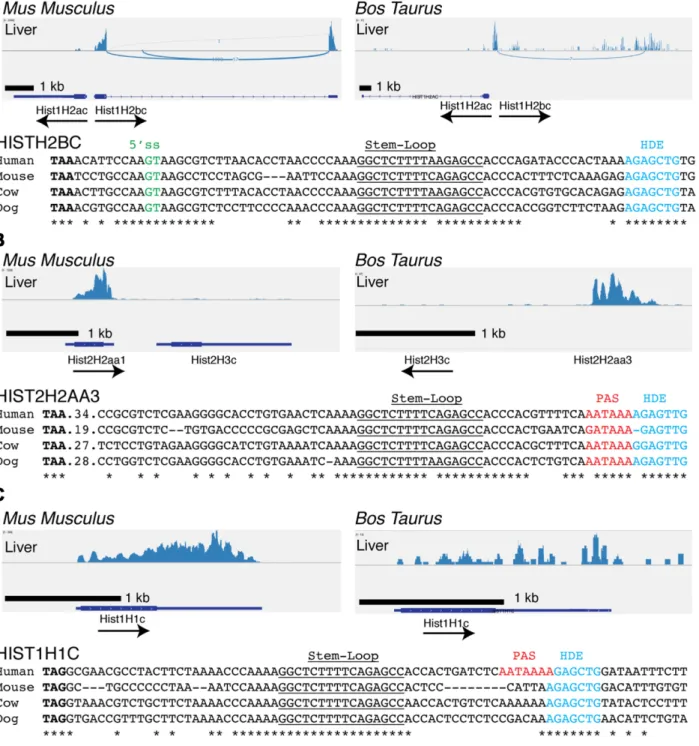
We examined the expression of histone genes from the tis-sues of two other mammals, mouse and cow, using
sequenc-ing data obtained from Jason Merkin and Chris Burge (31) (GSE41637). These data contained only sequences from polyadenylated mRNAs, but were sufficient for us to iden-tify the replication-dependent histone mRNAs that were ex-pressed as polyadenylated mRNAs in these non-dividing tissues (Figure2). Since the mouse core histone genes are over 95% conserved at the nucleotide level (37,38), it was not possible to unambiguously map reads containing only coding region sequences to individual genes. However, the polyadenylated mRNAs could be mapped because of the unique sequences in the 3UTR before and after the stem-loop. Like the human histone genes, the cow histone genes have enough differences in the coding regions to assign se-quencing reads to individual histone genes. Although the histone gene cluster was not completely annotated in the cow genome, we determined that its organization was the same as the mouse and human cluster and identified the or-thologues of each of the genes expressed in humans. Note that the naming convention for all human genes has the hu-man genes capitalized (e.g. HIST1H2BC) and the ortholo-gous genes in other mammals (i.e. mouse and cow) genes in lower case (e.g. Hist1H2bc) (32).
The orthologues of the HIST1H2BC (Figure 2A), HIST2H2AA3 (Figure 2B) and HIST1H1C (Figure 2C) genes were expressed in both cow and mouse brain (data not shown) and liver (Figure2). The Hist1H2bd gene was also expressed in cow, but not in mouse where it is a pseudogene (32). The HIST1H2BK gene is the most highly expressed H2B gene in human tissues (Table1), but it’s orthologue, Hist1H2bk, is not expressed in mouse or cow tissues as the 5 splice site in the Hist1H2bk gene is less well conserved throughout evolution and is not conserved in mouse or cow (Supplementary Figure S2). As in humans, the Hist1H2bc gene is found in a gene pair with Hist1H2ac in mouse and cow. In humans, both mRNAs are spliced and polyadeny-lated, but expression of the Hist1H2ac gene was not de-tected in either mouse or cow (Figure 2A). The sequenc-ing depth for the mouse and cow tissues was lower than that for the human tissues. If the mouse and cow Hist1H2ac genes were expressed at similar levels to the human ortho-logue, we would have detected that expression, since the human HIST2H2AC gene expression was similar to the HIST2H2AA3 gene whose expression was readily detected in the mouse and cow samples. We certainly cannot rule out low levels of expression of the mouse and cow Hist1H2ac genes. Note that the histone HIST1H3E and HIST1H4H and HIST2H4 genes are expressed at very low levels in the human tissues (∼10% of the H2a and H2b genes, Table1). The sequencing depth in the cow and mouse tissues was not sufficient to determine whether or not they were expressed in these tissues, and only the much deeper TCGA sequenc-ing data compared to the Illumina data, allowed us to quan-tify their expression.
Nucleic Acids Research, 2016, Vol. 44, No. 19 9195
The sequence of the HIST1H2BK 3UTR in primates was similar to the human HIST1H2BC and HIST1H2BD genes, but was totally dissimilar in other mammals (Supplemen-tary Figure S2C).
The mouse Hist2h2aa1 and cow Hist2H2aa3 genes are orthologues of the human HIST2H2AA3 gene, and have a conserved polyadenylation site in the HDE (Figure 2B, red). This gene is also expressed as a polyadenylated mRNA in mouse round spermatids (39). The HIST1H1C gene was also expressed in tissues from all three species (Figures1C and2C). In humans, the polyA signal is immediately after the stem-loop, within the HDE (Figures1C and2C). How-ever, in mice the polyA site is more than 1 kB 3of the stem-loop (25,40,41).Thus, the expression of the HIST1H2BC, HIST1H2BD, HIST2H2AA3 and HIST1H1C genes has been conserved throughout mammalian evolution, and they function in mammals to provide histone mRNAs expressed in terminally differentiated cells in addition to expressing non-polyadenylated mRNAs that are cell-cycle regulated in growing cells.
Validation of polyadenylated histone mRNA expression in adult mouse liver
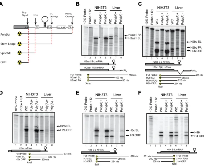
To confirm the expression of the mouse histone mRNAs detected by high-throughput sequencing we analysed his-tone gene expression in actively growing cultured fibrob-lasts (NIH3T3 cells) and liver tissue from 72-week-old mice. The liver is a relatively homogenous tissue with hepatocytes making up more than 70% of the cells. In healthy adult liv-ers, the vast majority of these hepatocytes are quiescent, only re-entering the cell cycle after injury. Unlike the hu-man histone genes, the coding regions of the mouse his-tone genes are highly conserved (>95%) at the nucleotide level (37). Because of the conserved ORF sequences, we can use an S1 nuclease mapping assay to simultaneously analyse both the mRNA expressed from a specific gene, as well as the mRNAs from all the other histone genes encoding that histone protein (26,42). A probe labelled in the coding re-gion will protect all the histone mRNAs to a rere-gion close to the stop codon or initiation codon, as well as a band from the specific gene extending to the 3end or the 5end of the transcript, depending on whether one is probing the 3 or the 5end of the mRNA (Figure3A). This highly sensitive method allows for mapping of multiple isoforms of mRNA from a single sample. Further, since all of the radioactivity is at the end of the probe, the relative intensity of the bands gives the relative amounts of the transcripts. A generalized schematic is shown in Figure 3A. In each experiment be-low, the different protected fragments for a given probe are diagrammed below the autoradiogram.
Total RNA from NIH3T3 cells and mouse liver was fractionated into polyA− and polyA+ RNA
us-ing oligo-d(T) cellulose. Orthologues of human histone HIST1H2BC (Hist1H2bc), HIST2H2AA3 (Hist2H2aa1) and HIST1H4H (Hist1H4h), which were expressed in hu-man tissues, and the HIST2H3C (Hist2H3c) gene, adjacent to the Hist2H2AA3 gene, which was not expressed in hu-man tissues, were analysed by S1 nuclease mapping, using 5
g of total RNA from exponentially growing NIH3T3 cells and 50g of total liver RNA (Figure3). In actively
prolifer-ating NIH3T3 cells, most of the 3S1 probes map two pro-tected fragments: one extending to the 3end of the mRNA ending in the stem-loop and a second fragment, mapping near the stop codon, due to the high level of conservation between the open reading frames of all mouse histone genes of a given class (26). This band is indicated by the name of that class of histone protein (e.g. H3.1 or H2a.1).If the hi-stone mRNA is spliced or polyadenylated then the probe is protected up to the point where the mRNA sequence di-verges from the probe (Figure3A; e.g. at the 5splice site in Hist1H2bc, before the 3end of the mRNA or at the polyA addition site in Hist2H2aa1).
The S1 nuclease protection assays confirmed the low levels of histone mRNAs in liver and there were eas-ily detectable amounts of the polyadenylated forms of Hist2H2aa1 (Figure 3B, lane 7) and Hist1H2bc (Figure
3C, lane 7) mRNAs. There were no polyadenylated mR-NAs detected in the NIH3T3 cells. There is some non-polyadenylated histone mRNA detected in the liver that gives the same pattern of S1 nuclease protected fragments as the NIH3T3 cell mRNA. This likely arises from a low level of expression of the histone mRNAs expressed in growing cells. Note that the non-polyA mRNA expressed in mouse liver is greatly reduced compared to NIH3T3 cells, and we used 10 times more RNA (50 g versus 5 g) for assay of liver RNA, compared to neonatal mice or growing 3T3 cells. We assayed the same RNAs with probes for Hist1H2ac (Figure3D), Hist2H3c (Figure3F) and Hist1H4h (Figure
3E) genes. There was no polyadenylated mRNAs detected from these genes in mouse, consistent with the sequencing data.
Differential transcription of individual histone genes in the adult mouse liver
Nucleic Acids Research, 2016, Vol. 44, No. 19 9197
Figure 3.Expression of histone mRNAs is mouse fibroblasts cells and mouse liver. (A) Schematic of generalized S1 nuclease protection assay performed on mouse histone mRNA. A generalized histone mRNA is depicted indicating possible 5splice sites (5’ss), stem-loop cleavage sites (S-L cleavage) or poly(A) cleavage sites. S1 nuclease protection probes will protect a given mRNA to the point that cleavage or splicing occurs (indicated below). Note that due to the high degree of conservation in mice, all of the mRNAs for a specific histone class (i.e. H2B.1 or H2A.2) will be protected up to the stop codon. (B–F) Total cell RNA from NIH3T3 cells and mouse liver was fractionated into polyA+ and polyA−fractions. Total RNA (whole cell (WC)) and poly(A)+and poly(A−) RNA were probed in an S1 nuclease protection assay. Five micrograms of total 3T3 cell RNA and 50g of liver RNA was analysed using the S1 nuclease protection assay of a 3labelled fragment at the indicated restriction enzyme site for (B) Hist2H2aa1, (C) Hist1H2bc, (D) Hist1H2ac, (E) Hist2H3c or (F) Hist2H4H. The probe used for histone H4H was labelled at the 5end of the indicated restriction site. For each experiment, below autoradiogram, possible mRNA isoforms are indicated as well as protected fragments and sizes for S1 nuclease probes.
In mouse liver, all the histone genes that express polyadenylated mRNAs (Table1) contained sequence reads corresponding to active transcription (e.g. Hist1H2bc (Fig-ure4A) and Hist2h2aa1 (Figure4C)) with prominent peaks near the start site of transcription, and small amounts of transcription detected in the gene body, characteristic of rel-atively lowly expressed genes (46). In contrast, these genes were highly expressed in actively growing mESC (Figure4, lower blue panels) or MEFs (data not shown), with tran-scribing polymerases detected throughout the gene charac-teristic of highly active genes, as well as a peak of activ-ity near the 3 end where the polymerases would be ter-minating. In the liver there were genes that were not ac-tive as detected by GRO-Seq, (e.g. Hist2H3c), while the ad-jacent gene was transcribed (e.g. Hist1H2aa1, Figure4C).
The Hist1H2ac gene gave a GRO-Seq signal similar to the Hist1H2bc gene (Figure4B), even though mRNA was only produced from the Hist1H2bc gene. Note that it is not pos-sible using GRO-Seq to distinguished a gene expressed at a low level, from a gene that has a stalled polymerase and does not express mature mRNAs, since in a low expressed gene, the polymerase is stalled at the promoter most of the time. Surprisingly, there were many genes that were active based on the GRO-Seq signal, but for which we did not detect as accumulating histone mRNAs by RNA-Seq or the S1 nuclease protection assay, e.g. Hist1H2ac, (Figure
his-Figure 4. Paused polymerases are present on many histone genes in mouse liver. We analysed the GRO-Seq data for mouse liver (43) and from mouse embryonic stem cells (mESC) and mouse embryo fibroblasts (44)(not shown). The data for selected histone genes are shown in panelsA–Ffor liver (top) and mESC (bottom). (A) GRO-Seq reads for Hist1H2bc demonstrate a low level of transcription in mature mouse liver and in mESC. In the liver there is a large peak of paused polymerase and a low level of transcription within the gene. Note that characteristic of the highly expressed genes in the mESC, there is a large peak at the 3end of the gene, but smaller peak at the 5end of the gene. (B) GRO-Seq reads at the 5end of the Hist1H2ac suggest engagement of polymerase at the promoter in both liver and mESC. However, Hist1H2ac mRNA does not accumulate in mouse liver (Table1). Note that reads throughout the ORF and 3UTR of the gene are absent in liver in contrast to mESC where it is actively transcribed. (C) GRO-Seq reads for the Hist2H2aa1 gene shows a low level of expression in the liver, and a high level of expression in the mESC. In contrast Hist2H3c is not expressed in the liver (there is no paused polymerase) but shows active transcription in the mESC cells. (D–F) (D) Hist4H4, (E) Hist1H4h and (F) Hist1H3e have a paused polymerase in the liver and active transcription in the mESC cells.
tone genes in non-dividing tissues were ‘active’ but did not accumulate RNA. Whether this is due to a lack of process-ing signals, resultprocess-ing in rapid degradation of the transcript, or failure of the polymerase to enter productive elongation is not clear. Release of paused polymerases is clearly a major regulated event on many genes in mammalian cells (45,47). We verified these findings by analysing RNA Polymerase II ChIP-Seq data sets performed by Sun et al. on mouse liver and brain (not shown) (48). These data confirmed data found by GRO-Seq; however, experimental limitations of their ChIP-Seq only define a region∼400 nts at which RNA Pol II was engaged. Therefore, these data were not as pre-cise, but were consistent with GRO-Seq data.
Differentiation, but not exit from the cell cycle, results in ex-pression of polyadenylated histone mRNAs
The switch to production of polyadenylated histone mR-NAs derived from a few genes could result from exiting the cell cycle, or could be part of the process of terminal differ-entiation. To distinguish between these two processes mouse NIH3T3 fibroblasts were arrested either by serum starva-tion or 3T3-L1 fibroblasts were arrested and induced to ter-minally differentiate into adipocytes. We arrested NIH3T3 cells by serum starvation (Figure5A), causing them to enter a ‘G0’ state. Arrested cells can be stimulated to re-enter the cell cycle by addition of 10% FBS, and they enter S-phase 12–16 h later. There is a large change in histone mRNA levels between cells stimulated to enter S-phase and the ar-rested cells (49). Upon arrest by serum starvation, levels of Hist1H2bc and other H2B.1 mRNAs were reduced as has been previously reported (Figure5B) (49). SLBP protein is
barely detectable (Figure5C, Lane 1), while SLBP mRNA is reduced only 4- to 5-fold (Figure5D), characteristic of the post-transcriptional regulation of SLBP during the cell cycle (9). Serum starvation of NIH3T3 cells leads to a 90% reduction of Hist1H2bc levels as determined by comparing the intensity of the protected bands from 5 micrograms of RNA from growing cells with 50 micrograms of RNA from the arrested cells (see Figure5E, cf. lanes 2 and 6). There was no detectable polyadenylated mRNA expressed from the H2bc gene in the arrested cells (Figure5E, compared to the abundant polyadenylated H2bc mRNA in the same amount of liver mRNA (Figure5E, lane 1). Note that the ratio of the other H2b.1 mRNAs (the protected fragment at the termination codon) and the specific Hist1H2bc mRNA ending at the stem-loop did not change upon serum starva-tion. SLBP protein levels rapidly increase as cells re-enter S-phase (Figure5D, Lanes 2–6) with the maximum number of cells in S-phase 16–20 h after serum stimulation. There-fore, the reduction of SLBP levels is not itself a determinant for the switch from histone mRNAs ending in a stem-loop to those ending in a polyA tail.
Nucleic Acids Research, 2016, Vol. 44, No. 19 9199
Figure 5. Differentiation but not cell cycle arrest activates polyadenylation of histone mRNA. (A) NIH3T3 cells were arrested in G0 by serum starvation and stimulated to reenter the cell cycle by the addition of 10% FBS as previously described (7). (B) Five micrograms of total cell RNA from the indicated time points was analysed by S1 nuclease mapping for the Hist1H2bc mRNA. Lane 7 is 12 h after addition of serum and lane 8 is 30 h after addition of serum. Serum starvation did not result in production of spliced Hist1H2bc mRNA (see also panel E). (CandD) Levels of SLBP protein (panel C) and mRNA (panel D) were determined in cells serum starved for 6 days (lanes 1) and over 20 h after restimulation with serum (lanes 2–6) by Western blotting and RNase protection assays, respectively. Levels of SLBP protein reduced>20-fold in cells serum-starved for 6 days, while levels of SLBP mRNA (panel D) were only reduced 4-fold. The levels of both SLBP protein and SLBP mRNA increased starting 8 h after re-addition of 10% serum. The * (panel C) indicate cross-reacting proteins whose levels don’t change. (D) The RNase protection assay protects a 276 nt fragment of SLBP mRNA. The markers are 350 nt and 242 nt DNA fragments. (E) S1 nuclease protection assays of Hist1H2bc mRNA in 50gs of mouse liver RNA (lane 1), 5 or 50gs of RNA from growing NIH 3T3 cells (lanes 2,3), 5gs of RNA from cells starved for 4 days (lane 4) and 5gs or 50gs of RNA from cells serum starved for 7 days (lanes 5 and 6). Note that the most abundant H2b mRNA in liver is the polyadenylated H2b mRNA. (EandF) Hist2Haa1 mRNA (panel E) and Hist1H2bc mRNA (panel F) were analysed by S1 nuclease protection assay in actively growing pre-adipocyte 3T3-L1 cells and differentiated adipocyte 3T3-L1 cells. Lane 1: 5g 3T3-L1 RNA; Lane 2: 5g neonatal (P1) mouse RNA; Lane 3, 50g 14 week liver RNA; Lane 4: 50g 72 week liver RNA; Lane 5: 5g pre-adipocyte RNA; lane 6. 50g adipocyte RNA. Results demonstrate the production of polyadenylated (E) or spliced and polyadenylated (F) mRNA after differentiation.
not differentiated. To test whether growth arrest or dif-ferentiation of the cells led to polyadenylation of histone mRNA, we compared histone gene expression in serum-starved 3T3 fibroblasts and 3T3-L1 fibroblasts differen-tiated into adipocytes (50). Like the NIH3T3 cells, the pre-adipocyte cells expressed solely stem-loop isoforms of Hist2H2aa1 and Hist1H2bc (Figure5F and G, lane 5). Af-ter seven days of differentiation, the cells had exited the cell cycle and differentiated into adipocytes as indicated by the presence of oilRED droplets in the cells (Supplemen-tary Figure S3A), decrease of expression of Pref-1, and in-duction of expression of Ppar2␥ (51)(Supplementary Fig-ure S3B). There was a large reduction in histone mRNA levels in the differentiated cell cultures compared to the pre-adipocytes (Supplementary Figure S3C, cf. lane 1 and 3). We used 10 times more RNA when probing adipocyte versus pre-adipocytes. As reported by Kariet al. (24), af-ter differentiation, adipocytes began producing polyadeny-lated isoforms of these two mRNAs (Figure5F and G, cf.
lanes 5 and 6). The detection of this band was not due to the increased loading of mRNA, as we did not detect it in pre-adipocyte RNA when 50g was analysed (Supplemen-tary Figure S3C, lane 2). Less polyadenylated mRNA was produced compared to the mRNA from the same genes in mouse liver, and there was still some normally processed Hist2H2aa1 and Hist1H2bc mRNA produced, likely as a result of not all cells entering terminal differentiation. Thus, the process of terminal differentiation may result in alter-ing the pathway of histone mRNA expression, to activate expression of H2A and H2B mRNA isoforms that are nor-mally expressed in terminally differentiated adult tissues.
Adult tissues express specific proteins and mRNAs required for histone mRNA biosynthesis
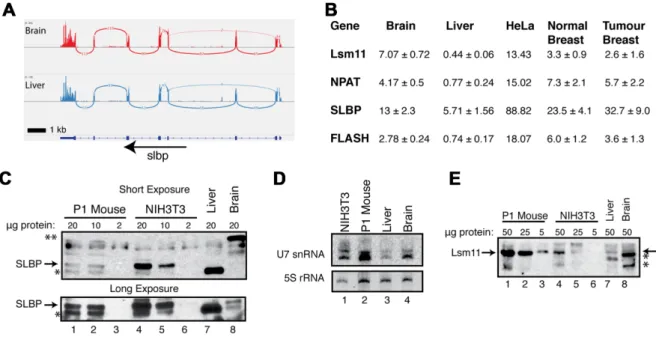
associ-ated with histone mRNA in all steps of histone mRNA metabolism; Lsm10 and Lsm11, proteins found only in the Sm ring of U7 snRNP (52,53); NPAT, a factor required for histone gene expression (54–56) that is concentrated in the Histone Locus Body (HLB), and FLASH, which binds to the Lsm11 protein and is essential for histone pre-mRNA processing (57). FLASH is also involved in FAS-casp8 de-pendent apoptosis, but this may be connected to its role in the HLB, since an aspect of this process is re-localization of FLASH from the HLB to the mitochondria (58). Pre-viously, the Sch ¨umperli lab showed that human tissues ex-pressed SLBP mRNA based on a tissue Northern blot, but did not determine if SLBP protein was present (59). We con-firmed that SLBP mRNA is expressed in mouse (Figure6A) and in human (Supplementary Figure S4E) liver and brain. In addition Lsm10, Lsm11, FLASH and NPAT mRNAs are expressed in both human (Supplementary Figure S4) and mouse (Supplementary Figure S5) liver and brain, as well as all the other human tissues in the Illumina BodyMap Project.
We also quantified the expression data of these factors in mouse liver and brain and in the TCGA data from human breast cancer and the adjacent normal tissue (Figure6B). Both the normal and tumour tissues expressed the mRNAs for all the factors involved in histone mRNA biosynthesis. There were increased levels of SLBP mRNA relative to the expression of the other factors in the tumour compared to the normal tissue. Lsm11, NPAT and FLASH mRNAs were also expressed in normal tissue, at a similar level in both normal and tumour tissue, with the normal tissue slightly higher. The mRNAs for all the factors were expressed at higher levels in cultured HeLa cells.
The expression of many proteins, including SLBP, is reg-ulated post-transcriptionally (9), and the levels of mRNA do not necessarily reflect the amount of protein present. In cultured cells, SLBP protein is tightly cell-cycle regulated by proteolysis and translational regulation, and there are only small changes in SLBP mRNA level, while U7 snRNP is present throughout the cell cycle (9). We analysed the amounts of SLBP protein, U7 snRNA and Lsm11 protein in mouse liver. SLBP was readily detected in lysates from ex-ponentially growing NIH3T3 fibroblasts and from neonatal (P1) mice (Figure6C, lanes 1–6). Consistent with SLBP be-ing present in high levels only in S-phase cells, SLBP was not detected in the liver lysate by Western analysis, and was present at very low levels, if at all, in the brain lysate (long exposure; Figure 6C, lanes 7,8, lower panel). There was a prominent band detected by the␣-SLBP antibody in the liver and in the NIH3T3 lysate which migrated slightly faster than SLBP (indicated by (*) in Figure 6C) and an-other band in brain lysate migrating much slower (indicated by **). However, these cross-reacting bands do not bind the histone stem-loop in an RNA affinity purification as-say, while SLBP does (Supplementary Figure S6), indicat-ing that they are cross-reactindicat-ing proteins.
We also assayed for both U7 snRNA by Northern blot-ting (Figure 6D) and the U7 snRNP specific protein, Lsm11, by Western blotting (Figure 6E), in mouse liver, brain, NIH3T3 cells and the neonatal (P1) mouse. The lev-els of U7 snRNA and the U7 core protein Lsm11 in the P1 mouse were similar to those in NIH3T3 cells, consistent
with many of the cells in the mouse rapidly proliferating. The amount of U7 snRNA in mouse liver and brain was lower than the P1 mouse (Figure6D, lanes 2–4), but, un-like SLBP, was still readily detectable and Lsm11 was also detectable at low levels (Figure6E, lanes 7 and 8). The mR-NAs for U7 snRNP components Lsm10 and Lsm11 were also present in adult tissues (Figure6B; Supplementary Fig-ures S4 and S5). These results strongly suggest that there is U7 snRNP present in terminally differentiated cells.
DISCUSSION
Polyadenylation of histone mRNAs allows for expression of histones outside of S-phase
Following terminal differentiation, it is necessary to replace histones as they are lost, since the proper ratio of DNA and histone proteins is critical for genomic stability (60,61). The average age of many epithelial cells in the human body is several years with neuronal cells living even longer (62). In these cells, there must be a mechanism to replace lost histones that have a half-life of weeks to months (11,12). In continuously growing cells, there are small amounts of replication-dependent histone mRNAs expressed in G1 cells (7) and in fibroblasts arrested in G0 there are also small amounts of histone mRNA processed at the stem-loop (Fig-ure5B) (49).
In the data from the Illumina BodyMap project, in which total RNA (ribominus) was sequenced and hence both polyA+ histone mRNA as well as any normally processed histone mRNAs were sequenced, we did not detect his-tone mRNA processed at the stem-loop from hishis-tone genes that are normally actively expressed (e.g. HIST2H3C or HIST1H1E, Figure1), although a subset of histone genes expressed polyadenylated RNAs. In contrast, ENCODE data from ribominus RNA samples from growing cells there are large amounts of replication dependent histone mRNAs expressed and very small amounts of polyadenylated his-tone mRNAs were expressed (33,63).
A variety of mechanisms might be utilized for synthesis of different histone proteins in terminally differentiated tis-sues. One obvious mechanism to provide replacement hi-stone is to utilize different genes to encode the replace-ment histones. This is the case for the histone H3 genes, but not the other core histone genes. Previously, H3.3 (H3F3A, H3F3B) had been identified as a replacement histone that accumulates as a large fraction of the histone H3 in non-dividing cells over an extended period of time (months to years) (64), as well as playing important functions in gene regulation in growing cells. The H3.3 protein is expressed from genes that produce a spliced polyadenylated mRNA that is not linked to the replication dependent histone gene clusters (65,66). Histone H1Ohas been suggested to
func-tion as a replacement histone for H1 proteins, and again, this gene is expressed as a polyadenylated mRNA lacking a stem-loop and is not linked with the replication-dependent histone gene cluster (67,68).
Nucleic Acids Research, 2016, Vol. 44, No. 19 9201
Figure 6.Expression of Histone Specific factors in mammalian tissues. (A) Sashimi plot demonstrating continued expression of SLBP mRNA in mouse brain (top) and liver (bottom) from (31) (B) RNA-Seq data from 3 replicates of mouse liver and mouse brain (31) and 4 sets of paired human normal breast and breast tumour tissue were analysed for levels of expression of Lsm11, NPAT, SLBP and FLASH. For the 4 human and 3 mouse samples, the average plus standard deviation is indicated. Note the expression levels of all the factors except SLBP were slightly higher in normal tissue. The expression levels of SLBP were higher in the tumour tissue (P=0.06) in a Student’st-test. (C) Analysis of SLBP by Western blotting in cells and tissues from neonatal (P1) mouse (lanes 1–3), 3T3 cells (lanes 4–6) and mouse liver (lane 7) and brain (Lane 8). The amount of protein loaded is indicated above each lane. Tissue specific cross-reacting bands are indicated (*, liver and **, brain) (D) Analysis of U7 snRNA levels in cells and tissues. Twenty five micrograms of total cell RNA from 3T3 cells, a neonatal (P1) mouse, liver and brain were resolved on a 15% urea-polyacrylamide gel and transferred to a nylon membrane. Membranes were probed for U7 snRNA and visualized by exposure to phosphor screen. The gel was stained with ethidium bromide and photographed to determine the relative amounts 5S rRNA. (E) Analysis for Lsm11 by Western blotting in cells and tissues. The indicated amounts of the same extracts used in panel C are shown. Note the high levels of Lsm11 in the P1 mouse (arrow) consistent with the high amount of U7 snRNA in the P1 mouse.
of those in the replication-dependent clusters. We propose that the small subset of the replication-dependent genes that are expressed as polyadenylated mRNAs in termi-nally differentiated cells encode the replacement H2A, H2B and H4 histone proteins. We found both RD-histone H2A and RD-histone H2B genes expressed in all tissues. Most of these mRNAs contain both a stem-loop and a polyA tail, similar to the H2A.X mRNA expressed outside of S-phase. The exceptions are the HIST1H2BC, HIST1H2BD and HIST1H2BK mRNAs, in humans, and Hist1H2bc and Hist1H2bd mRNAs in other mammals. These spliced H2B mRNAs have a conserved structure, using a 5 donor site before the stem-loop, resulting in an mRNA that does not contain the stem-loop and is not a target for nonsense mediated decay. The 3 UTR and 5 splice site are con-served among all these H2B genes, even though they encode slightly different proteins. The other expressed human H2B gene, HIST2H2BE, is found in the HIST2 cluster, and this gene encodes a polyadenylated mRNA that still contains the stem-loop, similar to HIST1H1C or HIST2H2AA3.
Recently, it has been shown that over a six-month period in mouse brain, that histone H3.1, H3.2 and H4 are signif-icantly more stable than histone H2A and H2B (12). This observation suggests that the H2A-H2B protein dimer has a shorter half-life than the H3-H4 tetramer (12). Therefore, H2A and H2B genes would need to be expressed at higher levels to compensate for the increased turnover. There is an isolated RD-H4 gene on human chromosome 11, but this
HIST4H4 gene although conserved as an isolated gene in mammals, is expressed only as a stem-loop RNA (32), and is not expressed in non-dividing tissues. We identified two pos-sible replacement histone H4 genes, which were expressed at very low levels (∼5- to 10-fold less than the H2A and H2B genes), in all human tissues we analysed. Since there have been no other histone H4 genes or cDNAs identified other than the RD-histone H4 genes, these two genes likely pro-vide the replacement histone H4 protein. The depth of se-quencing in the cow and mouse samples was not sufficient to detect a similar level of expression from the H4 genes in these species.
Histone H3.3 has long been recognized as a replace-ment histone, which replaces most of the histone H3 in ter-minally differentiated cells. However, a significant amount of the H3.2 protein remains in non-dividing cells in the mouse (14) (see Figure7). We identified one RD-histone H3 gene, HIST1H3E, which is expressed at low levels in terminally differentiated human cells, similar to the level of the HIST1H4H gene. This is consistent with the possibil-ity that there is a small amount of ‘replacement’ histone H3 encoded by this replication-dependent gene.
Figure 7. Expression of Histone proteins in mouse liver. Total histones were isolated from purified nuclei were prepared from the livers of mice from 1 week before birth (−1) to 30 weeks of age. The histones were re-solved on Triton-X/Acid/Urea Polyacrylamide gels (6 mM Triton-X 100, 5% Acetic Acid, 6M Urea) and stained with Amido Black. ‘T’ is histones from mouse thymus that shows a similar pattern as mouse embryos. The H2A.1, H2B.2 and H3.1 and H3.2 proteins decrease over time, while the H2A.2, H2B.1, H3.3 and H10proteins accumulate with time. The mul-tiple bands for H3.2, H3.3 and H4 proteins are acetylated forms of his-tones. These changes in protein levels are consistent with the core histone genes that continue to be expressed in tissues. Reproduced from Zweidler, A. (1984) In: Stein, G., Stein, W. and Marzluff, W. F. (eds.),Histone genes: structure, organization and regulation. John Wiley and Sons, New York, pp. 373–395 with permission of John Wiley & Sons Ltd. CopyrightC 1984 by
John Wiley & Sons, Inc.
genes (all the histone H4 genes encode the same protein). There are increases in the relative amounts of the H2A.2 protein (encoded by the Hist2H2aa1 gene), the H2B.1 tein (encoded by the Hist1H2bc gene), and the H3.2 pro-tein, (encoded by the Hist1H3h gene) in mouse liver dur-ing the first 30 weeks. At the same time the H3.3 and H1O
proteins increase. Thus, the RD-histone genes we have iden-tified encode the proteins that accumulate in adult mouse non-dividing tissues.
Polyadenylation of histone mRNAs may be a feature of ter-minal differentiation
Knockdown of a number of factors in RNA biosynthesis by RNAi in cultured human cells leads to polyadenylation of a small percentage of histone mRNA. These include knock-down of NELF, a transcription elongation inhibitor (19);
ARS2, a factor required for efficient cell cycle progression (20) that is also present in the NEXT complex that binds to nascent capped transcripts (69); CDK9 (21), a transcrip-tion elongatranscrip-tion factor; and RNF20 and RNF40, which are involved in H2B ubiquitination (21). In many of these ex-periments the primary assay is measurement of changes of polyadenylated histone mRNA by qRT-PCR of oligo(dT) primed cDNA. Results are often reported as ‘fold-increase’ and large fold-increases in polyadenylated histone mRNAs are often observed using this technique. In the cases where the expression of the polyadenylated histone mRNAs rela-tive to normally processed histone mRNAs have been mea-sured (19,20,22), the amount of poly(A)+ histone mRNA is in the range of 2–3% of the properly processed RNA. The large increases reported reflect the fact that the amount of polyadenylated histone mRNA under normal conditions is extremely low. It is likely that all of these treatments disrupt the coupling between transcription and histone 3end pro-cessing, resulting in a small amount of RNA, which is not processed normally and may then be polyadenylated. The HIST2H2AA3 mRNA and several histone H2B mRNAs which are expressed in tissues are among those affected. In at least one case, the polyadenylated histone mRNA formed is not the same as the polyadenylated histone mRNAs ex-pressed in tissues. Knockdown of SLBP resulted in expres-sion of a novel histone H4 mRNA detected by Northern blotting (23). This RNA was derived from the HIST1H4J gene, which is not expressed in human tissues.
The switch in histone gene expression is likely a conse-quence of terminal differentiation, and not simply a con-sequence of cells stopping cell division. When 3T3 fibrob-lasts are arrested cells in G0 by serum starvation, we do not detect expression of polyadenylated histone mRNAs over the 7 day serum starvation. When we differentiated the mouse 3T3-L1 fibroblasts over 7 days, polyadenylated Hist1h2bc and Hist2h2aa1 mRNAs were expressed, consis-tent with activation of this pathway in terminally differen-tiated cells, and not simply as a result of serum starvation. Our data contrasts with the report of Pirngruberet al. that serum starvation of HTC116 cells resulted in polyadenyla-tion of HIST1H2BC mRNA (70), and other pharmacolog-ical treatments that also arrest cells show a similar result. In their study, they reported a 3-fold increase in polyadeny-lated HIST1H2BC in response to serum starvation, but they did not measure the absolute amount of polyadenylated mRNAs expressed. In subsequent experiments, Kariet al. (24) reported that there are increases in the same polyadeny-lated H2B and H2A mRNAs that we observed are expressed in human tissues, when human fibroblasts differentiate into adipocytesin vitro. They did not compare these results with the effect of serum starving these fibroblasts.
Nucleic Acids Research, 2016, Vol. 44, No. 19 9203
senescent cells, which may represent another type of termi-nal differentiation.
How does the switch to polyadenylated histone mRNAs oc-cur?
A combination of factors likely determines how his-tone mRNAs switch their 3 end formation and which genes remain expressed. In actively growing cells, at the G1/S-phase transition, CycE/Cdk2 phosphorylates NPAT (54,55,72,73) resulting in a high rate of expression of pro-cessed histone mRNAs. In cultured mammalian cells, there is a significant rate of histone gene transcription outside of S-phase (49) or when histone RNA levels are rapidly re-duced by inhibiting DNA replication (74,75). Surprisingly, the available GRO-Seq data from Fanget al. (43) show that in mouse liver most histone genes (about 80%) have stalled RNA polymerase II at their promoters including the genes that express polyadenylated histone mRNAs. Many of these genes do not express histone mRNA in tissues.
One could imagine two different extremes for the state of these genes. First, only the genes expressed in tissues release RNA polymerases at a low rate from their promoters into productive elongation or second, many of the RD-histone genes with stalled polymerases at their promoters are tran-scribed at a low level but do not accumulate any mRNA because there are not effective splicing/polyadenylation sig-nals. Comparing the GRO-Seq data from growing cells with mouse liver and brain, it is clear that a major regulatory step is at the level of transition of the RNA polymerase into processive elongation after synthesis of the short tran-script. Genes that are expressed as polyadenylated mRNAs also have a stalled polymerase at their promoter, and the re-lease of this polymerase into productive elongation is likely tightly controlled resulting in production of low levels of polyA mRNA. The importance of regulation at this step has been emphasized recently by studies of Adelmanet al. (47,76). The levels of some of these mRNAs (HIST1H3E, HIST2H4, HIST1H4H and HIST1H2AC) may be further reduced by NMD, since they are all spliced with an intron in the 3UTR more than 100 nts 3of the stop codon. Both the HIST1H2B mRNAs, and the HIST2H2AA3 mRNAs that are expressed are not sensitive to NMD and accumulate at significantly higher levels that the other transcripts that are spliced.
Some of the histone genes, e.g. the Hist2H3c gene adja-cent to the expressed Hist2H2aa1 gene, are not expressed in tissues (do not have a stalled polymerase at the promoter), although the Hist2H3c gene is expressed at high levels in cultured mouse cells (75). In growing cultured cells, the his-tone genes do not show a strong GRO-Seq peak at the pro-moter, but show transcription throughout the gene with a major peak 3of the processing site, consistent with the high level of RNA polymerase present at histone genes (77,78).
The expression of components of the histone locus body, NPAT and FLASH, in terminally differentiated cells sug-gest that they play a role in the expression of histone genes in tissues, supporting a basal level of transcription of the histone genes, likely by forming a histone locus body. In normally growing fibroblasts (54,56) and ES cells (79,80), the HLB persists throughout the cell cycle, as it does in
Drosophila, both in replicating and non-replicating cells (81–83). SLBP mRNA was also expressed in the tissues. It was expressed at higher levels in cultured cells compared to the other mRNAs, and at higher levels in human breast tu-mours relative to the adjacent normal tissue. In contrast to the components of U7 snRNP, the level of SLBP protein in the liver and brain is dramatically reduced despite expres-sion of the SLBP mRNA, as it is in cultured cells in G1. The reduction of SLBP protein is unlikely to be the deter-minant for the switch to polyadenylated histone mRNA in differentiated cells. This is most clearly demonstrated by ar-rest of fibroblasts in a G0 state, which reduces the levels of SLBP protein, but does not cause result in the accumula-tion of polyadenylated histone mRNA, and the failure of the HIST1H4J gene to be expressed in tissues, although it is expressed as a polyadenylated mRNA in HeLa cells when SLBP is knocked down (23).
In summary, we report a subset of histone genes express polyadenylated histone mRNAs (many of which are also spliced) in terminally differentiated tissues. These mRNAs likely encode the replacement core histones which turn over very slowly in these cells, which are very long-lived. The pro-teins expressed from these genes encode the same histone variants proteins shown to accumulate in mouse tissues by Fred Zweidler years ago, replacing some of the histone pro-teins originally present in these tissues.
SUPPLEMENTARY DATA
Supplementary Dataare available at NAR Online.
ACKNOWLEDGEMENTS
The authors would like to thank Dr Jessica Sorrentino and Dr Norman Sharpless for providing frozen dissected mouse liver. We would like to thank Dr Jason Merkin and Dr Chris Burge for sharing data prior to publication and Dr Nancy Kedersha for critical reading of the manuscript.
FUNDING
National Institutes of Health (NIH) [GM29832 to W.F.M., GM67113 to Y.X., CA130795 to M.L.W.]; Summer Un-dergraduate Research fellowships from the UNC Office of Undergraduate Research [to C.H.C., A.G. and B.W.]; NSF Graduate Research Fellowship [DGE-1144081] and NIH BD2K Fellowship [T32 CA201159] to J.D.W. Funding for open access charge: National Institutes of Health (NIH) [GM29832 to W.F.M., GM67113 to Y.X., CA130795 to M.L.W.].
Conflict of interest statement.None declared.
REFERENCES
1. Marzluff,W.F., Wagner,E.J. and Duronio,R.J. (2008) Metabolism and regulation of canonical histone mRNAs: life without a poly(A) tail.
Nat. Rev. Genet.,9, 843–854.
2. Dominski,Z., Yang,X.C. and Marzluff,W.F. (2005) The
polyadenylation factor CPSF-73 is involved in histone-pre-mRNA processing.Cell,123, 37–48.
4. Gick,O., Kr¨amer,A., Keller,W. and Birnstiel,M.L. (1986) Generation of histone mRNA 3ends by endonucleolytic cleavage of the pre-mRNA in a snRNP-dependent in vitro reaction.EMBO J.,5, 1319–1326.
5. Cotten,M., Gick,O., Vasserot,A., Schaffner,G. and Birnstiel,M.L. (1988) Specific contacts between mammalian U7 snRNA and histone precursor RNA are indispensable for the in vitro 3RNA processing reaction.EMBO J.,7, 801–808.
6. Schaufele,F., Gilmartin,G.M., Bannwarth,W. and Birnstiel,M.L. (1986) Compensatory mutations suggest that base-pairing with a small nuclear RNA is required to form the 3end of H3 messenger RNA.Nature,323, 777–781.
7. Harris,M.E., B ¨ohni,R., Schneiderman,M.H., Ramamurthy,L., Sch ¨umperli,D. and Marzluff,W.F. (1991) Regulation of histone mRNA in the unperturbed cell cycle: Evidence suggesting control at two posttranscriptional steps.Mol. Cell. Biol.,11, 2416–2424. 8. Zheng,L.X., Dominski,Z., Yang,X., Elms,P., Raska,C.S.,
Borchers,C.H. and Marzluff,W.F. (2003) Phosphorylation of SLBP on two threonines triggers degradation of SLBP, the sole cell-cycle regulated factor required for regulation of histone mRNA processing, at the end of S-phase.Mol. Cell. Biol.,23, 1590–1601.
9. Whitfield,M.L., Zheng,L.X., Baldwin,A., Ohta,T., Hurt,M.M. and Marzluff,W.F. (2000) Stem-loop binding protein, the protein that binds the 3end of histone mRNA, is cell cycle regulated by both translational and posttranslational mechanisms.Mol. Cell. Biol.,20, 4188–4198.
10. Hancock,R. (1969) Conservation of histones in chromatin during growth and mitosis in vitro.J. Mol. Biol.,40, 457–466.
11. Bondy,S.C. (1971) The synthesis and decay of histone fractions and of deoxyribonucleic acid in the developing avian brain.Biochem. J.,
123, 465–469.
12. Toyama,B.H., Savas,J.N., Park,S.K., Harris,M.S., Ingolia,N.T., Yates,J.R. 3rd and Hetzer,M.W. (2013) Identification of long-lived proteins reveals exceptional stability of essential cellular structures.
Cell,154, 971–982.
13. Kohler,S.J., Williams,N.I., Stanton,G.B., Cameron,J.L. and Greenough,W.T. (2011) Maturation time of new granule cells in the dentate gyrus of adult macaque monkeys exceeds six months.Proc. Natl. Acad. Sci. U.S.A.,108, 10326–10331.
14. Zweidler,A. (1984) In: Stein,G, Stein,W and Marzluff,WF (eds).
Histone genes: structure, organization and regulation. John Wiley and Sons, NY, pp. 373–395.
15. Panyim,S. and Chalkley,R. (1969) A new histone found only in mammalian tissues with little cell division.Biochem. Biophys. Res. Commun.,37, 1042–1049.
16. Paull,T.T., Rogakou,E.P., Yamazaki,V., Kirchgessner,C.U.,
Gellert,M. and Bonner,W.M. (2000) A critical role for histone H2AX in recruitment of repair factors to nuclear foci after DNA damage.
Curr. Biol.,10, 886–895.
17. Bonner,W.M., Mannironi,C., Orr,A., Pilch,D.R. and Hatch,C.L. (1993) Histone H2A.X gene transcription is regulated differently than transcription of other replication-linked histone genes.Mol. Cell. Biol.,13, 984–992.
18. Mannironi,C., Bonner,W.M. and Hatch,C.L. (1989) H2A.X. a histone isoprotein with a conserved C-terminal sequence, is encoded by a novel mRNA with both DNA replication type and polyA 3 processing signals.Nucleic Acids Res.,17, 9113–9126.
19. Narita,T., Yung,T.M., Yamamoto,J., Tsuboi,Y., Tanabe,H.,
Tanaka,K., Yamaguchi,Y. and Handa,H. (2007) NELF interacts with CBC and participates in 3end processing of replication-dependent histone mRNAs.Mol. Cell,26, 349–365.
20. Gruber,J.J., Olejniczak,S.H., Yong,J., La Rocca,G., Dreyfuss,G. and Thompson,C.B. (2012) Ars2 promotes proper replication-dependent histone mRNA 3end formation.Mol. Cell,45, 87–98.
21. Pirngruber,J., Shchebet,A., Schreiber,L., Shema,E., Minsky,N., Chapman,R.D., Eick,D., Aylon,Y., Oren,M. and Johnsen,S.A. (2009) CDK9 directs H2B monoubiquitination and controls
replication-dependent histone mRNA 3-end processing.EMBO Rep.,10, 894–900.
22. Kohn,M., Ihling,C., Sinz,A., Krohn,K. and Huttelmaier,S. (2015) The Y3** ncRNA promotes the 3end processing of histone mRNAs.Genes Dev.,29, 1998–2003.
23. Sullivan,K.D., Mullen,T.E., Marzluff,W.F. and Wagner,E.J. (2009) Knockdown of SLBP results in nuclear retention of histone mRNA.
RNA,15, 459–472.
24. Kari,V., Karpiuk,O., Tieg,B., Kriegs,M., Dikomey,E., Krebber,H., Begus-Nahrmann,Y. and Johnsen,S.A. (2013) A subset of histone H2B genes produces polyadenylated mRNAs under a variety of cellular conditions.PLoS One,8, e63745.
25. Wang,Z.F., Sirotkin,A.M., Buchold,G.M., Skoultchi,A.I. and Marzluff,W.F. (1997) The mouse histone H1 genes: Gene
organization and differential regulation.J. Mol. Biol.,271, 124–138. 26. Graves,R.A., Wellman,S.E., Chiu,I.M. and Marzluff,W.F. (1985)
Differential expression of two clusters of mouse histone genes.J. Mol. Biol.,183, 179–194.
27. Student,A.K., Hsu,R.Y. and Lane,M.D. (1980) Induction of fatty acid synthetase synthesis in differentiating 3T3-L1 preadipocytes.J. Biol. Chem.,255, 4745–4750.
28. Gerin,I., Bommer,G.T., Lidell,M.E., Cederberg,A., Enerback,S. and Macdougald,O.A. (2009) On the role of FOX transcription factors in adipocyte differentiation and insulin-stimulated glucose uptake.J. Biol. Chem.,284, 10755–10763.
29. Dominski,Z., Yang,X.C., Purdy,M. and Marzluff,W.F. (2003) Cloning and characterization of the Drosophila U7 small nuclear RNA.Proc. Natl. Acad. Sci. U.S.A.,100, 9422–9427.
30. Dominski,Z., Yang,X., Kaygun,H. and Marzluff,W.F. (2003) A 3 exonuclease that specifically interacts with the 3end of histone mRNA.Mol. Cell,12, 295–305.
31. Merkin,J.J., Chen,P., Alexis,M.S., Hautaniemi,S.K. and Burge,C.B. (2015) Origins and impacts of new mammalian exons.Cell Rep.,10, 1992–2005.
32. Marzluff,W.F., Gongidi,P., Woods,K.R., Jin,J. and Maltais,L.J. (2002) The human and mouse replication-dependent histone genes.
Genomics,80, 487–498.
33. Brooks,L. 3rd, Lyons,S.M., Mahoney,J.M., Welch,J.D., Liu,Z., Marzluff,W.F. and Whitfield,M.L. (2015) A multiprotein occupancy map of the mRNP on the 3end of histone mRNAs.RNA,21, 1943–1965.
34. Yang,L., Duff,M.O., Graveley,B.R., Carmichael,G.G. and Chen,L.L. (2011) Genomewide characterization of non-polyadenylated RNAs.
Genome Biol.,12, R16.
35. Zhang,J., Sun,X., Qian,Y., LaDuca,J.P. and Maquat,L.E. (1998) At least one intron is required for the nonsense-mediated decay of triosephosphate isomerase mRNA: a possible link between nuclear splicing and cytoplasmic translation.Mol. Cell. Biol.,18, 5272–5283. 36. Nagy,E. and Maquat,L.E. (1998) A rule for termination-codon
position within intron-containing genes: when nonsense affects RNA abundance.Trends Biochem. Sci.,23, 198–199.
37. DeBry,R.W. and Marzluff,W.F. (1994) Selection on silent sites in the rodent H3 histone gene family.Genetics,138, 191–202.
38. Taylor,J.D., Wellman,S.E. and Marzluff,W.F. (1986) Sequences of four mouse histone H3 genes: implications for evolution of mouse histone genes.J. Mol. Evol.,23, 242–249.
39. Moss,S.B., Ferry,R.A. and Groudine,M. (1994) An alternative pathway of histone mRNA 3end formation in mouse round spermatids.Nucleic Acids Res.,22, 3160–3166.
40. Cheng,G.H. and Skoultchi,A.I. (1989) Rapid induction of
polyadenylated H1 histone mRNAs in mouse erythroleukemia cells is regulated by c-myc.Mol. Cell. Biol.,9, 2332–2340.
41. Cheng,G.H., Nandi,A., Clerk,S. and Skoultchi,A.I. (1989) Different 3-end processing produces two independently regulated mRNAs from a single H1 histone gene.Proc. Natl. Acad. Sci. U.S.A.,86, 7002–7006.
42. Liu,T.J., Liu,L. and Marzluff,W.F. (1987) Mouse histone H2A and H2B genes: four functional genes and a pseudogene undergoing gene conversion with a closely linked functional gene.Nucleic Acids Res.,
15, 3023–3039.
43. Fang,B., Everett,L.J., Jager,J., Briggs,E., Armour,S.M., Feng,D., Roy,A., Gerhart-Hines,Z., Sun,Z. and Lazar,M.A. (2014) Circadian enhancers coordinate multiple phases of rhythmic gene transcription in vivo.Cell,159, 1140–1152.


![Poly[triaquatris(μ 4 pyridine 3,5 dicarboxylato)didysprosium(III)]](data:image/gif;base64,R0lGODlhAQABAIAAAP///wAAACH5BAEAAAAALAAAAAABAAEAAAICRAEAOw==)