INTERNATIONAL RESEARCH JOURNAL OF PHARMACY
www.irjponline.com
ISSN 2230 – 8407
Research Article
COMPARATIVE ANALYSIS OF LIGAND BINDING MODES OF PPAR-
γ
FULL AND PARTIAL AGONISTS
Nagashree K S, Praveen T. Krishnamurthy *
Department of Pharmacology, JSS College of Pharmacy, Ooty (JSS Academy of Higher Education and Research), Ooty,
Tamil Nadu, India
*Corresponding Author Email: [email protected]
Article Received on: 30/03/19 Approved for publication: 14/05/19
DOI: 10.7897/2230-8407.1009266
ABSTRACT
Clinically used thiazolidinediones such as rosiglitazone and pioglitazone control hyperglycemia by acting as full agonist on PPARγ receptors. Unfortunately, they suffer with serious side effects such as weight gain, fluid retention, congestive heart failure, bladder cancer etc. It is reported that, these effects are attributed to full activation of PPARγ receptors by these ligands. Newer approaches like development and selective PPAR modulators or partial agonists have been extensively researched to overcome or reduce these side-effects. In this research paper, we have carried out a comparative analysis of binding agonists. The results show that PPARγ full agonists form H-bond interactions with TYR473, His 449, His 323 whereas partial agonists form H-bond interaction with Ser 342, Ser 289 and Arg 280. The above findings, therefore, may help in choosing PPAR-γ leads with partial agonistic activity.
Keywords: rosiglitazone, pioglitazone, PPARγ receptors
INTRODUCTION
Thiazolidinedione’s (TZDs) or glitazones are insulin sensitizers1
which act by increasing the trans activation activity of PPARγ2.
Peroxisome Proliferator Activated Receptors (PPARs) are transcription factors3, that can be turned on or off by binding to
small lipophilic compounds due to their pleiotropic effect4. They
act by coordinating the activities of multiple pathways involved in metabolism instead of acting through one major target like one enzyme or one pathway5. This unique property of PPARs has
created lot of interest for their possible use in a complex metabolic disorder such as type 2 diabetes mellitus (T2DM).
Further, TZDs reverse insulin resistance without causing hypoglycemic effect which is major side effect of most widely used antidiabetic drugs such as sulfonylureas. They reduce hepatic output of glucose and increase peripheral uptake, leading to reducing both pre-load and afterload on the beta cell6. Thus,
providing an excellent rationale for the use of glitazones in T2DM.
Unfortunately these clinically used glitazones / Thiazolidinedione (TZDs) such as Troglitazone, Rosiglitazone and Pioglitazone suffer with some serious side effects such as idiosyncratic hepatotoxicity, fluid retention, Cardiac heart failure, bone fracture, bladder cancer, weight gain etc.,7-10
TZDs, such Troglitazone, the first TZD approved as antidiabetic agent for clinical use in 1997 by USFDA, unfortunately caused fatal idiosyncratic hepatotoxicity and withdrawn from the market in the year 2000. Later rosiglitazone and pioglitazone were got approved for clinical use in US by 199911. Later Rosiglitazone
was also banned in many countries. In May 2011, US FDA imposed several restrictions on its prescribing and use as a result of its ability to increase the risk of heart failure in susceptible
individuals. Pioglitazone, unlike rosiglitazone, did not attract the same degree of controversy with regard to cardiovascular risks. However recently concerns were raised on the apparent risk of bladder cancer with pioglitazone as a result FDA has updated the label of pioglitazone12. Troglitazone alone faced hepatotoxicity
not all other glitazones this may be due to it is derivative of quinone metabolite which caused13.
Therapeutic effects and side effects of TZDs coincide with each other such a way that increase in dose increases efficacy and also degree of side effect14,15. One of the reasons for the failure of
these clinically used glitazones is, their time of development. Even though TZDs are well known and proven for their glucose lowering activity in 1988,16 but it was only in the year 1995 their
target, PPARγ (a regulatory master of adipogenesis) was identified17. This indicates that these drugs were developed when
there was very little scientific data available on structure and the transcriptional mechanisms of the target peroxisome proliferator activated receptors (PPARs).
Recent advancements in PPAR ligands
Recent advances in the understanding the structure and function of PPARs, however, have led to more rationalized approaches towards the discovery of glitazones. These include the development PPAR- dual agonists, PPAR-pan agonists and Selective PPAR-γ modulators (SPPARγMs) or partial agonists18.
SPPARγMs or Partial agonists
SPPARγMs provide a target oriented therapeutic profile by maintaining the desired therapeutic benefits and at the same time have minimal adverse effects due to their inability to fully activate the receptor as that of a full agonist19. SPPARMs are reported to
PPAR receptors and thus selectively activating the genes responsible for insulin sensitization, adipogenesis, fluid retention and bone remodeling20.
SPPARγMs hypothesis is based on recruitment of certain differential receptor binding andco-factor recruitment/displacement which specifically selective tissue and their expression in favorable target cells. This concept is from similar approach which successfully shown in case of selective estrogen receptor modulators (SERMs)21. Tamoxifen and
raloxifene due to their specifically selective tissue gene regulation they behave differently in different tissue. In bone and cardiovascular tissues they act as agonist whereas in breast tissue as antagonists22,23.
Side effects of PPARγ full agonist such as weight gain or fluid retention, may occur through full agonism24 and due to their
substantial portion of the inhibition of the phosphorylation at Ser27325. Thus, an effective partial agonist of PPARγ would have
a weak transactivation activity and high phosphorylation inhibitory activity on phosphorylation at Ser27326. These
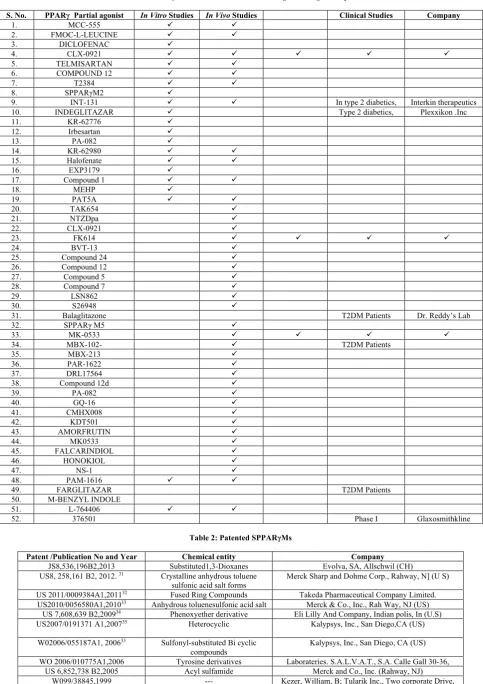
compounds could, therefore, provide the same therapeutic benefits without the associated side effects. Many SPPARγMs are in the pipeline and many have shown promising activities without the side effects related to PPARY activation (Table 1).
The differential effect of SPPARγMs have been observed in vitro, where they have shown low adipogenesis and increased glucose uptake, which in turn translate in vivo into insulin sensitization with little or no weight gain. Notably, some of them have even shown no fluid retention or plasma volume expansion, validating the SPPARγMs concept of retaining insulin sensitization while avoiding the side effects27 and few of them which are in pipeline
are listed in Table 1.
During last decade, a major investment was made by pharmaceutical industry to develop SPPARγMs. This increased interest on SPPARγMs has drastically increased the number of patent application and interest of the pharmaceutical industry on SPPARγMs fetching their wings towards patenting the novel SPPARγMs which reported with significant enhanced insulin sensitivity without having any serious adverse events. Some of the companies hold patent on SPPARγMs which are on pipeline which reported with significant enhanced insulin sensitivity without having any serious adverse effects listed in Table 2.
PPARs belong to the nuclear hormone receptor superfamily consisting of more than 48 receptors but with very distinct in their function they share a common structure consisting of 5 conserved regions or domains. These include the N-terminal A/B domain (LBD), a medial DNA binding domain (DBD), hinge region and the C-N terminal ligand binding domain. The N-terminal A/B domain is reported to least conserved region, showing significant variations in length between the receptors belonging to this superfamily. This region, reported to contain a weak ligand independent transcriptional activation function (AF-1), is often a site for posttranslational medications that can affect receptor activity. The C 7 region is reported to contain the DBD with 2 zinc finger motifs and is highly conserved among the superfamily members. The D region functions as a hinge and allows the C and E domains to swivel slightly to accommodate multiple
Structure of PPARγ
The LBD is folded into a single domain with 13 helices (H) and 4 stranded B sheets (S1 to S4). In contrast to other NRs, PPARS LBD contain extra helix H2’and the helices H10 and H11 are one continuous helix. The ligand binding site is reported to be a large Y shaped cavity; this cavity is enclosed by helices H2’, H3, H4, H5, H7, H10/H11, H2 and B strands s3 and s4. The c terminal helix H12 is positioned closer to the LBD and it is known as AF-2 and reported to extend from the surface of protein and the branches into two arms. The ARM-I extends towards AF-2 (H12 helix) and I is found to be substantially polar. The 4 polar residues of Arm-I is reported to be highly conserved isotypes Ser289, His 323, His 449, Tyr 473 in PPARγ. These residues are reported to take part in the hydrogen bonding interactions with the natural ligands and with synthetic ligands like TZDS. The hydrogen bonding between the Tyr 473 of H12 (AF-2) helix and ligand play an important role by holding the AF-2 region in active conformation, which allows Coactivators binding.
Protein ligand interactions play central role in biology and as we know biological processes are often depend on protein–ligand binding events28. At present they are several number of protein
structures in the Protein Data Bank this increase in number which has open the door for researchers to use data and analyze according to their need. Thus, we made an effort to list and know which types of interactions are formed between ligands and proteins of PPARγ receptor. In this article, effort been made to analyze and compare different protein ligand binding interactions with amino acid residues of PPARγ full and partial agonists.
The aim in this survey is to check and list different interactions reliably in a representative group of protein–ligand crystal structures which will help to design new PPAR gamma partial agonists which have different interactions than full agonists.
MATERIALS AND METHODS
95X-ray crystal structures of protein–ligand complexes from the Protein Data Bank (PDB) (https://www.rcsb.org) were taken and protein - ligand interactions were listed. Among them 60 are full agonists and 35 are partial agonists. Protein–ligand interactions were listed from pose view Image of 2D interaction diagram of particular PDB ID and were analyzed for their type of interactions.
We listed following different interactions between each ligand and receptor among full and partial agonists.
• Hydrogen bonding • (π-π) and
• Hydrophobic interactions with were analyzed and listed.
calculated by considering distribution of each amino acid residue in how many ligandsv/s number of ligands x 100. Further common and different residues among full and partial agonists were listed and % is calculated. Later comparative observation of amino acid residues interactions among full and partial agonists were listed and studied.
RESULTS OF ANALYSIS
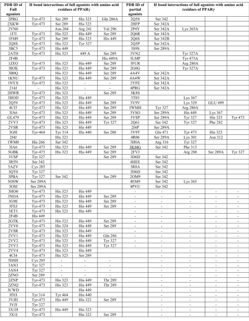
Hydrogen bonding interactions in LBD of PPARγ full and partial agonists and percentage distributions of various amino acid residues were listed in Table 3, 4 and 5, Figure 1 and 2, respectively. After comparative analysis, we observed that among 60 full agonists, 20 different amino acid residues were interacted with hydrogen bond and 17 in case of 35 partial agonists (Table 3). Only 5 amino acid residues were common in case of both partial agonists and Full agonists (Table 4).
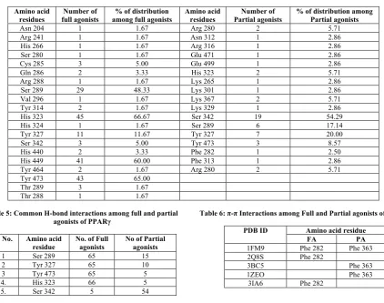
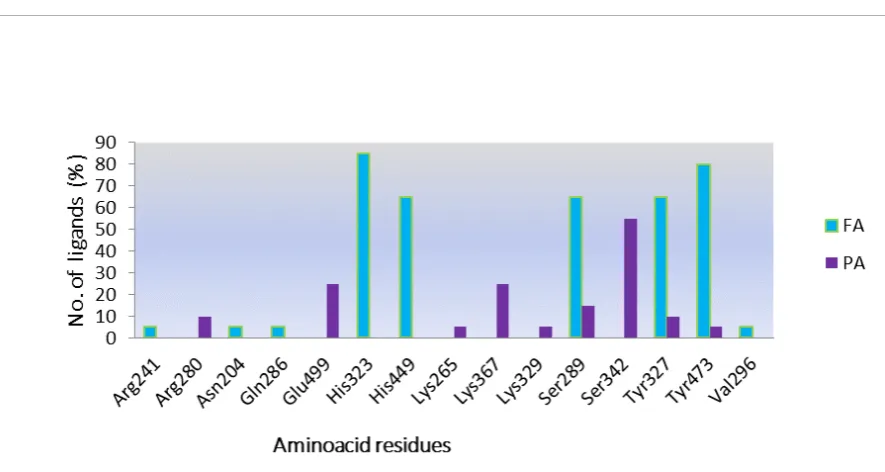
In full agonists, residues like Tyr 327, His 449, His 323 were found to be common residues which interacted through hydrogen bond formation. Whereas, In case of Partial agonist, residues like Ser 342, Ser 289 and Arg 280 are found to be common residues. When we compared both full and partial agonist interactions very few common interactions like Ser 289, Tyr 327, Tyr 473, Arg 280 His 449 were found. Even though common interactions were found but distributed only in one or two ligands among 35 partial agonists.
When we calculated the individual % distribution among 60 full agonist, 66.6 % of the full agonists were interacted with His 323 and 65% with Tyr 473, followed by His 449- 60% and Ser 289-48.3%. Whereas among 40 partial agonists Ser 342 interacted among 54 % and Ser 289-17% .Whereas in case of % distribution of H bond interactions partial agonist we can see very few common interactions as that of full agonist in case of partial agonists among 35 partial agonisted,19 ligands i.e 54% are binding with Ser 342 as amino acid residue. Remaining 15 full and Partial have different amino acid residues but only 5% of them has interactions with Tyr-473, His 323-5.7%, His 449-2.85%, Ser 289- 17% (Table 4 and Figure 2).
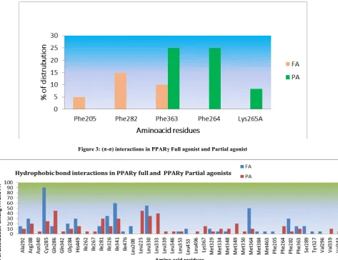
π-π interactions
We can list 5 amino acid residues among full and 3 in partial agonists which formed π-π interactions. Few (π-π) interactions are seen in both which are listed in Table 6 and Figure 3. Among 6 amino acid residues, only Phe 363 amino acid residue is common.
Hydrophobic interactions
From the analysis we observed that, 46 amino acid residues were interacted 60 full agonists which formed hydrophobic interactions and 33 in among 35 Partial agonists. When we compared it found that 17 i.e. > 50% hydrophobic interactions were found common. Among 60 full agonists Cys 285, Ile 341, Leu 330, Met 364 are mostly interacted amino acid residues through hydrophobic interactions; whereas in case of Partial agonists Gln 286, Leu 225, 330 and 333 are mostly interacted one. Ile 476, Leu 208, 453, Met
384, 463, Phe 205, Tyr 327, Val 296 are different amino acid residues which were distributed only in Full agonists (Table 7 and Figure 4).
DISCUSSION
Protein ligand interactions play central role in biology and as we know biological processes are often depend on protein–ligand binding events. In this comparative analysis, effort been made to list such Protein ligand interactions among full and partial PPARγ agonists.
Full agonist binds to PPARγ helix H12, stabilizing the agonist conformation through a direct hydrogen bond to Tyr 473, allowing H12 to dock against H3 and H11 helix29. Since there was
no change in conformation upon binding these various ligands, full agonists may function by directly stabilizing the AF-2 co-activators binding site, while partial agonists only stabilize regions away from H12, leaving H12 in a highly dynamic state. This differential stabilization may also transmit to regions of the receptor away from AF-2, such as the β sheet, suggesting a distinct co-activators binding surface, consistent with these findings that regions outside the multifunctional binding sequence motifs contribute to receptor binding.
It was found from this comparative analysis, Few of the partial agonists have shared common interactions as that of full agonists but none of them occupied and stabilized AF2 region as full agonists like rosiglita zone does (His 323, Tyr 473 and His 449), which is one the reason for their full agonistic activity instead majority ˃ 50% partial agonists interacted with Ser 342. Further single common residues were found in π-π interactions. Number of amino acid residues of which formed hydrophobic interactions is more in case of 35 partial agonists when compared to 60 full agonists. An increase in the number of hydrophobic atoms in the active core of drug -target interface further increases the biological activity of the drug lead30. With this we conclude that
among 60 full agonists, almost 80% of the full agonist interacts with His 323, and Tyr 473 which may be reason for full agonism and influences adverse effect; whereas, alternatively, partial agonists make hydrogen bond with other residues in the proximity such as Ser 342, Glu 499, Arg 280, Lys 367, Tyr 327 or Ser 289.
CONCLUSION
Table 1: SPPARγMs which are under various stages of drug development
S. No. PPARγ Partial agonist In Vitro Studies In Vivo Studies Clinical Studies Company
1. MCC-555 ü ü
2. FMOC-L-LEUCINE ü ü
3. DICLOFENAC ü
4. CLX-0921 ü ü ü ü ü
5. TELMISARTAN ü ü
6. COMPOUND 12 ü ü
7. T2384 ü ü
8. SPPARγM2 ü
9. INT-131 ü ü In type 2 diabetics, Interkin therapeutics 10. INDEGLITAZAR ü Type 2 diabetics, Plexxikon .Inc
11. KR-62776 ü
12. Irbesartan ü
13. PA-082 ü
14. KR-62980 ü ü
15. Halofenate ü ü
16. EXP3179 ü
17. Compound 1 ü ü
18. MEHP ü
19. PAT5A ü ü
20. TAK654 ü
21. NTZDpa ü
22. CLX-0921 ü
23. FK614 ü ü ü ü
24. BVT-13 ü
25. Compound 24 ü
26. Compound 12 ü
27. Compound 5 ü
28. Compound 7 ü
29. LSN862 ü
30. S26948 ü
31. Balaglitazone T2DM Patients Dr. Reddy’s Lab
32. SPPARγ M5 ü
33. MK-0533 ü ü ü ü
34. MBX-102- ü T2DM Patients
35. MBX-213 ü
36. PAR-1622 ü
37. DRL17564 ü
38. Compound 12d ü
39. PA-082 ü
40. GQ-16 ü
41. CMHX008 ü
42. KDT501 ü
43. AMORFRUTIN ü
44. MK0533 ü
45. FALCARINDIOL ü
46. HONOKIOL ü
47. NS-1 ü
48. PAM-1616 ü ü
49. FARGLITAZAR T2DM Patients
50. M-BENZYL INDOLE
51. L-764406 ü ü
52. 376501 Phase I Glaxosmithkline
Table 2: Patented SPPARγMs
Patent /Publication No and Year Chemical entity Company
JS8,536,196B2,2013 Substituted1,3-Dioxanes Evolva, SA, Allschwil (CH) US8, 258,161 B2, 2012. 31 Crystalline anhydrous toluene
sulfonic acid salt forms
Merck Sharp and Dohme Corp., Rahway, N] (U S)
US 2011/0009384A1,201132 Fused Ring Compounds Takeda Pharmaceutical Company Limited.
US2010/0056580A1,201033 Anhydrous toluenesulfonic acid salt Merck & Co., Inc., Rah Way, NJ (US)
Table 3: H-bond interactions with amino acid residues among Full agonists and partial agonists
PDB ID of Full agonists
H bond interactions of full agonists with amino acid
residues of PPARγ PDB ID of partial agonists
H bond interactions of full agonists with amino acid residues of PPARγ
2PRG Tyr-473 Ser 289 His 323 Gln 286A 2Q5S Ser 342 2XKW Tyr-473 Ser 289 His 323 2HFP Ser 342A
2VN0 Asn 204 Arg 241 Val 296 2P4Y Ser 342A Lys 265A 1I7I Tyr-473 His 323 His 449 Ser 289 2Q6R Ser 342A
1FM9 Tyr-473 Ser 289 His 323 His 449 2Q6S Ser 342B 2Q8S Tyr-473 His 323 Tyr 327 2Q5P Ser 342A 3BC5 Tyr-473 His 449 3S9S Ser 289A
2ATH Tyr-473 His 323 449 A Ser 289 3VN2 Tyr 327A
2F4B His 449A 3LMP Tyr 473A
1ZEO Tyr-473 His 323 His 449 Ser 289 3FUR Arg 280A 3B3K Tyr-473 His 323 His 449 Ser 289 2G0G Tyr 327A 3B0Q His 323 His 449 Ser 289 4A4V Ser 342A
1KNU Tyr-473 His 323 His 449 Ser 289 4A4W Ser 342A 1NYX Tyr-473 His 323 - - 2YFE Ser 342A 214J - His 323 - - 4PRG Ser 342A 2HWR Tyr-473 His 323 - Ser 289 3K8S
3HOD Tyr-473 His 323 His 449 3T03 Lys 367
2Q59 Tyr-473 His 323 His 449 Ser 289 3V9V Lys 329 GLU 499 4CI5 Tyr-473 His 323 His 449 Ser 289 3WMH Tyr 327 Arg 280A
3IA6 Tyr-473 His 323 His 449 Ser 289 3VSO Ser 289A His 449 Lys 367
GL479 Tyr-473 His 323 His 449 Ser 289 3VSP Ser 289A Tyr 327 His 323 Tyr 473 2VV3 Tyr-473 His 323 His 449 Tyr 327 2Q61 Ser 342 Tyr 327 Phe 282
2VSR Tyr-473 His 323 His 449 2I4P Ser 289A
3G8I Tyr-464 Tyr 314 His 440 Ser 280 3V9T Glu 471 Tyr 473 His 323 2I4J His 323 4R06 Ser 289A Lys 301 Asn 312 1WM0 His 266 Ser 342 3H0A Arg 316 Tyr 327
3IA6 Tyr-473 His 323 His 449 Ser 289 3KMG Ser 342 Phe 313
3GBK Tyr-473 His 323 His 449 Ser 289 2FVJ Arg 288 Ser 289A Tyr 327 3VSP Tyr 327 Ser 289 3D6D Ser 342
3R5N Ser 342 4HEE Ser 342
5AZV Cys 285 3R8A Ser 342
3QT0 Tyr 327 3D6D Ser 342
3PBA Tyr 327 Ser 342 Ser 289 2OM9 Ser 342
3OSW Ser 289A 4EM9 Ser 342 Lys 265
3OSI Ser 289A 4PVU Ser 342
3HO0 Tyr-473 His 323 His 449 - - - - -
3NOA Tyr-473 His 323 His 449 Ser 289 - - - - - 3G9E Tyr-473 His 323 His 449 Ser 289 - - - - - 3FEJ Tyr-473 His 323 His 449 Ser 289 - - - - -
3ET3 Tyr-473 His 323 His 449 - - - - -
2F4B His 449 - - - - -
2GTK Tyr-473 His 323 His 449 Ser 289 - - - - - 2VV0 Tyr-473 His 324 His 449 Ser 289 - - - - -
2VSR Tyr-473 His 323 His 449 - - - - -
2VV1 Tyr-473 His 323 His 449 Gln 286 - - - - - 2VV2 Tyr-473 His 323 His 449 Tyr 327 - - - - - 2VV3 Tyr-473 His 323 His 449 Tyr 327 - - - - -
2VV4 Tyr-473 His 323 His 449 - - - -
4CI4 Tyr-473 His 323 Ser 289 - - - -
5DSH Cys 285 - - - - - - -
3AN3 Tyr 327 - - - -
3AN4 Tyr 327 - - - -
2ZNO Ser 289 - - - -
2ZNP Tyr-473 His 323 His 449 Thr 289 - - - - 2ZNQ Tyr-473 His 323 His 449 Thr 289 - - - -
3CWD His 449 - - - -
3FEI Tyr 314 Tyr 464 His 440 - - - -
3VJH Tyr-473 His 449 His 323 Ser 289 - - - -
3VJI Tyr 327 - - - -
3X1H Tyr-473 His 449 His 323 - - - -
Table 4: Percentage distribution of amino acid residues among full and partial agonists
Amino acid
residues full agonists Number of among full agonists % of distribution Amino acid residues Partial agonists Number of % of distribution among Partial agonists
Asn 204 1 1.67 Arg 280 2 5.71
Arg 241 1 1.67 Asn 312 1 2.86
His 266 1 1.67 Arg 316 1 2.86
Ser 280 1 1.67 Glu 471 1 2.86
Cys 285 3 5.00 Glu 499 1 2.86
Gln 286 2 3.33 His 323 2 5.71
Arg 288 1 1.67 Lys 265 1 2.86
Ser 289 29 48.33 Lys 301 1 2.86
Val 296 1 1.67 Lys 367 2 5.71
Tyr 314 2 1.67 Lys 329 1 2.86
His 323 45 66.67 Ser 342 19 54.29
His 324 1 1.67 Ser 289 6 17.14
Tyr 327 11 11.67 Tyr 327 7 20.00
Ser 342 3 5.00 Tyr 473 3 8.57
His 440 2 3.33 Phe 282 1 2.50
His 449 41 60.00 Phe 313 1 2.86
Tyr 464 2 1.67 Arg 280 2 5.71
Tyr 473 43 65.00 Thr 289 3 1.67 Thr 288 1 1.67
Table 5: Common H-bond interactions among full and partial agonists of PPARγ
S. No. Amino acid residue
No. of Full agonists
No of Partial agonists 1 Ser 289 65 15 2 Tyr 327 65 10 3 Tyr 473 65 5 4. His 323 66 5 5. Ser 342 5 54
Table 6: π-π Interactions among Full and Partial agonists of PPARγ
PDB ID Amino acid residue
FA PA
1FM9 Phe 282 Phe 363 2Q8S Phe 282
3BC5 Phe 363
1ZEO Phe 363
3IA6 Phe 282
Table 7: Hydrophobic interactions and their % distribution among full and partial agonists
Hydrophobic Amino acid residues
No of Full agonist interacted
% No of Partial agonist interacted
%
Cys 285 53 88.33333 23 57.5
Gln 286 7 11.66667 5 12.5
Gly 284 13 21.66667 5 12.5
His 449 18 30 3 7.5
Ile 262 0 0 1 2.5
Ile 281 9 15 10 25
Ile 326 12 20 8 20
Ile 341 31 51.66667 19 47.5
Ile 343 2 3.333333 0 0
Ile 476 1 1.666667 0 0
Leu 208 0 0 0 0
Leu 225 0 0 2 5
Leu 330 23 38.33333 23 57.5
Leu 333 0 0 1 2.5
Leu 353 3 5 2 5
Leu 453 4 6.666667 0 0
Leu 247 1 1.666667 0 0
Leu 255 2 3.333333 2 5
Leu 321 1 1.666667 1 2.5
Leu 469 0 0 2 5
Met 329 4 6.666667 3 7.5
Met 334 2 3.333333 2 5
Met 348 4 6.666667 9 22.5
Met 330 2 3.333333 0 0
Val 296 1 1.666667 0 0
Val 339A 2 3.333333 3 7.5
Val 332 2 3.333333 0 0
Tyr 473 2 3.333333 0 0
Tyr 327 2 3.333333 0 0
Ser 342 2 3.333333 0 0
Ser 289 7 11.66667 0 0
Cys 276 3 5 0 0
Cys 275 2 3.333333 0 0
Val 341 2 3.333333 0 0
Val 343 0 0 1 2.5
Figure 1: H-bond interactions with different amino acid residues and their distribution among 60 PPARγ Full agonists
Figure 3: (π-π) interactions in PPARγ Full agonist and Partial agonist
Figure 4: Hydrophobic interactions in PPARγ Full agonist and Partial agonist
ACKNOWLEDGEMENT
The authors would like to thank Department of Science and Technology – Fund for Improvement of Science and Technology Infrastructure in Universities and Higher Educational Institutions (DST-FIST), New Delhi for their infrastructure support to our department.
REFERENCES
1. Krishnamurthy Praveen T, Joghee Nanjan Chandrasekar M, Joghee Nanjan M. Novel Glitazones with Diverse Peroxisome Proliferator Activated Receptor Modulatory Potential. Current Bioactive Compounds 2013; 9: 221-234. 2. Nanjan M, Mohammed M, Kumar BP, et al. Thiazolidinediones as anti diabetic agents: a critical review. Bio organic chemistry 2018; 77: 548-567.
3. Tyagi S, Gupta P, Saini AS, et al. The peroxisome proliferator-activated receptor: A family of nuclear receptors role in various diseases. Journal of Advanced Pharmaceutical Technology and Research 2011; 2: 236.
5. Mankovsky B, Kurashvili RB. Glitazones: Beyond glucose lowering! Diabetes and Metabolic Syndrome: Clinical Research and Reviews 2007; 1: 197-207.
6. Derosa G, Maffioli P. Effects of thiazolidinediones and sulfonylureas in patients with diabetes. Diabetes technology and therapeutics 2010; 12: 491-501.
7. Piccinni C, Motola D, Marchesini G, et al. Assessing the association of pioglitazone use and bladder cancer through drug adverse event reporting. Diabetes care 2011; 34: 1369-1371.
8. Pinaire JA, Miller AR, Gregoire FM. Development of Synthetic Modulators of PPARs: Current Challenges and Future Opportunities. PPAR research 2008; 2008: 7. 9. Cheung BM. Behind the rosiglitazone controversy. Expert
review of clinical pharmacology 2010; 3: 723-725. 10. Ikeda T. Drug-induced idiosyncratic hepatotoxicity:
prevention strategy developed after the troglitazone case. Drug metabolism and pharmacokinetics 2011; 26: 60-70. 11. Yki-Järvinen H. Thiazolidinediones. New England Journal
of Medicine 2004; 351: 1106-1118.
14. Lago RM, Singh PP, Nesto RW. Congestive heart failure and cardiovascular death in patients with pre diabetes and type 2 diabetes given thiazolidinediones: a meta-analysis of randomized clinical trials. The Lancet 2007; 370: 1129-1136.
15. Higgins LS, DePaoli AM. Selective peroxisome proliferator-activated receptor γ (PPARγ) modulation as a strategy for safer therapeutic PPARγ activation. The American Journal of Clinical Nutrition 2010; 91: 267S-272S.
16. Fujiwara T, Yoshioka S, Yoshioka T, et al. Characterization of new oral anti diabetic agent CS-045: studies in KK and ob/ob mice and Zucker fatty rats. Diabetes 1988; 37: 1549-1558.
17. Lehmann JM, Moore LB, Smith-Oliver TA, et al. An anti diabetic thiazolidinedione is a high affinity ligand for peroxisome proliferator-activated receptor γ (PPARγ). Journal of Biological Chemistry 1995; 270: 12953-12956. 18. Thaggikuppe Krishnamurthy P, Joghee Nanjan
Chandrasekar M, Joghee Nanjan M. Newer approaches to the discovery of glitazones. Mini-Reviews in Organic Chemistry 2013; 10: 66-72.
19. Horita S, Nakamura M, Satoh N, et al. Thiazolidinediones and edema: recent advances in the pathogenesis of thiazolidinediones-induced renal sodium retention. PPAR Research 2015; 2015.
20. Kubota N, Terauchi Y, Miki H, et al. PPARγ mediates high-fat diet–induced adipocyte hypertrophy and insulin resistance. Molecular cell 1999; 4: 597-609.
21. Martinkovich S, Shah D, Planey SL, et al. Selective estrogen receptor modulators: tissue specificity and clinical utility. Clinical interventions in aging 2014; 9: 1437. 22. Shang Y, Hu X, DiRenzo J, et al. Cofactor dynamics and
sufficiency in estrogen receptor–regulated transcription. Cell 2000; 103: 843-852.
23. Smith CL, O’malley BW. Co-regulator function: a key to understanding tissue specificity of selective receptor modulators. Endocrine reviews 2004; 25: 45-71.
24. Garcia-Vallvé S, Guasch L, Tomas-Hernández S, et al. Peroxisome proliferator-activated receptor γ (PPARγ) and ligand choreography: Newcomers Take the Stage: Mini
perspective. Journal of Medicinal Chemistry 2015; 58: 5381-5394.
25. Wang L, Waltenberger B, Pferschy-Wenzig E-M, et al. Natural product agonists of peroxisome proliferator-activated receptor gamma (PPARγ): a review. Biochemical Pharmacology 2014; 92: 73-89.
26. Amato AA, Rajagopalan S, Lin JZ, et al. GQ-16, a novel peroxisome proliferator-activated receptor γ (PPARγ) ligand, promotes insulin sensitization without weight gain. Journal of Biological Chemistry 2012; 287: 28169-28179. 27. Cho N, Momose Y. Peroxisome proliferator-activated
receptor γ agonists as insulin sensitizers: from the discovery to recent progress. Current topics in medicinal chemistry 2008; 8: 1483-1507.
28. Du X, Li Y, Xia Y-L, et al. Insights into protein–ligand interactions: mechanisms, models, and methods. International Journal of Molecular Sciences 2016; 17: 144. 29. Bruning JB, Chalmers MJ, Prasad S, et al. Partial agonists activate PPARγ using a helix 12 independent mechanism. Structure 2007; 15: 1258-1271.
30. de Freitas RF, Schapira M. A systematic analysis of atomic protein–ligand interactions in the PDB. Medchemcomm 2017; 8: 1970-1981.
31. Leyes AE. Crystalline salt form of an anti diabetic compound: Google Patents; 2012.
32. Tawaraishi T, Imoto H, Cho N. Fused ring compounds as partial agonists of PPAR-gamma: Google Patents; 2011. 33. Leyes AE. Novel crystalline salt form of an anti diabetic
compound: Google Patents; 2010.
34. Winneroski LL, Xu Y, York JS. Pheno xyether derivatives as PPAR modulators: Google Patents; 2009.
35. Bennett D, Severance D, Semple J. Heterocyclic modulators of PPAR: Google Patents; 2007.
Cite this article as:
Nagashree K S and Praveen T.Comparative analysis of Ligand binding modes of PPAR-γ full and partial agonists. Int. Res. J. Pharm. 2019;10(9):85-93
http://dx.doi.org/10.7897/2230-8407.1009266
Source of support: Department of Science and Technology – Fund for Improvement of Science and Technology Infrastructure in Universities and Higher Educational Institutions (DST-FIST), New Delhi, Conflict of interest: None Declared