THE EFFECT OF MEN’S LACROSSE EQUIPMENT ON CHEST COMPRESSION AND VENTILATION DELIVERY
Mikaela Paige Davis
A thesis proposal submitted to the faculty at the University of North Carolina at Chapel Hill in partial fulfillment of the requirements for the degree of Master of Arts in the Department of
Exercise and Sport Science (Athletic Training).
Chapel Hill 2017
ABSTRACT
Mikaela Paige Davis: The Effect of Equipment on the Ability of Athletic Trainers to Deliver Cardiopulmonary Resuscitation Men’s Lacrosse Athletes
(Under the direction of Meredith Petschauer)
Background: Current management guidelines for the care of equipped athletes in the
case of cardiac emergencies are unclear regarding the decision to remove protective sports equipment prior to the delivery of cardiopulmonary resuscitation (CPR). It has been shown that the presence of football equipment decreases the effectiveness of CPR delivery. Our objective was to determine the effect of men’s lacrosse equipment on performing chest compressions and delivering adequate ventilations on patient simulators. Hypothesis: Conditions with more men’s lacrosse equipment left in place would decrease chest compression and ventilation efficacy for athletic trainers. Methods: Twenty-six certified athletic trainers participated in three different compression conditions and six different ventilation conditions using human patient simulators. Data for chest compressions (mean compression depth, compression rate, percentage of correctly released compressions, and percentage of adequate compressions) and ventilations (total
TABLE OF CONTENTS
ABSTRACT ... iii
CHAPTER 1 ...1
Clinical Significance ...3
Research Questions & Hypotheses ...3
CHAPTER 2 ...5
Introduction ...5
Epidemiology ...6
Normal and Pathological Anatomy ...6
Pathological Conditions Precipitating Cardiac Emergencies ...9
Hypertrophic Cardiomyopathy ... 9
Coronary Artery Anomalies ... 10
Commotio Cordis ... 10
Marfan Syndrome ... 11
Long QT Syndrome ... 11
Sickle Cell Trait ... 12
Current Management Guidelines ...12
Cardiopulmonary Resuscitation Guidelines ... 12
Airway Opening Techniques ... 13
Guidelines for Athletic Trainers ... 14
Airway Access Techniques ...15
Lacrosse Airway Access Guidelines ... 15
Men’s Lacrosse Equipment Removal as Compared to Football ... 16
Cardiopulmonary Resuscitation on Patient Simulators ... 17
Significance of the Study ...18
CHAPTER 3 ... 20
Study Design ...20
Participants ...20
Instrumentation ...22
Protocol ...23
Chest compressions ... 24
Ventilations ... 25
Data Analysis ...26
CHAPTER 4 ... 27
Introduction ...28
Materials and Methods ...30
Study Design and Participants ... 30
Instrumentation ... 30
Procedures ... 31
Results ...34
Chest Compression Conditions ... 34
Pocket Mask vs. Bag Valve Mask ... 35
Ventilation Conditions ... 35
Discussion ...36
Chest Compressions ... 36
Ventilations ... 37
Limitations ... 39
Conclusion ...40
TABLES AND FIGURES ... 41
CHAPTER 1
INTRODUCTION
Men’s lacrosse is a rapidly growing sport in the United States, experiencing a 41% expansion in collegiate teams since 1988 (Dick, 2007). With this increase in participation, there is a growing need for establishing evidence-based guidelines for proper emergency management of catastrophic injuries, particularly cardiac injuries. Sudden cardiac death is the most common athletic related death in young athletes, representing 64 of the 182 deaths of National Collegiate Athletic Association athletes from 2001 to 2011 (B. J. Maron, Haas, Murphy, Ahluwalia, & Rutten-Ramos, 2014). Athletic trainers, as on-site medical providers and first responders to injuries, are often the primary party responsible for initial management of cardiac emergencies. Early cardiopulmonary resuscitation (CPR), defibrillation, and access to advanced medical care greatly improve cardiac emergency outcomes (Drezner, 2007). Thus, it is imperative the
responding athletic trainer is able to quickly and successfully access an athlete’s chest and airway to initiate rescue breathing, chest compressions, or both.
CPR guidelines, which recommends a ventilation rate of <12 breaths per minute to minimize the
impact of positive pressure ventilation on blood flow (Field et al., 2010).
There is currently no definitive evidence-based guideline for how to manage a cardiac episode in equipment-laden athletes such as men’s lacrosse. There are, however,
recommendations for equipment removal during other catastrophic injuries, such as cervical spine injury scenarios. The current National Athletic Trainers Association (NATA) position statement on preventing sudden death in sport states that access to the chest and airway must be established in equipment laden cervical spine injured athletes in case CPR or an AED must be administered for vital life support (Douglas J. Casa, 2012). Additionally, the current NATA position statement on acute management of the cervical spine injured athlete recommends that removal of the equipment be deferred with three exceptions, one being that the equipment prevents airway or chest access (Swartz, 2009).
definitive recommendation regarding lacrosse equipment removal in cardiac emergency
situations, some athletic trainers may choose to remove equipment in order to allow unobstructed chest access, while others may choose to leave equipment in place, in order to save time,
potentially resulting in less effective CPR. This study aims to determine the appropriate course of action in these cardiac emergency scenarios by examining CPR effectiveness in different equipment conditions. Therefore, the purpose of this study is to compare the efficacy of
cardiopulmonary resuscitation in different men’s lacrosse equipment scenarios, and to use these data to determine whether equipment should be removed or left in place when managing lacrosse cardiac emergencies.
Clinical Significance
One of the major roles of athletic trainers is to act as first responders to emergency situations. Successful athlete outcomes depend on athletic trainers having proper preparedness in emergency protocols and procedures. For this reason, this study aims to determine best practices in the case of cardiac emergencies in lacrosse equipment laden athletes. Both chest compressions and rescue breathing will be assessed. The current investigation will equip athletic trainers with empirical evidence to determine best practices for safely removing, or not removing, equipment in the event of emergency situations in which chest compressions or rescue breathing are necessary.
Research Questions & Hypotheses
depth?
Hypothesis 1: There will be an effect of equipment condition on compression depth, compression rate, percentage of correctly released compressions, and percentage of compressions that reached an adequate depth such that compressions will be deeper and delivered at a higher rate in conditions without chest pads compared to conditions with the chest pads lifted up. Additionally, compression depth will be greater and delivered at a higher rate in conditions with chest pads lifted up compared to chest pads left on.
Research Question 2: What is the effect of men’s lacrosse equipment conditions (helmet off, facemask flipped up, facemask removed) on athletic trainers’ ability to deliver quality
ventilations individually (pocket mask) or in pairs (2-person bag-valve mask), as measured by ventilation volume, ventilation rate, and percentage of ventilations achieving adequate volume?
CHAPTER 2
LITERATURE REVIEW
Introduction
Sudden cardiac arrest is a risk to athletes and access to immediate and appropriate medical care is essential in the management of this condition. According to the 2013
participation study, there are 319 sanctioned men’s lacrosse teams (11,722 total student athletes) in the National Collegiate Athletic Association (National Collegiate Athletic Association, 2013). Lacrosse is a fast-paced collision sport which increases injury risk, including risk for cardiac emergencies. Athlete care for cardiac emergency situations in nonequipment laden sports is relatively straightforward; however, in men’s lacrosse the added component of equipement worn over the chest and face complicates the treatment of cardiovascularly compromised athletes by potentially limiting access to the airway and chest for administration of cardiopulmonary
resuscitation. Currently, there are no explicit guidelines for the management of equipment-laden athletes during a cardiac emergency.
Epidemiology
Sudden cardiac death is the most common source of athletic related death in young athletes, representing 64 of the 182 deaths of NCAA athletes from 2001 to 2011 (B. J. Maron et al., 2014). Sudden cardiac death is defined as an unexpected, non-traumatic death from cardiac causes, signaled by an abrupt loss of consciousness within 1 hour of the onset of acute symptoms (Lateef, 2000). The incidence of sudden cardiac death in NCAA student-athletes is rare, with 1 case per every 43,770 athletes annually (Harmon, Asif, Klossner, & Drezner, 2011). The most common cause of cardiac related deaths is hypertrophic cardiomyopathy, but other mechanisms include: coronary artery anomaly, coronary artery disease, aortic rupture, arrhythmogenic right ventricular cardiomyopathy, dilated cardiomyopathy, myocarditis, long QT syndrome, mitral valve prolapse, and acute myocardial infarction (B. J. Maron et al., 2014). Lacrosse is the sport with the third highest risk of SCD, behind basketball and swimming, with an overall incidence rate of 1 in 23,357 athletes (Harmon et al., 2011).
Normal and Pathological Anatomy
CPR," 2006). The pumping action of the heart is regulated by the heart’s electrical system that is responsible for the timing of the contraction of each chamber (Rhodes, 2008).
Cardiac arrest (CA) is a clinical syndrome defined as the “cessation of cardiac mechanical activity, as confirmed by the absence of signs of circulation (Varvarousis et al., 2015). Cardiac arrest occurs when the heart stops beating normally, due to an interruption of the heart’s electrical system. The American Heart Association reports that 166,000 Americans experience out-of-hospital cardiac arrest each year (Sasson, Rogers, Dahl, & Kellermann, 2010). Other estimates put the number of cardiac arrest cases even higher at over 300,000 within the United States and Canada (Graham Nichol, 2008). Sudden cardiac death can be caused by several different interruptions of electrical activity. Ventricular fibrillation causes a rapid unsynchronized contraction of the ventricles. Atrial fibrillation is a fast irregular contraction of the atria of the heart. Ventricular tachycardia results in an extremely rapid contraction of the ventricles. In some cases, the electrical system of the heart fails entirely causing no contraction of the heart to occur at all, a conditions known as asystole (Rhodes, 2008).
In the event of a cardiac emergency in sport, quick access to CPR and defibrillation is crucial to increasing survival rates (Nolan, Perkins, & Soar, 2015). In their 2015 consensus statement, the European Resuscitation Council state that “expeditious response with initiation of immediate resuscitation at the side of a collapsed player remains crucial for survival, and chest compressions should be continued until the automated external defibrillator (AED) has been fully activated. They recommend the onsite medical team respond to a non-contact collapsed player on the field of play with AED and defibrillation within a maximum of 2 min from
determine the relationship between time to recall and survival rates As time to shock increased, survival rate decreased from 71.1% with shock occurring within 2 minutes, to 63.4% at 4
minutes, and to 20.9% at 12 minutes (Blom et al., 2014). This recommendation of early AED use is supported by another study for the American Heart Association by Larsen that found that for each minute that access to CPR and defibrillation is delayed, a victim’s chance of survival is reduced about 7 to 10% (Larsen, 1993).
When the heart fails to beat normally, oxygen is not being delivered to the systems of the body and body systems begin to die. This lack of oxygenation affects some systems more
quickly than others. Specifically, the brain is most quickly affected. Postanoxic coma is the leading cause of death among people who have survived a cardiac arrest. Specifically, the
condition hypoxic-ischemic encephalopathy, which is brain injury due to asphyxia and can begin to happen within just a few minutes of cardiac arrest (Taccone, Crippa, Dell'Anna, & Scolletta, 2015). This is highlighted by the fact that there is a significant linear association between a good neurologic outcome in CA patients and emergency system arrival time (p<0.0001) and patients with an EMS response time longer than 10 minutes are prone to more significant declines (Bur et al., 2001).
During chest compressions, the practitioner is attempting to mimic the pumping action of the heart manually. By compressing the heart, the practitioner forces the blood to pump to the body, rather than the now failed electrical system causing the heart muscle to contract
CPR is important to mimic the normal heartbeat and effectively circulate blood. Compression rates that are too low do not allow adequate oxygen to be delivered to the body’s systems;
however, when compression rates are too fast, the compressions are often not deep enough or the chest is not being allowed to fully recoil between compressions. Both of these scenarios result in ineffective compressions being performed.
The second element involved in CPR is the delivery of ventilations. During ventilations the practitioner is introducing oxygen into the body that is then circulated by the action of the chest compressions. Without the correct administration of ventilations there can be too little oxygen that is available to be delivered to the body’s systems. However, too much volume or frequency of ventilation can cause vomiting and further injury to the patient. It is crucial that both chest compressions and ventilations be performed correctly, at the appropriate rates and volumes, for the most successful patient outcomes.
Pathological Conditions Precipitating Cardiac Emergencies
There are a number of conditions that may predispose an athlete to a cardiac emergency. These predisposing conditions are detailed below.
Hypertrophic Cardiomyopathy
Hypertrophic cardiomyopathy (HCM) is the most common cause of sudden cardiac death in young athletes accounting for almost half (21 out of 47) of confirmed cardiac related sudden deaths in NCAA athletes between the years of 2001 and 2011 (B. J. Maron et al., 2014).
during an aerobically demanding sport, such as lacrosse. Coronary Artery Anomalies
The second most frequent cause of sudden cardiac death in young athletes is congenital coronary artery anomalies, which account for between 12 and 20% of sudden cardiac death cases (B. Maron, 2003). Most patients with coronary artery anomalies are asymptomatic and may die suddenly at the first manifestation of their abnormality. In those individuals who are
symptomatic, complaints are rarely of typical angina and are more often of nonspecific
symptoms such as syncope, dyspnea on exertion, and palpitations. (Graham et al., 2005) The vast majority of these symptoms are related to exertion.
In the case of coronary artery anomalies, the problem occurs when myocardial ischemia, or blockage of blood flow to the heart muscle, is precipitated by exercise. This ischemia may lead to a disruption of the normal electrical activity of the heart, causing a fatal arrhythmia in athletes with coronary artery abnormalities.
Commotio Cordis
Commotio cordis is another cardiac condition that must be considered when discussing management of cardiac conditions in athletes. Commotio cordis is defined as cardiac arrest resulting from non-penetrating, blunt chest blow produced by projectiles or bodily contact, in the absence of underlying cardiac disease. The trauma occurs at a point during the cardiac cycle in which the heart is vulnerable, the electrical signal in the heart is disrupted and virtually
Mueller, 2013). Marfan Syndrome
Marfan syndrome has an estimated incidence in the population of 1 in 5,000-7,000 (Marsalese, 1989). Marfan syndrome is a genetic condition characterized clinically by a diverse constellation of abnormalities variable in severity and involving primarily the ocular, skeletal, and cardiovascular organ systems (DePaepe, 1996). Cardiovascular manifestations of Marfan Syndrome that can lead to SCD include progressive dilatation of the aortic root or descending aorta which are predisposed to dissection and rupture (Marsalese, 1989) as well as associated risks of mitral valve prolapse with associated mitral regurgitation or possible left ventricular systolic dysfunction (Yetman, 2003). Any of these complications of Marfan syndrome can result in SCD and the need for emergency cardiopulmonary resuscitation.
Long QT Syndrome
The incidence of cases of SCD due to Long QT Syndrome (LQTS) in young athletes is estimated to be anywhere from 0.5% to 8% (Corrado, 2006; Puranik, Chow, Duflou, Kilborn, & McGuire, 2005). The term congenital long QT syndrome (LQTS) includes a constellation of inherited disorders caused by cardiac ion channel mutations, which produce prolonged
ventricular repolarization and a tendency to experience ventricular tachycardia (Kapetanopoulos, Kluger, Maron, & Thompson, 2006). Ventricular tachycardia can progress to ventricular
fibrillation, which is a fatal arrhythmia. Long QT Syndrome may manifest itself as heart
syndrome should be excluded from participation in competitive sports, except for sports with low dynamic component (golf, billiards, bowling, cricket, curling, and riflery) (Mitchell, Haskell, Snell, & Van Camp, 2005).
Sickle Cell Trait
Sickle cell trait is a condition in which an individual inherits one normal and one mutated hemoglobin gene, resulting in the sickling of red blood cells during exertion. Sickle cell trait is present in 8% of the African American population in the United States (Harris, Haas, Eichner, & Maron, 2012). A study examining the Unites States Sudden Death in Athletes Registry found that of 2,462 total athlete deaths over a 31 year period, 23 of them occurred in association with sickle cell trait, meaning 0.9% of all athlete deaths and 3.3% of all African American athlete deaths were associated with sickle cell trait (Harris et al., 2012). While not yet conclusively proven, this evidence supports the idea that sickle cell trait may well be linked to sudden cardiac death, and sickle cell trait should be considered in the conversation and planning of sudden cardiac death.
These conditions are not the only pathologies that can lead to SCD in athletes, merely the more common causes. Other possible causes of SCD include: arrhythmogenic right ventricular dysplasia, myocarditis, conduction abnormalities, aortic stenosis, idiopathic concentric left ventricular hypertrophy and, possibly, mitral valve prolapse (O'Connor, 1998).
Current Management Guidelines
Cardiopulmonary Resuscitation Guidelines
compressions per minute (Field et al., 2010). This represents a change in wording from previous guidelines, in which the recommendations were a depth of 1.5-2 inches and a compression rate of approximately 100 compressions per minute. The AHA continues to recommend a
compression to breath ratio of 30 compressions to 2 breaths (Field et al., 2010). When administering rescue breathing, the AHA recommendation is that breaths occur at a rate of 1 breath every 6 to 8 seconds, with each breath taking approximately 1 second to administer. The AHA guidelines do not offer a specific volume recommendation when giving rescue breaths, instead recommending that the practitioner look for a visible chest rise as an indicator of
effective ventilation (Hazinski, 2010). The European Resuscitation Council has recommended a tidal volume of 0.5 to 0.6 liters during ventilations (Baskett & Zideman, 2005). This volume has been shown to decrease risk of gastric inflation; however, it is also associated with a lower lung tidal volume (Wenzel, 1998). It is important to attempt to minimize gastric inflation due to the risks of vomiting during CPR. This volume guideline is echoed in The Textbook of Emergency Cardiac Care and CPR which states that “a tidal volume of 500 to 600 mL is recommended during CPR because it is likely that this is the minimal volume required to produce chest rise in a victim with no advanced airway” (John M. Field, 2012).
Airway Opening Techniques
practitioner wants to minimize head and neck motion while providing care. This maneuver involves the practitioner positioned above the head of the victim, placing one hand on either side of the their face, hooking their fingers under both sides of the victim’s jaw, and pulling the jaw forward in order to open the airway with no neck motion needed (American Red Cross, 2010). Guidelines for Athletic Trainers
The National Athletic Trainers’ Association (NATA) has published a position statement on preventing sudden death in sport. This statement recommends that, in any athlete who is
collapsed and unresponsive, cardiac emergency should be suspected (Douglas J. Casa, 2012). Additionally, if normal pulse and/or breathing are absent, Emergency Medical Services should be activated and CPR should be performed immediately until an automated external defibrillator can be retrieved (Douglas J. Casa, 2012). There is not a specific guideline for the management of equipment in the case of sudden cardiac episode; however, in the management of cervical spine injury, a provision is included in the position statement for the equipment-laden athlete that states that the primary acute treatment goals in equipment-laden athletes are to ensure that the cervical spine is immobilized in neutral and vital life functions are accessible (Douglas J. Casa, 2012). The position statement recommends that removal of helmet and shoulder pads in any equipment intensive sport should be deferred until the athlete has been transported to an
emergency medical facility except in three specific circumstances, those being that the helmet is not properly fitted to prevent movement of the head independent of the helmet, the equipment prevents neutral alignment of the cervical spine, or the equipment prevents airway or chest access (Douglas J. Casa, 2012).
place during the administration of cardiac emergency care, likely because sufficient evidence is not available to make an evidence-based recommendation.
While information surrounding the effect of lacrosse equipment on CPR is unknown, a few studies have been conducted with football equipment. In 2014, Waninger et al. studied whether CPR can be performed over football protective equipment. This study found that chest compressions, when performed over football shoulder pads, had a 15% decrease in compression depth (Waninger et al., 2014). No significant difference was found in chest compression rate or chest recoil (Waninger et al., 2014). This study offers some evidence that athletic protective equipment may prevent the chest access needed to adequately perform CPR; however, this study was limited in scope to compression-only CPR. Further research is needed to determine whether a helmet or facemask interferes with the administration of ventilations.
Airway Access Techniques
Lacrosse Airway Access Guidelines
In helmeted sport, during cardiac emergencies it is essential that an unobstructed airway be established in order to effectively administer cardiopulmonary resuscitation. The current guidelines for men’s lacrosse athletes, per US Lacrosse Sport and Safety, is that the facemask of an injured athlete should be removed prior to transportation in emergency vehicle so that the athlete’s face and airway may be accessed without interference from the facemask in order to perform, CPR, administer oxygen, and other life-saving tasks. It is the current guideline that all other equipment, shoulder pads and helmet, should be left in place until taken off upon arrival to hospital (Lacrosse Helmet Facemask/Chinguard Removal Hints for Certified Athletic Trainers). Lacrosse Facemask Removal
cordless screwdriver, facemask extractor, or even pruning shears (Lacrosse Helmet
Facemask/Chinguard Removal Hints for Certified Athletic Trainers). In a 2013 study, Bradney and Bowman found that of the available tools, the cordless screwdriver is the fastest and easiest to use for men’s lacrosse facemask removal (Bradney & Bowman, 2013). This study found that not only was the cordless screwdriver the fastest facemask removal tool, but it was also given the lowest rate of perceived exertion by subjects in the study (Bradney & Bowman, 2013).
Therefore, the cordless screwdriver is the most beneficial tool for facemask removal purposes and is the tool being utilized in this project.
Men’s Lacrosse Equipment Removal as Compared to Football
Football equipment removal and airway access have been far more heavily researched than men’s lacrosse. For this reason, many current men’s lacrosse equipment removal guidelines are highly based off research in football equipment laden athletes. Currently, the guidelines for football airway access, and thus for men’s lacrosse, indicate that equipment should be left in place and only the facemask removed for airway access (Douglas J. Casa, 2012).
This guideline stems from research that has found that complete helmet removal without shoulder pad removal places the c-spine in a disadvantageous position (Decoster et al., 2012) Thus, it is not recommended that the helmet be removed to gain access to the airway in a football equipment-laden athlete. It is problematic to extend this recommendation to cardiorespiratory emergencies in which the positioning of the cervical spine is irrelevant in emergency
This shoulder pad discrepancy is notable due to the lordosis that is subsequently caused in a football helmet removed situation and is the basis for the recommendation that the helmet remain on; this phenomenon has been shown to be not as notable in individuals equipped with lacrosse shoulder pads (Higgins, 2010).
These differences between equipment designs between the three equipment-laden sports warrant further investigation to most optimally manage emergencies in each sport. The
disparities present between men’s lacrosse and football mean it may be problematic to generalize findings in football to lacrosse as well. Additionally, all current recommendations are centered on care for a cervical spine injured athlete. As many sudden cardiac emergencies occur without cervical spine injury, there is a need for more research for dealing with lacrosse equipment in the case of an athlete with isolated cardiovascular compromise.
Cardiopulmonary Resuscitation on Patient Simulators
A number of devices have been developed that provide guidance or measurement of CPR performance for use in training, research, or clinical setting. These devices are widely used and generally accepted as an appropriate tool for CPR research, as use of human subjects in this area poses many challenges to researchers. These devices can range from mannequins that provide feedback on CPR metrics, to systems that obtain data during actual resuscitations via defibrillator pads, pressure sensors and accelerometers placed on the patients’ chests (Abella, 2005). A study compared two of these methods of CPR analysis when it compared chest compression metrics as measured by the mannequin-based Laerdal Skill Reporter versus those measured by
adequate depth, or the number of compressions exhibiting leaning between the LSR and the Phillips Q-CPR devices (Davey, Whatman, & Dicker, 2015). However, there were significant differences present in the measurement of duty cycle and also the depth of compressions between the 2 devices with the Phillips Q-CPR device measuring lower depth of compression and duty cycle compared with the Laerdal device (Davey et al., 2015).
Significance of the Study
As previously stated, proper emergency planning and early access to CPR and
defibrillation are crucial to positive outcomes in cardiovascular emergencies. In general, CPR tends to be unsuccessful, with studies showing that the survival to hospital admission is 23.8% of patients and survival to hospital discharge was only 7.6% of patients (Sasson et al., 2010).
However, this same study shows that cardiac emergency survival rates increased in instances where a bystander (6.4%-13.5% survival rate) or EMS (4.9%-18.2% survival rate) witnessed the cardiac event, causing earlier access to CPR and emergency services (Sasson et al., 2010). For this reason, it is crucial that athletic trainers perform the most effective CPR in order to give cardiac injured athletes the best possible chance of survival.
There is a limited amount of research currently available that specifically pertains to men’s lacrosse airway and chest access. Current guidelines instruct on-field personnel to remove the facemask as the sole means of gaining airway access, and to leave on both the helmet and chest pads (Swartz, 2009). Additionally, it has been found that athletes frequently are
wearing improperly fitted helmets (Petschauer, 2010). Both of these findings warrant complete helmet removal, for obtaining airway access in an emergent situation (Julian E. Bailes, 2007). As the research currently stands, all emergency equipment removal studies and subsequent
compromise.
The missing piece of this equation, that this study will aim to address, is the effect that equipment has on the administration of cardiac emergency procedures, both chest compressions and ventilations. The purpose of this study is to determine if chest compression and ventilation efficacy is affected by the equipment condition of the athlete. We hypothesize that both
CHAPTER 3
METHODOLOGY Study Design
This cross-sectional cohort study will investigate athletic trainers in chest compression and ventilation scenarios. These procedures will be performed during equipped, semi-equipped, and no equipment conditions. Specifically, the chest compression scenarios will include 4 lacrosse equipment combinations: 1) fully equipped (pads on, helmet on), 2) pads lifted, helmet and facemask on, 3) pads lifted, helmet on, facemask off, and 4) no equipment. Compression depth, compression rate, percent of compressions reaching appropriate depth, and percentage of compressions released fully will be the four primary dependent variables. The ventilation scenarios will include 3 equipment scenarios: 1)facemask removed, helmet and chinstrap on 2) facemask and chinstrap removed, helmet on, and 3) no equipment. Each of these equipment scenarios will be performed in 2 different rescuer scenarios: 1) 1-person giving ventilations via pocketmask and 2) 2-person giving ventilations via bag-valve mask. Frequency of ventilations attempted, ventilation volume (in milliliters), and percentage of attempted ventilations delivering adequate volume will be our primary outcome measures.
Participants
Our group has completed a similar study (IRB: 13-1617) examining the effects of
for a more conservative approach to the sample size estimate. We used G*Power 3.1.9.2
(options: F test, ANOVA: Fixed effects, omnibus, one-way design with alpha = 0.05 and power = 95%). The effect size observed from the previous study was 0.98 in detecting a difference in chest compression depth (a primary outcome of the current study) between 6 equipment conditions. Using this effect size, with one fewer condition, we will need 24 total participants. The effect size for detecting a difference in ventilation volume (a primary outcome of the current study) across six equipment conditions was 1.47 in the previous study. To observe this effect size with 95% power requires 18 subjects.Given these sample size estimates a total of 26 participants will be recruited into this study in order to obtain an adequate effect size with 95% power due to the fact that lacrosse chest pads are significantly thinner than football pads and likely more subjects will be needed to observe the same effect as the previous study. Testing was
counterbalanced, with half of the participants completing chest compression scenarios first and the other half completing ventilation scenarios first. The condition order within each scenario will be randomized for each participant. Twenty-six certified athletic trainers (ATs) (female = 18; age =24.85 ± 6.78y; AT experience = 2.08 ± 1.62y; equipped sport experience =0.88 ±1.62y) participated in our study. Participants were excluded if they had any current upper extremity injury, neuromuscular disorder, or general unfitness that may influence their ability to provide proper emergency care. Exclusion was determined by verbally asking subjects whether they have any of these limitations. If they answered “yes” to having an upper extremity injury,
completed a demographic questionnaire (age, height, mass, years of AT experience, years experience working with equipped sports) and were permitted to ask questions regarding their participation in the study. All participants completed all the procedures described during a single data collection session lasting approximately 1 hour.
Instrumentation
The lacrosse helmet used in testing scenarios was a Cascade R (Cascade Lacrosse, Liverpool, NY). The helmet facemask is attached via three screws: one on either side of the jaw and one at the forehead. The helmet also features a chinstrap that attaches via four snap fasteners, two on either side of the helmet. The facemask and chinstrap can be removed independently of one another, and in this study the facemask was removed to allow access to the face and airway, but the chinstrap remained in place to keep the helmet attached to the head. The lacrosse chest pads used in testing were STX Cell III Liner shoulder pads (STX, Baltimore, MD). They attach via one Velcro strap on either side of the athlete’s chest attaching the front pad to the back pad. The pads feature shoulder padding and a soft pad covering both the chest and the upper back. These equipment choices are representative of typically worn men’s lacrosse equipment.
reaching an adequate depth, percentage of compressions released completely, number of ventilations, ventilation volume, and ventilations reaching an adequate volume.
Protocol
Each study participant was assigned a unique participant ID number after completing consent and confidentiality forms. Participants were asked to provide demographic information related to age, height and mass, years of athletic training experience, and years of athletic training experience working with equipment intensive sports. A member of the research team provided each participant with a familiarization session, to ensure each individual was
introduced and comfortable with the patient simulator technology, athletic equipment, and breathing apparatuses that were to be used during data collection. Participants completed a CPR training session in which they were able to view their CPR performance in real time on a tablet wirelessly connected to the simulator to confirm appropriate rates and depths of compressions and ventilations. Participants were instructed to perform 100-120 chest compressions per minute at least 2 inches in depth and perform ventilations at 8-10 breaths per minute with each
ventilation lasting 1 second. Participants were instructed to open the airway from the cephalic position using a jaw thrust maneuver. Subjects were allowed to practice until they felt
the participant demonstrated proficiency and was rated by the Laerdal SkillReporter as an ‘Advanced’ performer in the Basic Life Support skills necessary to complete the data collection scenarios, they were block randomized to one of the following task scenarios: 1) chest
compression, or 2) ventilation. If subjects were not graded as an “Advanced Resucuer” by the SkillReporter software on their first testing attempt, they were permitted to practice with feedback again, and then were allowed one more attempt at the 30-second test to achieve “Advanced Rescuer” status.
Chest compressions
We evaluated chest compression outcome measures in four different equipment conditions performed in a randomized order: 1) lacrosse pads on, 2) lacrosse pads lifted with helmet on and facemask in place, 3) pads lifted with helmet on and facemask removed, and 4) no equipment. Prior to beginning the trial, the primary researcher read each participant a script highlighting that they have just observed an athlete collapse suddenly with no evident
mechanism of injury (i.e. no collision event precipitating collapse). Participants were told that the athlete was unresponsive and had no pulse, and there was no reason to suspect any other traumatic injury (head, neck, etc.). The participant was instructed to begin chest compressions as soon as possible after entering the room. Participants were instructed to perform compressions on the simulator in the equipment condition in which the simulator was found, and perform no equipment removal themselves. Participants were instructed to limit any interruptions in chest compressions for two minutes. Regardless of the equipment condition of the simulator,
completed this process until all three conditions had been completed in counterbalanced order. If the participant was randomly assigned to begin with the compression conditions, they then proceeded to the ventilation portion of the study upon completion.
Ventilations
We evaluated ventilation outcome measures in three lacrosse equipment conditions: facemask removed with chinstrap attached, facemask and chinstrap removed, and helmet removed. For each equipment condition, ventilations were administered using two breathing methods: 1-person pocket mask and 2-person bag valve mask.
Prior to entering the ventilation room, participants were read a standardized script explaining that they had witnessed an athlete suddenly collapse and that the athlete was not breathing, but did have a pulse and did not require chest compressions. The subject then entered the room and began giving ventilations, in the manner to which they were instructed (1-person pocket mask or 2-person bag valve mask). In the 2-person bag valve mask condition the research assistant was responsible for compressing the bag while the subject sealed the mask and
completed the jaw thrust. Participants were instructed to give care in the cephalic position and open the airway via jaw thrust maneuver.
Ventilation outcome measures were measured in six different equipment conditions performed in a randomized order: a) Facemask removed, chinstrap attached, 1-person
assigned to begin with the ventilation conditions, they then proceeded to the compression portion of the study upon completion.
Data Analysis
All data were analyzed using Levene’s test of equal variance and normality was assessed with q-q plots to determine if parametric analysis is appropriate. We used a within-subject analyses of variance (ANOVA) employing equipment condition as our independent variable. Separate ANOVAs were performed for each outcome measure for the compression conditions:
compression depth (in millimeters), compression rate (compressions per minute), percentage of correctly released compressions, and percentage of compressions that reached an adequate depth, and the ventilation conditions: frequency of ventilations attempted, ventilation volume (in
CHAPTER IV
MANUSCRIPT
Background: Current management guidelines for the care of equipped athletes in the
case of cardiac emergencies are unclear regarding the decision to remove protective sports equipment prior to the delivery of cardiopulmonary resuscitation (CPR). It has been shown that the presence of football equipment decreases the effectiveness of CPR delivery. Our objective was to determine the effect of men’s lacrosse equipment on performing chest compressions and delivering adequate ventilations on patient simulators. Hypothesis: Conditions with more men’s lacrosse equipment left in place would decrease chest compression and ventilation efficacy for athletic trainers. Methods: Twenty-six certified athletic trainers participated in three different compression conditions and six different ventilation conditions using human patient simulators. Data for chest compressions (mean compression depth, compression rate, percentage of correctly released compressions, and percentage of adequate compressions) and ventilations (total
delivery are compromised in equipment-laden conditions in the sport of men’s lacrosse. As a result, in the case of a men’s lacrosse athlete requiring CPR, the shoulder pads must be lifted or cut away to expose the chest, and the facemask and chinstrap must be removed to access the airway.
INTRODUCTION
Sudden cardiac death is the most common athletics-related death in young athletes, representing 64 of the 182 deaths of National Collegiate Athletic Association athletes from 2001 to 2011 (Maron, Haas, Murphy, Ahluwalia, & Rutten-Ramos, 2014). Athletic trainers are often the primary party responsible for initial management of cardiac emergencies. Early CPR, defibrillation, and access to advanced medical care greatly improve cardiac emergency survival (Drezner, 2007). Thus, NATA guidelines indicate that responders should begin CPR as quickly as possible after identifying a sudden cardiac arrest, and that good quality compressions should be delivered while EMS is summoned and an AED is retrieved (Casa et al., 2012). However, current recommendations do not specifically address the unique situations that exist when performing compressions and ventilations on patients in equipment intensive sports, specifically the sport of men’s lacrosse.
It has been previously found that protective sports equipment may decrease chest compression effectiveness (Waninger et al., 2014). This is of particular note because one measure of compression effectiveness, compression depth, significantly impacts cardiac arrest survival, with survivors of cardiac arrest being more likely to have received deeper compressions during out-of-hospital cardiac care (Vadeboncoeur et al., 2014).
keeping the helmet and shoulder pads in place for ensuing emergency transport (Swartz et al., 2009). While removing the facemask does allow for airway exposure with less motion than helmet removal (Swartz, Mihalik, Beltz, Day, & Decoster, 2014), research has not established if the patient can be adequately ventilated during rescue breathing with the helmet and shoulder pads still in place. Additionally, for adequate stabilization of the cervical spine with the helmet left in place, the chinstrap must be kept on, and the presence of a chinstrap provides another potential barrier to delivering ventilations. To this point, all of these studies have focused on airway and chest access in football players. It was necessary to study this in men’s lacrosse athletes due to the differences in lacrosse equipment.
Human patient simulation is a rapidly growing technology used for medical education and training (Al-Rasheed et al., 2013; Villamaria et al., 2008). Patient simulator research can be used to answer clinical questions that are difficult to answer in human studies due the critical nature of resuscitation and cardiac emergency. Simulators can be used to compare various techniques, procedures, or protocols that otherwise would require complex randomization schemes impossible to carry out.
over the chest; and that ventilation volume would be significantly affected by the interference of the helmet and chinstrap preventing the practitioner from opening the airway.
MATERIALS AND METHODS
Study Design and Participants
Twenty-six certified athletic trainers (ATs) (female = 18; age =24.85 ± 6.78y; AT experience = 2.08 ± 1.62y; equipped sport experience =0.88 ±1.62y) participated in our cross-sectional study. Any AT with a history of upper extremity injury or a neurological condition resulting in tingling or loss of strength in his or her arms was excluded from participating. The University of North Carolina Institutional Review Board approved the use of human participants. All ATs provided written informed consent and completed all the procedures described herein during a single data collection session.
Instrumentation
We used the Laerdal® (Wappingers Falls, NY) Resusci Anne® Simulator with SimPad® with SkillReporter® feedback to collect CPR performance metrics. We fit the simulator with Cascade R® helmets and STX Cell Liner II® shoulder pads per manufacturer guidelines. The patient simulator’s jaw, head, and lungs are modeled to simulate human anatomy. We used the simulators as a means of capturing metrics related to administering chest compressions and ventilations during emergency simulations in different equipment scenarios. Simulators
embedded with SkillReporter measurement and feedback tools can reliably measure all of our variables of interest (compression depth and rate, percentage of compressions reaching adequate depth, percentage of compressions correctly released, ventilation volume and rate, and
Procedures
Orientation
Participants were asked to provide demographic information related to age, height and mass, years of athletic training experience, and years of athletic training experience working with equipment intensive sports. A member of the research team provided each participant with a familiarization session, to ensure each individual was introduced and comfortable with the patient simulator technology, athletic equipment, and breathing apparatuses that were to be used during data collection. Participants completed a CPR training session in which they were able to view their CPR performance in real time on a tablet wirelessly connected to the simulator to confirm appropriate rates and depths of compressions and ventilations. Participants were instructed to perform 100-120 chest compressions per minute at least 2 inches in depth and perform ventilations at 8-10 breaths per minute with each ventilation lasting 1 second. Participants were instructed to open the airway from the cephalic position using a jaw thrust maneuver. Subjects were allowed to practice until they felt comfortable that they were regularly performing CPR within the correct parameters, as seen on the SkillReporter feedback screen. There was no specific amount of time that subjects were instructed to practice. Once subjects had a chance to become familiar with mannequin and CPR feedback equipment, they were instructed to perform correct rate and depth of compression without any feedback for 30 seconds in order to confirm adequate compression performance. Next, subjects were instructed to perform
the following task scenarios: 1) chest compression, or 2) ventilation. If subjects were not graded as an “Advanced Resucuer” by the SkillReporter software on their first testing attempt, they were permitted to practice with feedback again, and then were allowed one more attempt at the 30-second test to achieve “Advanced Rescuer” status.
Chest compressions
We evaluated chest compression outcome measures in three different equipment
conditions performed in a counterbalanced order: 1) fully equipped with both lacrosse pads and helmet on, 2) lacrosse pads lifted with helmet on and facemask removed and 3) no equipment. Prior to beginning the trial, the primary researcher read each participant a script highlighting that they have just observed an athlete collapse suddenly with no evident mechanism of injury (i.e. no collision event precipitating collapse). Participants were told that the athlete was unresponsive and had no pulse, and there was no reason to suspect any other traumatic injury (head, neck, etc.). The participant was instructed to begin chest compressions as soon as possible after approaching the victim in the equipment condition in which they were found. They were to continue chest compressions to the best of their ability until a member of the research team ended the trial with a verbal “stop” (at 2 minutes). The participant left the area and a member of the research team prepared the next equipment scenario on the simulator. Each participant completed this process until all three compression conditions were completed. If the participant was assigned to begin with the compression conditions, they then proceeded to the ventilation portion of the study.
Ventilations
chinstrap removed, and helmet removed. For each equipment condition, ventilations were administered using two breathing methods: 1-person pocket mask and 2-person bag valve mask.
Prior to beginning data collection, participants were read a standardized script explaining that they had witnessed an athlete suddenly collapse and that the athlete was not breathing, but did have a pulse. The participant then entered the room and began giving ventilations, in the manner to which they were instructed (1-person pocket mask or 2-person bag valve mask). In the 2-person bag valve mask condition the research assistant was responsible for compressing the bag while the participant sealed the mask and completed the jaw thrust. in the cephalic position.
Ventilation outcome measures were recorded in six different equipment conditions (Figure 2) performed in a random order. Each condition lasted for two minutes, beginning with the first ventilation delivered above the simulator’s minimum ventilation volume threshold. If the participant was assigned to begin with the ventilation conditions, they then proceeded to the chest compressions portion of the study upon completion.
Data Analysis
All data were analyzed using Levene’s test of equal variance and normality was assessed with q-q plots to determine if parametric analysis is appropriate. We used a within-subject analyses of variance (ANOVA) employing equipment condition as our independent variable. Separate ANOVAs were performed for each outcome measure for the compression conditions:
compression depth (in millimeters), compression rate (compressions per minute), percentage of correctly released compressions, and percentage of compressions that reached an adequate depth, and the ventilation conditions: frequency of ventilations attempted, ventilation volume (in
rank sum test. Post hoc t-tests were used to determine which conditions caused any significant finding. Wilcoxon rank sum tests were used for non-normal data. Tukey correction was used to adjust for multiple comparisons. All statistical analyses were performed using R.
RESULTS
Generally, the subjects exhibited a marginal performance at reaching the American Red Cross (ARC) recommended compression metrics. Even in the control condition, with a
completely unequipped mannequin, subjects only had, on average, 62.5 percent of chest
compressions reach an adequate depth, and 83 percent of compressions fully released. Subjects did average a chest compression depth (51.0 mm) and rate (110.3 compressions/second) that were contiguous with the ARC recommended depth of at least 50 mm and rate of 100-120 compressions per minute.
In ventilation metrics, subjects exhibited even more difficulty with consistently reaching ARC recommendations for care, with the percentage of adequate ventilations ranging anywhere from 83.5 percent in the unequipped bag valve mask condition, to only 26.3 percent in the fully-equipped pocket mask condition. Subjects did manage, in all conditions other than conditions in which the chinstrap remained in place, to perform ventilations on average at a volume that fell within the acceptable range of 400-700 mL. In all ventilation conditions, the average rate of ventilation fell within the acceptable range of 8 to 12 ventilations per minute.
Chest compression conditions
We found a significant main effect of chest pad condition on depth (F2,50=26.57,
compression rate (F2,50=1.602, p=0.212) or percentage of correctly released compressions (F2,23=3.179, p=0.051) the equipment conditions. Chest compression results summarized in Table 1.
Ventilation conditions
We observed an effect of equipment condition and method of ventilation delivery on ventilation volume (F2,125=17.79, p<0.001), percentage of adequate ventilations (F2,125=17.79, p<0.001), and percentage of ventilations with optimal volume (F2,125=43.05, p<0.001). In both pocket mask and bag valve mask scenarios the presence of the chinstrap resulted in lower mean ventilation volume and lower percentage of adequate ventilations than either the chinstrap removed or helmet removed conditions. For the pocket mask only, the chinstrap on condition also had a lower mean number of ventilations than the other two conditions (F= 7.194, p=0.027). When using the bag-valve mask, the number of ventilations was not statistically significant. For all three variables, the facemask and chinstrap removed condition and the helmet-removed condition were similar.
Pocket mask vs. bag valve mask
While not a focus of this study, during data analysis we noted that when comparing 1-person pocket mask to 2-1-person bag valve conditions, in general bag valve mask conditions resulted in a greater average ventilation volume than a pocket mask in the same equipment conditions. However, this difference was not found to be statistically significant in any conditions.
Additionally, when examining the percentage of ventilations reaching adequate volume, again the 2-person rescuer scenarios consistently provided a higher percentage of quality
mask was able to achieve an average of 83.5 percent of ventilations reaching adequate volume, while the bag valve mask was only able to achieve 61.6 percent of ventilations reaching adequate volume. However, when looking at pairwise comparisons, this difference was only statistically significant in one condition, being the “chinstrap off” condition.
DISCUSSION
The findings of this study demonstrate that chest compression and ventilation delivery are compromised in equipment-laden conditions in men’s lacrosse. Compressing the chest over shoulder pads resulted in the lowest compression depth and smallest percentage of adequate compressions when compared to any of the other shoulder pad conditions. Additionally, administering ventilations with the helmet chinstrap still in place resulted in lower ventilation volume and a lower percentage of adequate ventilations in both one and two-rescuer scenarios, as well as a decreased total number of delivered ventilations in one-rescuer scenarios as recorded by the simulator.
Chest Compressions
This study finds that chest compression effectiveness is decreased when shoulder pads remain in place. This finding is supported by Del Rossi et al. (Del Rossi, Bodkin, Dhanani, Courson, & Konin, 2011) and Waninger et al. (Waninger et al., 2014) who found decreased efficacy of chest compressions when performed over football shoulder pads. This study is the first to examine chest compression effectiveness in men’s lacrosse equipment situations.
(Douglas J. Casa, 2012). Our data provide evidence that performing chest compressions over shoulder pads results in the lowest mean compression depths and the worst percentage of adequate compressions than any other condition studied. For this reason, our study finds that access to the chest is in fact prevented by the presence of lacrosse shoulder pads, and supports removal of lacrosse chest pads off the chest before performing CPR. There was no difference in any CPR metrics between the pads off and pads lifted conditions; therefore, simply lifting or cutting the pads off the front of the chest is sufficient to deliver quality chest compressions. This may be important when needing to get the chest compression started quickly in the case of cardiac arrest.
Notably, the difference between the mean compression depth in pads off and pads lifted conditions (51.0 mm and 51.3 mm respectively) and the mean compression depth in the pads on condition (45.2 mm) marked a distinction between the subjects reaching the ARC recommended depth of compression, in the pads off/lifted conditions, versus subjects not reaching the ARC recommended depth in the pads on condition. Also of note, Vadeboncoeur found in a study of out-of-hospital cardiac arrest patients, that survivors, on average, received chest compressions that were 5mm deeper than non-survivors (Vadeboncoeur et al., 2014), a difference that is nearly identical to the difference found in this study between the fully equipped condition and the other conditions. This is strong evidence that the removal of pads presents a clinically relevant
difference in the ability of practitioners to deliver quality chest compressions. Ventilations
to an emergency medical facility. The current position statement guidelines recommend equipment removal only in three specific circumstances, those being that: 1) the helmet is not properly fitted to prevent movement of the head independent of the helmet, 2) the equipment prevents neutral alignment of the cervical spine, or 3) the equipment prevents airway or chest access. Previous studies have already found that men’s lacrosse athletes often wear their helmets improperly fitted (Petschauer, 2010) and it has been shown that an equipped men’s lacrosse athlete has more cervical spine motion when spine boarded, than an unequipped men’s lacrosse athlete (Petschauer, 2010). Additionally, this study demonstrates that the presence of a buckled helmet does indeed prevent airway access as we found that ventilation volume and percentage of adequate ventilations were both lowest in the condition in which the helmet was left on and buckled. The average ventilation depth observed was within the acceptable range of 400 to 700 mL in every condition other than the conditions in which the chinstrap remained in place, regardless of method of ventilation delivery. For that reason, this study supports removal of the facemask and unbuckling of the helmet to access the airway of a men’s lacrosse athlete for administration of ventilations. As there was no difference in the efficacy of ventilations between the completely unequipped condition and the condition in which the facemask and chinstrap is removed, it is possible that the helmet can remain on and unbuckled and adequate ventilations will be delivered. This however, raises a dilemma in the case of a cervical spine compromised athlete, as the athlete’s head is now unsecured in the helmet.
Cervical Spine Injury Considerations
necessary, and leaving the helmet in place would facilitate inline stabilization to limit
unnecessary cervical spine motion while maintaining neutral alignment during transport (Swartz et al., 2014). However, when attempting to limit cervical spine motion, the helmet’s chinstrap must remain fastened. Otherwise, the head is free to move within the helmet, eliminating any effectiveness the helmet may have had in limiting cervical spine motion (Mihalik, Beard,
Petschauer, Prentice, & Guskiewicz, 2008). However, our study shows that a chinstrap being left in place provides a significant impediment to athletic trainers’ ability to administer ventilations during CPR. The reasons being: the chinstrap makes it difficult to maintain a proper seal with the bag-valve mask or pocket mask, and the chinstrap inhibits the opening of the patient’s airway by blocking the jaw’s movement during a jaw-thrust maneuver. Thus, the chinstrap should be removed to allow for adequate administration of ventilations.
Removal of the chinstrap limits the effectiveness the helmet would have on stabilizing the cervical spine. As a result, the helmet should be entirely removed in the case of suspected cervical spine injury to a men’s lacrosse athlete in which airway access must be established.
A final point to consider is that time is of critical importance in cardiac emergency scenarios. Therefore in these situations, complete removal of the helmet may prove to be a better option to quickly access the airway and begin care. Further research is needed to determine whether helmet removal or facemask removal allows for faster initiation of care.
Limitations
findings were before initiating care. Data collection was performed in an indoor laboratory environment, not on a playing field and all equipment needed to perform CPR was readily available at the side of the patient with no retrieval necessary. While this study represents a basic level of simulation, more intensive simulation would be open-ended and provide participants with less information prior to initiating a patient care simulation. Additionally, we only included one type of shoulder pad and one helmet/facemask combination. There are several different manufacturers and styles of men’s lacrosse equipment, however the equipment used in this study is fairly standard and representative of commonly used equipment. Similarly, we only
investigated CPR efficacy in scenarios involving an injured men’s lacrosse athlete, therefore continued study of other equipment intensive sports (ice hockey, football) is warranted. Additionally, this study did not have the subjects perform any equipment removal themselves, instead having them enter the scenario to find the simulator with equipment already in the condition of interest. Further study is warranted to examine the time that it takes to remove equipment, as it is of critical importance that care begins as quickly as possible in cardiac emergencies.
CONCLUSION
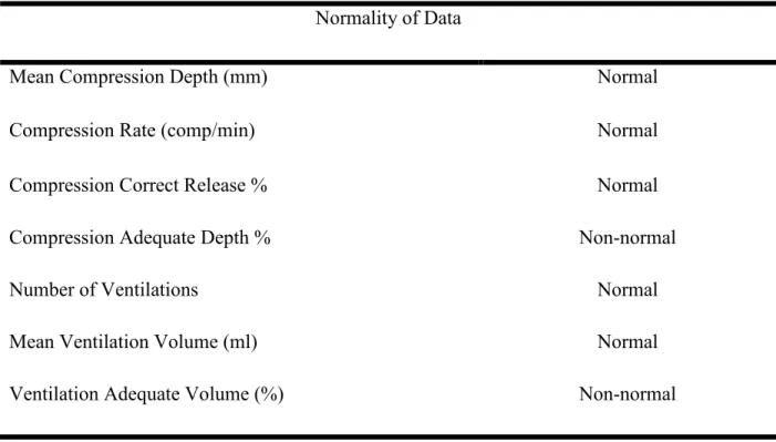
Table 1. List of dependent variables’ data normality. Normal data was assessed using within-subject ANOVAs. Non-normal data was assessed with Freidman’s rank sum test.
Normality of Data
Mean Compression Depth (mm) Normal
Compression Rate (comp/min) Normal
Compression Correct Release % Normal
Compression Adequate Depth % Non-normal
Number of Ventilations Normal
Mean Ventilation Volume (ml) Normal
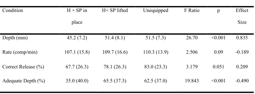
Table 2. Mean (standard deviation), F ratios, p values, and effect sizes for all compression condition outcome measures:
compression depth, compression rate, percentage of correctly released compressions, and percentage of adequate (correct)
compressions. Effect sizes compare the unequipped and pads lifted conditions to the fully equipment condition.
Compression Condition
Condition H + SP in
place
H+ SP lifted Unequipped F Ratio p Effect
Size
Depth (mm) 45.2 (7.2) 51.4 (8.1) 51.5 (7.3) 26.70 <0.001 0.835
Rate (comp/min) 107.1 (15.8) 109.7 (16.6) 110.3 (13.9) 2.506 0.09 -0.189
Correct Release (%) 67.7 (26.3) 78.1 (26.3) 83.0 (23.3) 3.179 0.051 0.209
Adequate Depth (%) 35.0 (40.0) 65.5 (37.3) 62.5 (37.0) 19.843 <0.001 -0.490
4
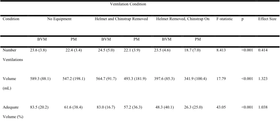
Table 3. Mean (standard deviation), F ratios, p values, and effect size for all ventilation condition outcome measures: total
ventilations attempted, mean ventilation volume, and percentage of ventilations delivering adequate volume (400-700mL).
Effect size compares the helmet removed and facemask and chinstrap removed conditions to the chinstrap in place condition,
collapsing across ventilation methods.
Ventilation Condition
Condition No Equipment Helmet and Chinstrap Removed Helmet Removed, Chinstrap On F-statistic p Effect Size
BVM PM BVM PM BVM PM
Number
Ventilations
23.6 (3.8) 22.4 (3.4) 24.5 (5.0) 22.1 (3.9) 23.5 (4.6) 18.7 (7.0) 8.413 <0.001 0.414
Volume
(mL)
589.3 (88.1) 547.2 (198.1) 564.7 (91.7) 493.3 (181.9) 397.6 (85.3) 341.9 (100.4) 17.79 <0.001 1.323
Adequate
Volume (%)
83.5 (20.2) 61.6 (38.4) 83.0 (16.7) 57.2 (36.3) 48.3 (40.1) 26.3 (25.0) 43.05 <0.001 1.038
4
REFERENCES
Abella, B. S. (2005). Quality of Cardiopulmonary Resuscitation During In-Hospital Cardiac Arrest. Journal of the American Medical Association, 293(3), 305-310.
Al-Rasheed, R. S., Devine, J., Dunbar-Viveiros, J. A., Jones, M. S., Dannecker, M., Machan, J. T., . . . Kobayashi, L. (2013). Simulation intervention with manikin-based objective metrics improves CPR instructor chest compression performance skills without
improvement in chest compression assessment skills. Simul Healthc, 8(4), 242-252. doi: 10.1097/SIH.0b013e31828e716d
Anatomy and Physiology: Understanding the Importance of CPR. (2006). American Heart Association.
Baskett, P., & Zideman, D. (2005). European Resuscitation Council Guidelines for Resuscitation. Resuscitation, 67, S1-S2. doi: 10.1016/j.resuscitation.2005.10.001 Beesems, S. G., & Koster, R. W. (2014). Accurate feedback of chest compression depth on a
manikin on a soft surface with correction for total body displacement. Resuscitation, 85(11), 1439-1443. doi: 10.1016/j.resuscitation.2014.08.005
Blom, M. T., Beesems, S. G., Homma, P. C., Zijlstra, J. A., Hulleman, M., van Hoeijen, D. A., . . . Koster, R. W. (2014). Improved survival after out-of-hospital cardiac arrest and use of automated external defibrillators. Circulation, 130(21), 1868-1875. doi:
10.1161/CIRCULATIONAHA.114.010905
Bradney, D. A., & Bowman, T. G. (2013). Lacrosse helmet facemask removal. J Athl Train, 48(1), 47-56. doi: 10.4085/1062-6050-48.1.02
Bur, A., Kittler, H., Sterz, F., Holzer, M., Eisenburger, P., Oschatz, E., . . . Laggner, A. N. (2001). Effects of bystander first aid, defibrillation and advanced life support on neurologic outcome and hospital costs in patients after ventricular fibrillation cardiac arrest. Intensive Care Med, 27(9), 1474-1480. doi: 10.1007/s001340101045
Casa, D. J., Guskiewicz, K. M., Anderson, S. A., Courson, R. W., Heck, J. F., Jimenez, C. C., . . . Walsh, K. M. (2012). National athletic trainers' association position statement: preventing sudden death in sports. J Athl Train, 47(1), 96-118.
Corrado, D. (2006). Trends in Sudden Cardiovascular Death in Young Competitive Athletes After Implementation of a Preparticipation Screening Program. Journal of the American Medical Association, 296(13), 1593-1601.
Davey, P., Whatman, C., & Dicker, B. (2015). Comparison of Chest Compressions Metrics Measured Using the Laerdal Skill Reporter and Q-CPR: A Simulation Study. Simul Healthc, 10(5), 257-262. doi: 10.1097/SIH.0000000000000105
Decoster, L. C., Burns, M. F., Swartz, E. E., Murthi, D. S., Hernandez, A. E., Vailas, J. C., & Isham, L. L. (2012). Maintaining neutral sagittal cervical alignment after football helmet removal during emergency spine injury management. Spine (Phila Pa 1976), 37(8), 654-659. doi: 10.1097/BRS.0b013e31822da067
Del Rossi, G., Bodkin, D., Dhanani, A., Courson, R. W., & Konin, J. G. (2011). Protective athletic equipment slows initiation of CPR in simulated cardiac arrest. Resuscitation, 82(7), 908-912. doi: 10.1016/j.resuscitation.2011.02.022
DePaepe, A. (1996). Revised Diagnositc Criteria for the Marfan Syndrome. American Journal of Medical Genetics, 62, 417-426.
Dick, R. (2007). Descriptive Epidemiology of Collegiate Men's Lacrosse Injuries: National Collegiate Athletic Association Injury Surveillance System, 1988-1989 Through 2003-2004. Journal of Athletic Training, 42(2), 255-261.
Douglas J. Casa, P., ATC, FNATA, FACSM; Kevin M. Guskiewicz, PhD, ATC, FNATA, FACSM; Scott A. Anderson, ATC; Ronald W. Courson, ATC, PT, NREMT-I, CSCS; Jonathan F. Heck, MS, ATC|; Carolyn C. Jimenez, PhD, ATC; Brendon P. McDermott, PhD, ATC; Michael G. Miller, PhD, EdD, ATC, CSCS; Rebecca L. Stearns, MA, ATC; Erik E. Swartz, PhD, ATC, FNATA; Katie M. Walsh, EdD, ATC. (2012). National Athletic Trainers’ Association Position Statement- Preventing Sudden Death in Sports. Journal of Athletic Training, 47(1), 96-118.
Drezner, J. (2007). Interassociation Task Force recommendations on emergency prepreparedness and management of sudden cardiac arrest in high school and collegiate athletic programs: a consesus statement. Journal of Athletic Training, 2007(42), 143-158.
Field, J. M., Hazinski, M. F., Sayre, M. R., Chameides, L., Schexnayder, S. M., Hemphill, R., . . . Vanden Hoek, T. L. (2010). Part 1: executive summary: 2010 American Heart
Association Guidelines for Cardiopulmonary Resuscitation and Emergency Cardiovascular Care. Circulation, 122(18 Suppl 3), S640-656. doi:
10.1161/CIRCULATIONAHA.110.970889
Frederick O. Mueller, K. L. K., Leah M. Cox, Robert C. Cantu. (2013). Catastrophic Sport Injury Research 31st Annual Report: Fall 1982-Spring 2013. National Center for Catastrophic Sport Injury Research.