Schultz, Noxon, Sisley, Martinez. In Portable Biosensors This paper is a postprint of a paper submitted
and Point-of-Care Systems; 2017 to and accepted for publication in Portable
Biosensors and Point-of-Care Systems and is subject to Institution of Engineering and Technology Copyright. The copy of record is available at the IET Digital Library.
Paper-based
diagnostic
devices
Spencer
A.
Schultz
1,
Isabelle
C.
Noxon
1,
Tyler
A.
Sisley
1and
Andres
W.
Martinez
12.1
Introduction
Paper, broadly defined as any porous hydrophilic membrane, has served as a
platformfordiagnosticassaysformillennia.Oneoftheearliestrecordedexamples
of a paper-based assay comes from c. 77 A.D. when Pliny the Elder, a Roman
naturalist, describeda qualitative testfor detecting the presence of adulterants in
verdigrisusingpapyrusimpregnatedwithanextractofgallnuts[1,2].Startingwith
simplequalitativecolorimetricassaysandmovingallthewaytothecurrentfieldof
paperfluidics,paperoffersseveraluniqueadvantagesasaplatformforconductinga
widevarietyof diagnosticassaysaswellasforcollecting,storingandtransporting
samples.Thischapterwillprovideanoverviewofexistingdiagnosticdevicesmade
primarilyoutofpaperandthenfocusonpaper-basedmicrofluidicdevices,thenext
generationofpaper-baseddiagnosticdevicesthatpromisestoextendtheuseofpaper
asamaterialforfabricatingdiagnosticdeviceswellintothefuture.
Paperisdefinedformallyasaflexiblesheetmadefrompressedcellulosicfibers
(Figure2.1)[3].TheprocessofmakingpaperwasdevelopedinChinaduringtheHan
Dynasty(206B.C.to220A.D.)[4].WrittenrecordscreditTsaiLun,anofficialinthe
HanCourt,withdeveloping themodernformofpaperas wellas thepapermaking
processin105A.D.[5].Althoughtheequipmentusedformakingpaperhasevolved
overtime,theessentialstepsofthepapermakingprocesshaveremainedunchanged:
first,adilutesuspensionofcellulosicfibersispreparedinwater;then,thefibersare
depositedontoascreenformingamat;and,finally,thematoffibersispressedand
driedtoproduceasheet ofpaper[5].Thecellulosicfibersusedtomakepapercan
comefrommanysources,butmostdiagnosticdevicesaremadewithpaperproduced
fromcottonfibersmadeprimarilyofcellulose(Figure2.2(a))[4].Manyothertypes
of porous hydrophilicmembranes have been developed over time and have been
incorporatedintodiagnosticdevices[3].Amongthese,porousmembranesmadefrom
nitrocelluloseareusedmostcommonlybecauseoftheirhighprotein-bindingaffinity
1DepartmentofChemistryandBiochemistry,CaliforniaPolytechnicStateUniversity,SanLuisObispo,
AN: 1579526 ; Kintzios, Spiridon E..; Portable Biosensors and Point-of-Care Systems
100 µm
Figure2.1 SEMmicrograph(400)ofthesurfaceofWhatmanNo.1 chromatographypaper
HO
OH
OH OH
OH
O
O O O O
O
O O
O O
O
O2N
O2N
O2N (a)
O2N O 2N
NO2
O
n
n O
O HO
(b)
Figure2.2 Chemicalstructuresofcellulose(a)andnitrocellulose(b)
(Figure2.2(b))[3].Anenormousdiversityofpaperswithvaryingchemical
compo-sition,thickness,andporesizeareavailable,andeachhasuniquecharacteristicsthat
couldbeharnessedforspecificapplicationsinpaper-baseddiagnosticdevices.
Paperhasmanyinherentcharacteristicsthatmakeitattractiveasaplatformfor
diagnostic assays.Paper isinexpensive andwidely available,so scientists across
the globe andthroughouthistoryhave beenable toworkwithpaper anddevelop
teststhatarecompatiblewithpaper[2,4,6].Paperistypicallywhiteandprovidesan
excellent background for assays that produce a colored product or a change in
color. Another attractive characteristic of paper is that it has a large
surface-to-volumeratio, soasmallvolumeof fluid(1mL)cantypicallyproduceaspoton
largesurface-to-volume ratioof paperalso allowsforreagentsandsamples tobe
driedandstoredonpaper-baseddevices.Papercanevenbemodifiedchemicallyin
ordertocovalentlybondreagentstothefibers[7].Paper canalsoserveasthe
sta-tionaryphaseforchromatographicseparationsofanalytes[8].Paperisdisposable—
one simple method for disposing paper-based tests is through incineration. Paper
wicks fluid bycapillaryaction, so fluids will movethrough apaper-based device
withoutanyexternalsourcesofpower.Andfinally,asmentionedpreviously,paper
canbeproducedfromawidevarietyoffibersandwithawidevarietyof
character-istics(e.g.,poresize,wickingspeed,proteinbindingability),whichcanbeharnessed
for specific applications and can even be combined in a single device to enable
particularcapabilities.
2.2
Current
paper-based
diagnostic
devices
Oneofthebest-knownexamplesofapaper-basedtestislitmuspaper,whichisused
todeterminewhetherasolutionisacidicorbasic[7].Scientificreportsdatingbackto
thenineteenthcenturydescribetheuseoflitmuspaperforanalyzingsamples,anditis
still sold commercially and used routinely by professional scientists and students
[6,9].Inadditiontolitmuspaper,ahugevarietyofothercolorimetrictestshavebeen
developedtodetectthepresenceofanalytesonpaper,butmostofthesetestshavenot
beendevelopedintocommercialproducts[2,10–12].Currently,therearetwoprimary
formsofcommerciallyavailablepaper-baseddiagnosticdevices:dipstickassaysand
lateral-flowassays(LFAs)[3].Athirdformofpaper-baseddevicesknownas
paper-basedarraysareusedprimarilytocollect,store,andtransportsamplesbutarealso
usedinacademicresearchtoconductassays.
2.2.1
Dipstick
devices
Dipstickassaysareoneofthefirstexamplesofpaper-baseddiagnosticdevicesfor
clinicaluse[4].Dipstickdevicescompriseoneormoresmalltestpadsofpaperthat
havebeen impregnatedwithreagentsfor specificcolorimetricassaysandthatare
adhered to aplastic strip, which serves as a handle for the device (Figure 2.3).
Dipstickassaysareperformedbyquicklyimmersingthedipstickintothesampleto
be tested and then observing the colorsthat are produced on the test pads. The
colors produced on the test pads can often be compared to a color-coded chart
providedwiththedipstickinordertomakeasemiquantitativedeterminationofthe
concentrationoftheanalyteinthesample[3].
The first pH dipstick test strips were patented and commercialized in the
1920s[4].ThesedevicescanbepurchasedtodeterminethepHofasolutionwithin
agiven range withvaryingamountsof sensitivity, bothof whichare determined
bythenumberoftestpadsonthedipsticksaswellasthespecificindicatorsusedon
thetestpads [13].The firstdipstickassaysforclinical useweredevelopedinthe
1950sand wereintroduced commercially inthe 1960s [3]. Theseinitial dipstick
assays were developed to quantify the concentration of glucose in urine by
harnessing enzymatically catalyzed reactions involving glucose oxidase and
AN: 1579526 ; Kintzios, Spiridon E..; Portable Biosensors and Point-of-Care Systems
Figure2.3 Dipstickdeviceforanalysisofwater[18]
test pads to detect or analyze glucose, bilirubin, ketones, nitrite, urobilinogen,
protein,blood,leukocytes,pH,andspecificgravity[3,15,16].Thesetestsallowfor
the diagnosis of a variety of conditions and diseases including kidney disease,
urinary tract infections, carbohydrate metabolism disorders, and liver disease
[4,15,16].Formoresophisticatedurinalysistests,thedipstickscanbeanalyzedbya
detector thatanalyzesthe colorof thetestpads inordertoimprovethe accuracy
andprecisionofthetests[3,17].Anothercommonapplicationfordipsticktestsis
for theanalysis ofwater samples.Dipstickdevicesfortestingdrinkingwaterand
swimming poolscanbeusedtoquicklydeterminepH,totalhardness, total
chlor-ine,totalbromine,freechlorine,totalalkalinity,andcyanuricacidlevels[18].
Themainadvantagesofdipstickdevicesarethattheyareinexpensive,easyto
use and provide immediate results. Dipstick tests are also simple to design and
manufacture.Thedisadvantagesof dipstickassaysarethattheyaretypically
qua-litativeorsemiquantitative,theyoftenhavelimitedsensitivityandselectivity,and
theycanonlybedevelopedforanalytesthatcanreacttoproduceacolorchangeorbe
linked to a reaction that produces a color change [6]. Dipstick assays often also
requirearelativelylarge volumeofsamplein ordertowetthe testpads.For these
reasons,dipstickdevicesaremostpracticalfortestingrelativelysimplesamples,such
as urineand water, thatareabundant andcontain relativelyhighconcentrationsof
analytes,suchasglucoseorchlorine.Inordertoovercomesomeofthelimitations
withdipstickassays,LFAsweredevelopedtoexpandtherangeofanalytesthatcould
bedetectedaswellasthesensitivityandselectivityoftheassays.
2.2.2
Lateral-flow
devices
Asthename suggests,lateral-flow devicesrelyonthecapillarywicking offluids
across aporousmembraneinordertodetectananalyte[19].Lateral-flowdevices
aremadetypicallyofseveraldifferenttypesofpaperthataretreatedwithdifferent
reagents andheld in contact with each other to create a single fluidic path that
wicksasamplefromasampleporttoanabsorbentpad,whichservesasa
waste-collection zone (Figure 2.4) [20]. In a typical LFA, a few drops of sample are
Sample port
Sample pad
Test line Control line
Housing
Absorbent pad Conjugate pad
Membrane
Figure2.4 Schematicdiagramofalateral-flowdevice.Reproduced by permissionofEMDMilliporeCorporation. 2002,2008EMD MilliporeCorporation
whichitinteractswithalabelingreagent—typicallyanantibodyboundtoacolored
particle—that binds specifically to the analyte. The labeled analyte then wicks
across amembrane containing acapture reagent—typically a secondantibody—
thatisimmobilizedonthedeviceinanarrowtestlineandthatcapturesselectively
the labeled analyte. Thisprocess generatesa colored lineor band on the device
whentheanalyteispresent.Alltheothercomponentsofthesampleaswellasany
excesslabelingreagentcontinuetowickalongthedeviceintothewastezone.Most
commercialdevicesalsoincludeacontrollinecontainingasecondcapturereagent
onthemembranethatbindsonlytothelabelingreagentandservestoconfirmthat
thecapturereagentsareworkingproperly[3].
LFAsare widelyused inlaboratories,hospitals, andinthehome wherethey
candetectconditionssuch as pregnancy,disease, druguse,contamination,
infec-tion,andorganmalfunction[3,21–23]. ThefirstLFAsweredevelopedinthe late
1960sandhavebecomeauser-friendly,robust,affordable,point-of-care platform
fordiagnostics[19].Thefirst commercialLFAsweredevelopedtodetecthuman
chorionicgonadotropin(hCG);ahighlevelof thisbiomoleculeisapositive
indi-cator for pregnancy [24]. This test was fast (1–3 min), simple to use even for
untrainedusers,required littleuser input,used smallsample sizes, andwas
sub-sequently made commercial by1988[24].The hCG testisnowused globally to
detectpregnancyandcanbepurchasedforlessthanUS$1[25].
ThethreemainfacetsofLFAsarethedeviceorplatform,samplepreparation,
andtheimmunochemicalreaction.TheLFAplatformiscomposedofasamplepad,
afiberglass conjugate padfor storageof the labeled reagent, amembrane thatis
typicallynitrocellulose, anabsorbent waste pad, andoften asmallplastic holder
AN: 1579526 ; Kintzios, Spiridon E..; Portable Biosensors and Point-of-Care Systems
brittleandflammable,itisthemembraneusedinmostLFAsbecauseitisporous,
wicks fluids by capillary action, and its surface has a high affinity for binding
proteins[4,23].Thisbindingcharacteristicisessentialforthedetectionofanalytes,
asLFAsrelyonselectivebindingandretentionofthelabeledanalyte.Fabrication
of commercial LFAs begins with treatment of the different components. The
sample padis pretreated,the fiberglass conjugate padisloaded withthe labeled
reagent,andthemembraneisprintedwithtestandcontrollinesofcapturereagents
andthenblockedfornonspecificadsorption[20].Eachcomponentisdried,andthe
deviceisassembled,cutintostrips,andplacedintocartridgesforpackaging[26].
The hCG urinetest does not require much sample preparation,but there are
many otherLFAsthatdetectbiological componentsfrom bloodplasma orserum
that dorequire treatment. Often samplepreparation includes collection of blood,
treatmentwithanticoagulants,separationofbloodcellsfromplasmaorserum,and
dilution of the sample [19]. Thesesteps are important, but are cumbersomeand
timeconsuming—especiallyatthepointofcareinwhichexpensiveequipmentfor
sample treatmentisdifficulttoobtainornotavailable[27].
The immunochemical reaction is arguably the most important component of
LFAs. There are two main types: immunoassays that rely on antibody–antigen
interactions, and nucleic acid-basedassays that relyon nucleicacid aptamers and
target moleculebindingor nucleicacidhybridization[19].These immunochemical
reactions rely heavily on the sensitive and specific interactions that consequently
determinethesensitivityandselectivityoftheassayitself[19].Thecomponentofthe
immunochemical reactionthatallowsforvisualdetectionisthecolorimetricprobe,
whichisconjugatedtoanantibody,protein,orDNAsequencetomakethelabeling
reagent.Theprobesaremostcommonlylatexbeads,goldnanoparticles,fluorescent
tags, quantumdots,andenzymes.The typeof colorimetricprobe isdeterminedby
several factors including the type of sample, the analyte, pH, and device storage
conditions.Immunochemicalreagentscanbesynthesizedagainstamyriadofproteins
and biomolecules. The heterogeneity of immunochemical combinations is part of
whatmakesLFAssuchawidelystudiedandversatileplatform[19].
ThemainadvantagesofLFAs,aswithmostotherpaper-baseddevices,arethat
theyareinexpensiveandeasytouse—the usertypicallyonlyhastoapply afew
dropsofsampletothedeviceandthenreadtheresultingcoloredlines,unlessmore
sophisticatedsamplepreparationisrequired[3].WhatsetsLFAsapartfromother
paper-baseddevices, likedipsticks,isthatbyharnessing the wickingcapabilities
of paper, LFAs perform what is effectively a chromatographic separation of the
analytefromtheothercomponentsofthesampleaswellasaconcentrationofthe
analytealongthetestline.Boththeseactions leadtohighlysensitiveandspecific
assays.Somemoresophisticated LFAs includeasecondstepinwhichan
ampli-ficationreagentisaddedtothedeviceinordertoenhancethecolorofthetestline
andfurtherimprovethesensitivityofthedevice[3].AnotheradvantageofLFAsis
thatas theyrely onimmunochemical reactionstodetectanalytes, thesamebasic
devicedesigncanbeusedtodetectahugerangeofanalytesbysimplychangingthe
labeling reagent and the capture reagent [19].On their own,LFAs are typically
withelectronicreadersthatanalyzetheintensityofcolorthatdevelops atthe test
lineorrelyonfluorescentlabelsinordertomake quantitativedeterminations[3].
2.2.2.1 Vertical-flowdevices
Vertical-flowdevicesrely onthesameanalyte-detectionprinciplesaslateral-flow
devicesandcanbeusedtoconductthesametypesofassays,but,insteadoflining
up the different components of the device horizontally as in LFAs, the various
componentsarestackedvertically.Theadvantageofstackingthelayersvertically
isthat the pathlengthfor the sample towick throughthe device issignificantly
reduced;however,theseassaystypicallyrequiremultiplestepsbytheuserinorder
tobeperformedcorrectly[3].
2.2.3
Paper-based
arrays
Paper-basedarraysareusedprimarilyforthecollection,storage,andtransportation
of samples. Paper-based arrays typically comprise a card marked with multiple
circularzonesthatcanbefilledwithliquidsamples.Thezonesareoftenpretreated
withreagents toimprove the stability ofthe analytes or todisinfectthe samples.
Samples,suchasblood,canbespottedonthearrayanddried,andthenthe array
canbe shippedtoacentral laboratorywherethe samples canbeeluted fromthe
arrayandanalyzed [28].Thisprocess allowsfor large numbersof samplestobe
collectedandstoredinaconvenientformatthatdoesnotrequirerefrigeration[29].
The sample collection can also be performed in the field with minimal
instru-mentation[29].
Onecommonapplicationforpaper-basedarraysisindriedbloodspot(DBS)
testing [28].This techniquewasdevelopedintheearly1900sbutgained
wide-spread use in the1960s and1970s when RobertGuthrie used DBSto develop
atechniqueforthesystematicscreeningofnewbornsformetabolicdiseases[30].
NowknownastheGuthrietest,newbornsaroundtheworldaretestedforaseries
of metabolic disorders and diseases by collecting their blood from a heal
prickon a DBScard[28].In fact, DBScan be used todetect awide range of
analytesanddiseasesincludingHIV,dengue,hepatitis,andsmallmoleculedrugs
[28,29,31].
Analternativeapplicationofpaper-basedarraysisforthesynthesisoflibrariesof
shortpeptidesorsmallmoleculesviaatechniqueknownasspotsynthesis[32–35].
Onceprepared,thesearrayscanbeusedtoconductawiderangeofassaysincluding
bindingassays,enzymeassays,andstudieswithlivingorganisms[36].
2.3
Paper-based
microfluidic
devices
In2007,theWhitesidesgroupatHarvardUniversitypublishedanarticledescribing
thefirstexampleofwhatwouldbecomeknownaspaper-basedmicrofluidic
devi-cesormicrofluidicpaper-basedanalytical devices(microPADs)(Figure2.5)[37].
Havingcontributedasubstantialbodyof worktothefield ofconventional
AN: 1579526 ; Kintzios, Spiridon E..; Portable Biosensors and Point-of-Care Systems
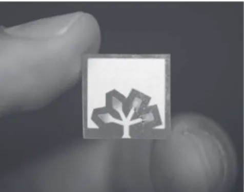
Figure2.5 Apaper-basedmicrofluidicdevicefordetectingglucose(leftzones) andprotein(rightzones)
Whitesides was looking to develop a new class of devices that married the
advantagesoflateral-flowanddipstickdevices,suchascapillarywickingandlow
cost, with the advantages of conventional microfluidic devices, such as low
volumesofsample,multiplexing,andcontrolledsequentialsteps.Theultimategoal
oftheWhitesidesgroupwastodevelopanewclassofultra-low-costpoint-of-care
diagnostic devices thatcould bedeployed toresource-limit settings andimprove
accesstohealthcaretopopulationsaroundtheglobe.
Paper was a natural choice as a material for fabricating this new class of
devices forallthesamereasonsmentioned inthe introduction.Themain
innova-tion describedintheinitialpublicationfromtheWhitesidesgroupwasthe
devel-opmentofamethodtopatternpaperintoanetworkofhydrophilicchannelsandtest
zonesboundedbyhydrophobicbarriers[37,40].Thechannelscouldwicksamples
and distributethem into the testzones whereassays wouldtakeplace. Although
examples of patterning paper usingwaxes had been described previously [2,41],
whatsettheworkfromtheWhitesidesgroupapartwasthefocusonthe
develop-ment of anew class of lowcost, verysimple to useportable diagnostic devices
[37,42].Thisworkcatalyzedasurgeofinterestinthedevelopmentofpaper-based
assaysandresultedintheestablishmentofthenewfieldofpaperfluidics[3,6,12].
2.3.1
Fabrication
of
paper-based
microfluidic
devices
Fabrication ofpaper-basedmicrofluidic devicesinvolvesthepatterningofapiece
of paper using hydrophobic inks to define hydrophilic channels and test zones
boundedbyhydrophobicbarriers[12].Severalmethodsfor patterningpaperhave
been developed since 2007, each with their own set of advantages and
dis-advantages [6,12].Althoughthe methodsare verydiverse interms of equipment
and inks, they can be classified broadly into two categories: indirect and direct
patterning.Inindirectpatterning,thefirststepoftheprocessinvolvescoatingthe
entirepieceofpaperwithahydrophobicinkorreagentsuchasawax,apolymer,or
aresin.In thesecondstep,thehydrophobicreagent isetchedawayselectivelyto
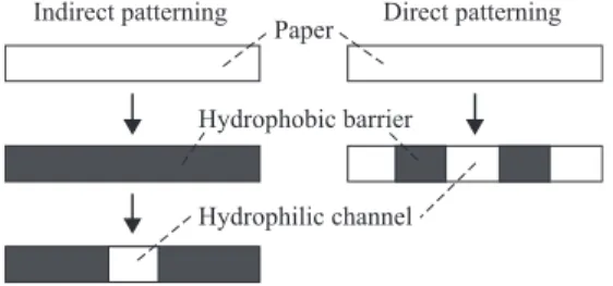
Indirect patterning Direct patterning Paper
Hydrophobic barrier
Hydrophilic channel
Figure2.6 Cross-sectionalschematicdiagramofindirectanddirectpatterning ofpaper
producehydrophilic channels.In directpatterning,thehydrophobicinkisprinted
directlyonthepaperinthedesiredpattern(Figure2.6).
Thefirstpublishedmethodforfabricatingpaper-basedmicrofluidicdeviceswas
anexampleofindirectpatterningandwasanadaptedformofphotolithography[37].
In this approach, thepaper is firstimpregnated with SU-8 photoresist,anegative
photoresist,anddried.ThepaperisthenexposedtoUVlightthroughaphotomaskin
order tocross-linkthe exposedportionsof the photoresist.Finally,theunexposed
portions of the photoresist are dissolved and removed from the paper using a
solventsuch as acetoneto yieldthe desiredpattern of channelsandtest zonesin
thepaper[37,40,42].Otherphotopolymers,inadditiontoSU-8,havealsoshownto
beusefulforpatterningpaperviaindirectpatterning[3].
Indirectpatterningwasalsoachievedusinganinkjetprinter[43].Theprocess
involvesfirstcoatingapieceofpaperwithpolystyrenetomakeithydrophobic.The
polystyreneisthenselectivelyremovedfromspecificareasofthepaperbyprinting
toluene ontothe paper usinganinkjet printer inthe desiredpattern. The toluene
washesthepolystyreneoutofthepaperandreturnsthewashedpapertoitsoriginal
hydrophilicstate.
A third example of indirect patterning using plasma treatment involves
impregnatingasheetofpaper withalkyl ketenedimer,areagent usedcommonly
forsizingpapertomake itmorehydrophobic[44].Thereagentisthenselectively
etched awaybyplacingthe paper betweentwometal stencilsandtreatingit ina
vacuum plasma reactor. The areas of the paper that are exposed to the plasma,
through the metal stencils, are made hydrophilic andthe areasof paper thatare
protectedbythestencilremainhydrophobic.
In direct patterning, hydrophobic agents are applied onto the paper in the
desired pattern. Hydrophobic agents are applied typically through conventional
printingtechnologies,suchasinkjetprinting,screenprinting,flexographicprinting,
andstamping,butcouldevenbeappliedbyhand.Eachtechniquehasitsownsetof
advantagesanddisadvantagesintermsofcost,resolution,throughput,and
techni-caldifficulty.Oneofthemostpopularmethodsofdirectpatterningiswaxprinting
[45–47]. In wax printing, asolid-ink printer is used toprint wax directly onthe
surfaceofpaperinanydesiredpattern.Theprintedpaperisthenheatedtoreflow
AN: 1579526 ; Kintzios, Spiridon E..; Portable Biosensors and Point-of-Care Systems
completed inlessthan5minfor anentiresheetof paper,andfeaturesasmallas
1 mm can be patterned reliably [46].The equipment required for wax printing,
namelythesolid-inkprinterandtheheater(e.g.,oven,hotplate,thermallaminator,
heatgun,andiron)canbepurchasedforlessthanUS$1,000,andtheprocessdoes
notrequireacleanroomorafumehood,sowaxprintingcanbeperformedbyusers
with limited resources or limited experience with fabrication [12]. Another
advantage of waxprinting isthat thereis virtually noproduction-cost penaltyfor
makingsmallbatchesofdevicesatatime,whichmakesitwellsuitedfor
laboratory-scaleprototyping.
In comparing indirect and direct patterning methods, indirect patterning
methodsaregenerallymoretimeconsumingastheyinvolveatleasttwosteps,but
oftenseveralmore.Indirectpatterningmethodsalsoconsumelargerquantitiesof
reagents and solvents as the entire piece of paper is treated and made
hydro-phobic. Direct patterning methodsare generallyfaster, less expensive, andless
technically challenging than indirect methods. One advantage of indirect
patterning,however,isthat ittends toallowforhigherresolutionpatterns tobe
produced. Specifically in the case of photolithography, the resolution of the
patternis controlled by the resolution of the exposure to UV light, so smaller
featurescanbeproducedusingthismethodcomparedtowaxprinting[12].Inone
instance,directandindirectmethodsofpatterningwerecombinedtopatternpaper
withTeflonin orderto generatemicroPADsthat werecompatible withorganic
solvents[48].
A third method of preparing microPADs is via shaping or cutting. In this
approachthe desired network of channels andtest zones iscut out of asheet of
paper,eithermanuallyorusingalasercutterorcuttingplotter[49,50].Thismethod
issimpletoperformanddoesnotrequireanyreagents.Thedevicesfabricatedvia
cuttingusuallyhavetobeencasedinplastictoprotectthepaper,providestructural
supporttothepaper,andfacilitatemanipulationofthedevices[12].
Once the patterning processis complete, devicescan be furthermodified to
addadditionalcapabilities.Onecommonpost-patterningstepistoencasedevices
inplasticusingeithertape,thermallaminationsheetsortoner[49,51,52].Another
optionistoaddadditionalfeaturestothedevicesuchaselectrodes.Electrodesare
typicallyprinted ontothe devices after they are patterned usingconductive inks
[53–57].Devicescanalsobemodifiedtocontrolthewickinginthedeviceeitherby
incorporating valves [58], adding reagents or shunts that slow down wicking
[59,60], or adding additional layers of paper or plastic to speed up wicking
[52,57,61].Finally,multiplelayersofpatternedpapercanbestackedontopofeach
other to produce three-dimensional (3D) microPADs (Figure 2.7) [62–65]. The
layersofpaper inthe3D devicecan eitherbe bondedtoeach otherpermanently
usingpermanentadhesivesortapes,ortheycanbeheldtogethertemporarilyusing
amanifoldorremovableadhesivesortapes.
Tocompletethefabricationprocess,reagentsfortheassaymustbeappliedto
the device.Typically reagentsare deposited onto microPADsfrom solution, and
the solutions are then allowed to dry leaving the reagent behind on the device.
1 cm Cellulose powder Channel 0.2 min 2 min 4 min
1 mm Tape Channel
Figure2.7 Topviewandcross-sectionalviewofathree-dimensional microPADassembledfromtwolayersofpatternedpaperand alayerofdouble-sidedtape.ReproducedbypermissionofThe NationalAcademyofSciencesfromReference62. 2008 NationalAcademyofSciences,USA
deposited using an inkjet printer or liquid-dispensing robot. More recently, a
methodforsoliddepositionofreagentsusingcustom-madepencilswasdescribed,
whichledtoimproved shelflifeofthereagentsonthedevices[66].
2.3.2
Applications
of
paper-based
microfluidic
devices
Initial research on paper-based microfluidic devices focused primarily on the
development of methods of fabricating devices. The first devices consisted
gen-erallyof asampleinletthatdirectedthe sampleinto onemorechannels andthen
into multipletestzones inwhichreagentsfor colorimetric assays weredeposited
(Figure2.5).Theseinitialdevicesrequired smallvolumesoffluid (20mL)and
could performmultiple assayssimultaneously fromasinglesample-addition step
[37,42].Asworkwithpaper-devicesexpanded,newapplicationsanduniqueassays
were developed, many of which were enabled by the unique capabilities of
microPADs.Thissection will highlightfourareasofapplications for microPADs
thatshowpromiseforfuturediagnosticdevices.
Colorimetric assays, assays that result in a color change in response to the
presenceofanalyte,remainoneofthemostcommontypesofassaysconductedon
microPADs [3]. Awide range of colorimetric assays have developedfor
micro-PADsincludingindicator-basedassays,enzymaticassays,sorbent-basedassays,as
wellas combinationsas theseapproaches such asenzyme-linkedimmunosorbent
assays (ELISAs) [3].These assays have beenshown tobe usefulfor detecting a
widerangeof analytesincludingions,smallmolecules,proteins,andDNA.
Con-current with the development of colorimetric assays on microPADs, several
AN: 1579526 ; Kintzios, Spiridon E..; Portable Biosensors and Point-of-Care Systems
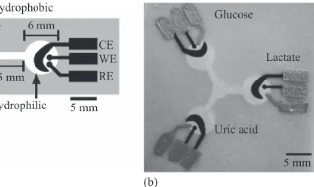
Hydrophobic
Glucose 6 mm
CE
WE Lactate
5 mm RE Hydrophilic 5 mm
(a) Uric acid
5 mm (b)
Figure2.8 Schematicofanelectrochemicalpaper-basedmicrofluidicdevice includingthreeelectrodes(a),andimageofanactualdevicefor detectingglucose,lactate,anduricacid(b).Adaptedwithpermission fromReference53. 2009AmericanChemicalSociety
developed. One promising approach is theuse of digital image colorimetry,in
whichadigitalimageoftheresultsofacolorimetricassayisobtained,andthen
thepixelintensityofthecolorismeasured[67,68].Theimagescouldbeobtained
using a scanner, a digital camera or, most interestingly in the context of
portablepoint-of-carediagnosticassays,acameraphone[42].Theresultscanbe
calibrated througheither externalcalibrationorstandard additionandcan
typi-callyproduce resultswithrelativeerrorsandrelative standarddeviationswithin
tenpercent[68].
As an alternative to colorimetric assays, paper-based assays that rely on
electrochemical detection offer many advantages, especially when it comes to
performing quantitative tests [53,55,56]. Electrochemical paper-based assays
have been demonstrated for a wide range of analytes with high accuracy and
precision, and with the abilityto detect analytes at femtomolar concentrations
[6,69,70].Theelectrodesforelectrochemicalassaysaretypicallyprinteddirectly
on themicroPADs(Figure 2.8),and anexternalreader is usedtypicallyto
per-formtheassay[6].
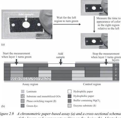
A third type of paper-based assays that eliminate the need for sophisticated
external readersarechronometricassays,inwhichthesignalfromtheassayisthe
time that it takes the sample to wick across a channel in a device (Figure 2.9)
[64,71–74].TheseassaysrelyontheuniquepropertiesofmicroPADstowickfluids
alongchannelsinpaperaswellastheabilitytopatternreagentsinmultiplelayers
ofpaperandthenassembletheselayersintoasingle3Ddevice.Inthesedevices,a
reagent that creates and impermeable or semi permeable barrier is added to a
channelinthedevice.Thepresenceoftheanalytetriggersachemicalreactionthat
resultsinthedegradationofthebarrier.So,whentheanalyteispresent,thesample
wicks acrossthe channelmorequickly thanwhen the analyte isabsent.
Chrono-metricassaysstandoutfortheirsimplicity,sensitivity,andlowlimitsofdetection,
Add sample
Wait for the left Measure the time to region to turn green appearance of color in the right region relative to the left
(a)
Start the measurement when layer 1 turns green
Add sample
Stop the measurement when layer 1 turns green
layers
1 2 3 4 5
Assay region Control region
Laminate Hydrophilic paper
Substrate and immobilized GOx Hydrophobic paper
Phase-switching reagent (2) Buffer containing MgCI2 (b) Green dye Enzyme substrate (1)
Figure2.9 Achronometricpaper-basedassay(a)andacross-sectionalschematic ofthelayersandcomponentsmakingupthedevice(b).Adaptedwith permissionfromReference74. 2013AmericanChemicalSociety
Inadditiontodevelopingspecifictypesofquantitativeassays,thepaperfluidics
communityhas devotedconsiderable efforttoward developingdevicescapableof
performing multiple sequential stepsin order to automatically perform multistep
assays,such as ELISAs or enzyme-inhibition assays.For example, a device that
performs a typical lateral-flow immunoassay and then automatically adds an
amplification reagent to enhance the intensity of the test line on the device and
improvethe limit of detectionof the assay was developedby designinga device
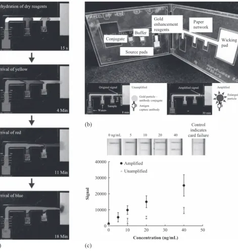
with multiple fluid inlets leading to a single-test zone (Figure 2.10) [50,75,76].
Alternatively,asimilartypeofassaywasachievedbycarefullydesigningaseriesof
channelsthatwouldwickfluidfromasinglesampleinletanddeliverittoatestzone
atdifferenttimes[77].Thedevelopmentoffluidicdiodesprovidedathirdoptionfor
achievingautomatedmultistepassaysonmicroPADs [78,79].Finally,paper-based
enzyme-inhibitionteststhatrequireincubationofanenzymewithasamplefollowed
byintroductionoftheenzymesubstratewereautomatedbyusingerodiblepolymeric
bridges,whichallowedfortheintroductionofsingle-usetimedshut-offvalvesinto
AN: 1579526 ; Kintzios, Spiridon E..; Portable Biosensors and Point-of-Care Systems
Rehydration of dry reagents
Gold
Paper enhancement
network reagents
Buffer
Conjugate Wicking
pad
15 s
Source pads
Arrival of yellow
Original signal Unamplified Amplified signal Amplified Enlarged Gold particle –
Test
control
particle antibody conjugate
Antigen
Sample
capture antibody 4 Min Water
8 min 30 min
(b) Control
indicates
Arrival of red 0 ng/mL 5 10 20 40
card failure
40000
11 Min
30000
Amplified
Unamplified
10000
0
0 10 20 30 40 50
18 Min Concentration (ng/mL)
(a) (c)
Figure2.10 Multisteppaper-basedassay.(a)Imagesofthedeviceperforming multiplesequentialdeliveryofreagentsusingdyesassample reagents.(b)ImageofactualdevicefordetectingMalaria. (c)Resultsofthetestshowingtheimprovedsignalobtainedusing themultistepdevice(amplified).Adaptedwithpermissionfrom Reference76. 2012AmericanChemicalSociety
2.4
Conclusions
Paperoffersseveraladvantagesasaplatformforconductingsimple,point-of-care
diagnosticassays.Current,commerciallyavailablepaper-baseddiagnosticdevices,
suchasdipstickassaysandLFAs,willcontinuetobeusedwidelyduetotheirlow
cost and simplicity. A newgeneration of paper-based devices is currently being
developed and promises to extend those applications of paper for point-of-care
diagnostics.
Signal
20000
Theconceptofmakingdiagnosticdevicesoutof paperisbeingadoptedbya
growingcommunityofresearchers.MicroPADswerefirstdevelopedasdiagnostic
devices for usein developingcountries butare now being developedtodetect a
wide rangeof analytes andcould beused tomonitorair,soil, andwater quality;
theycouldbeusedasdiagnosticdevicesforanimalsandplants;theycouldbeused
inhomehealthcaretodiagnose diseaseormonitordruglevels; andtheycouldbe
usedbythemilitaryandfirst-responderstoassessaperson’shealthstatusordetect
toxins, biohazards, or explosives. MicroPADs also have many potential
applica-tionsinbasicresearch.
Oneofthegreataspectsofpaper-basedmicrofluidic devicesisthatthereisa
verylowbarriertoentry,bothintermsofthecostofequipmentandthetechnical
expertisethatisrequiredtofabricatedevices.Apairofscissorsandapapertowelis
allthatisreallyneededtomakesimplepaper-baseddevices.So,muchlike
open-sourcesoftware,welookforwardtoseeingcontributionstothisfieldfromallkinds
ofscientistsfromallovertheworldandseeingwherewecangowiththissimple
yet-powerfultechnology.
References
[1] Pliny,B.J.,RileyH.T.TheNaturalHistoryofPliny.1857.VolVI.pp.140–1;
235–8;362–4.
[2] Yagoda H. ‘Applications of confined spot tests in analytical chemistry:
Preliminary paper’. Industrial Engineering Chemistry and Analytical
Edition.1937;9(2):79–82.
[3] YetisenA.K.,AkramM.S.,Lowe C.R.‘Paper-based microfluidic
point-of-carediagnosticdevices’.LabonaChip.2013;13(12):2210–51.
[4] Hu J., Wang S., Wang L., et al. ‘Advances in paper-based point-of-care
diagnostics’.BiosensorsandBioelectronics.2014;54:85–97.
[5] Hunter D.Papermaking: TheHistory andTechnique ofan Ancient Craft.
NewYork,NY:DoverPublicationsInc.;1978.
[6] Cate D.M., Adkins J.A., Mettakoonpitak J., Henry C.S. ‘Recent
develop-ments in paper-based micro fluidic devices’. Analytical Chemistry.
2015;87:19–41.
[7] Pelton R. ‘Bioactive paper provides a low-cost platform for diagnostics’.
TrAC–TrendsinAnalyticalChemistry.2009;28(8):925–42.
[8] BlockR.J.,DurrumE.L.,ZweigG.AManualofPaperChromatographyand
PaperElectrophoresis.NewYork:AcademicPress;1955.
[9] Davy J. ‘On gaseous compound of organic oxide and chlorine’.
Philoso-phicalTransactionsoftheRoyalSocietyofLondon.1812;102:144–51.
[10] FeiglF.,AngerV.,KoslowR.H.‘Spottestsininorganicanalysis’.Journalof
TheElectrochemicalSociety.1973;120:261C.
[11] Hoffman J.H.,WeaverJ.W.‘Qualitative spottests’,inAnalyticalMethods
foraTextile Laboratory.Research TrianglePark, NC:American
AN: 1579526 ; Kintzios, Spiridon E..; Portable Biosensors and Point-of-Care Systems
[12] MartinezA.W.,PhillipsS.T.,WhitesidesG.M.,CarrilhoE.‘Diagnosticsfor
thedevelopingworld:Microfluidicpaper-based analytical devices’.
Analy-ticalChemistry.2010;82(1):3–10.
[13] ‘EMD Millipore pH Indicator Strips’ [Online]. Available: http://www.
emdmillipore.com/Web-US-Site/en_CA/-/USD/ViewParametricSearch-Simple OfferSearch?SearchTerm=*&SingleResultDisplay=SFProductSearch&Search
ContextCategoryUUIDs=UJWb.qB.jtAAAAE_9A93.Lxj.[Accessed:
30-Jun-2016].
[14] Free A.H., Adams E.C., Kercher M.L., Free H.M., Cook M.H. ‘Simple
specifictestforurineglucose’.ClinicalChemistry.1957;3(3):163–8.
[15] Boyd J., Barratt J. ‘Interpretation and management of abnormal dipstick
urinalysis’.Medicine(Baltimore).2011;39:(6):312–16.
[16] RobertsJ.R.‘Urinedipsticktesting:Everythingyouneedtoknowurinalysis:
Acomprehensivereview’.EmergencyMedicineNews.2007;29:24–7.
[17] PeeleJ.D.,Gadsden R.H.,CrewsR.‘Semi-automatedvs.visualreadingof
urinalysisdipsticks’.ClinicalChemistry.1977;23(12):2242–6.
[18] ‘AquaChekPool&SpaTestStrips’ [Online].Available:
http://www.aqua-chek.com/pool-spa-owners/aquachek-select-7-in-1/.[Accessed:15-Jun-2016].
[19] BahadirE.B.,Sezgintu¨rkM.K.‘Lateralflowassays:Principles,designsand
labels’.TrACTrendsinAnalyticalChemistry.2016;82:286–306.
[20] EMD Millipore. Rapid Lateral FlowTest Strips. Billerica,MA: Millipore
Corporation;2008.
[21] ChanC.P.Y.,SumK.W., Cheung,K.Y.,etal.‘Developmentofa
quantita-tive lateral-flow assay for rapid detection of fatty acid-binding protein’.
JournalofImmunologicalMethods.2003;79:91–100.
[22] Posthuma-TrumpieG.A.,KorfJ.,VanAmerongenA.‘Lateralflow(immuno)
assay: Its strengths, weaknesses, opportunities and threats. A literature
survey’.AnalyticalandBioanalyticalChemistry.2009;393(2):569–82.
[23] SajidM.,Kawde A.N.,DaudM.‘Designs,formatsandapplications of
lat-eral flow assay: A literature review’. Journal of Saudi Chemical Society.
2015;19(6):689–705.
[24] WildD.TheImmunoassayHandbook,4thed.Amsterdam:ElsevierScience; 2013.
[25] ‘ClinicalGuardOne StepHCGPregnancy TestStrip’[Online].Available:
https://www.clinicalguard.com/one-step-hcg-pregnancy-test-strip-p-704.html.
[Accessed:28-Jun-2016].
[26] SharmaS.,Zapatero-RodriguezJ., EstrelaP.,O’KennedyR.‘Point-of-care
diagnostics in low resource settings: Present status and future role of
microfluidics’.Biosensors.2015;5(3):577–601.
[27] ChinC.D.,LinderV.,SiaS.K.‘Lab-on-a-chipdevicesforglobalhealth:Past
studiesandfutureopportunities’.LabonaChip.2007;7(1):41–57.
[28] LehmannS.,Delaby C.,VialaretJ., DucosJ., HirtzC.‘Current andfuture
useof‘‘driedbloodspot’’analysesinclinicalchemistry’.ClinicalChemistry
[29] Meesters R.J., Hooff G.P. ‘State-of-the-art dried blood spot analysis: An
overview of recent advances and future trends’. Bioanalysis. 2013;5
(17):2187–208.
[30] GuthrieR.,SusiA.‘Asimplephenylalaninemethodfordetecting
phenylk-etonuria in large populations of newborn infants’. Pediatrics. 1963;32:
338–43.
[31] StevensR.,PassK.,FullerS.,etal.‘Bloodspotscreeningandconfirmatory
tests for syphilis antibody’. Journal of Clinical Microbiology. 1992;30
(9):2353–8.
[32] FrankR.‘Spot-synthesis:Aneasytechniqueforthepositionallyaddressable,
parallelchemical synthesisonamembranesupport’.Tetrahedron.1992;48
(42):9217–32.
[33] Frank R. ‘The SPOT-synthesis technique: Synthetic peptide arrays on
membranesupports–Principlesandapplications’. Journalof
Immunologi-calMethods.2002;267(1):13–26.
[34] Blackwell H.E. ‘Hitting the SPOT: Small-molecule macroarrays advance
combinatorial synthesis’. Current Opinion in Chemical Biology. 2006;10
(3):203–12.
[35] KramerA., Reineke U., Dong L.,etal.‘Spotsynthesis: Observations and
optimizations’.JournalofPeptideResearch.1999;54(4):319–27.
[36] HilpertK., Winkler D.F.H., Hancock R.E.W. ‘Peptide arrayson cellulose
support:SPOTsynthesis,atimeandcostefficient methodfor synthesisof
large numbers of peptides in a parallel and addressable fashion’. Nature
Protocols.2007;2(6):1333–49.
[37] MartinezA.W.,PhillipsS.T.,ButteM.J.,WhitesidesG.M.‘Patternedpaper
asaplatformforinexpensive,low-volume,portablebioassays’.Angewandte
ChemieInternationalEdition.2007;46:1318–20.
[38] Xia Y., Whitesides G.M. ‘Soft lithography’. Annual Review of Materials
Science.1998;28(1):153–84.
[39] XiaY., Whitesides G.M.‘Soft lithography’. Angewandte Chemie
Interna-tionalEdition.1998;37(5):550–75.
[40] Martinez A.W., Phillips S.T., Wiley B.J., Gupta M., Whitesides G.M.
‘FLASH:Arapidmethodforprototypingpaper-basedmicrofluidicdevices.’
LabonaChip.2008;8(12):2146–50.
[41] Muller R.H., Clegg D.L. ‘Automatic paper chromatography’. Analytical
Chemistry.1949;21:1123–5.
[42] Martinez A.W., Phillips S.T., Carrilho E., Thomas S.W., Sindi H.,
Whitesides G.M. ‘Simple telemedicine for developing regions: Camera
phones and paper-based microfluidic devices for real-time, off-site
diag-nosis’.AnalyticalChemistry.2008;80(10):3699–707.
[43] Abe K., Suzuki K., Citterio D. ‘Inkjet-printed microfluidic multianalyte
chemicalsensingpaper’.AnalyticalChemistry.2008;80(18):6928–34.
[44] Li X., Tian J., Nguyen T., ShenW. ‘Paper-based microfluidic devices by
AN: 1579526 ; Kintzios, Spiridon E..; Portable Biosensors and Point-of-Care Systems
[45] LuR.,ShiW.,JiangL.,QinJ., LinB. ‘Rapidprototyping ofpaper-based
microfluidics with wax for low-cost, portable bioassay’. Electrophoresis.
2009;30(9):1497–500.
[46] CarrilhoE.,MartinezA.W.,WhitesidesG.M.‘Understandingwaxprinting:
Asimplemicropatterningprocessforpaper-basedmicrofluidics’.Analytical
Chemistry.2009;81(16):7091–5.
[47] LuY., Shi W., Qin J., Lin B. ‘Fabrication and characterization of
paper-basedmicrofluidicspreparedinnitrocellulosemembranebyWaxprinting’.
AnalyticalChemistry.2010;82(1):329–35.
[48] Deiss F., Matochko W.L., Govindasamy N., Lin E.Y., Derda R.
‘Flow-throughsynthesisonteflon-patternedpapertoproducepeptidearraysfor
cell-basedassays’.AngewandteChemieInternationalEdition.2014;53(25):6374–7.
[49] FentonE.M.,MascarenasM.R.,LopezG.P.,SibbettS.S.‘Multiplex
lateral-flow test strips fabricated by two-dimensional shaping’. ACS Applied
MaterialsandInterfaces.2009;1(1):124–9.
[50] Fu E., Ramsey S.A., Kauffman P., Lutz B., Yager P. ‘Transport in
two-dimensional paper networks’. Microfluidics and Nanofluidics. 2011;10
(1):29–35.
[51] Schilling K.M., Lepore A.L., Kurian J.A.,Martinez A.W. ‘Fullyenclosed
microfluidicpaper-basedanalyticaldevices’.AnalyticalChemistry.2012;84
(3):1579–85.
[52] CamplissonC.K.,SchillingK.M.,PedrottiW.L.,StoneH.A.,MartinezA.W.
‘Two-plychannels forfaster wickinginpaper-basedmicrofluidicdevices’.
LabonaChip.2015;15(23):4461–6.
[53] Dungchai W., Chailapakul O., Henry C.S. ‘Electrochemical detection for
paper-basedmicrofluidics’.AnalyticalChemistry.2009;81(14):5821–6.
[54] NieZ.,NijhuisC.A.,GongJ.,etal.‘Electrochemicalsensinginpaper-based
microfluidicdevices’.LabonaChip.2010;10(4):477–83.
[55] NieZ.,DeissF.,LiuX.,AkbulutO.,WhitesidesG.M.‘Integrationof
paper-basedmicrofluidic devices withcommercial electrochemicalreaders’. Lab
onaChip.2010;10(22):3163–9.
[56] AdkinsJ., Boehle K.,Henry C.‘Electrochemical paper-basedmicrofluidic
devices’.Electrophoresis.2015;36(16):1811–24.
[57] Renault C., Anderson M.J., Crooks R.M. ‘Electrochemistry in
hollow-channel paper analytical devices’. Journal of the American Chemical
Society.2014;136(12):4616–23.
[58] Jahanshahi-AnbuhiS.,HenryA.,LeungV.,etal.‘Paper-basedmicrofluidics
withanerodiblepolymericbridgegivingcontrolledreleaseandtimedflow
shutoff’.LabonaChip,2013;14(1):229–36.
[59] Lutz B., Liang T., Fu E., Ramachandran S., Kauffman P., Yager P.
‘Dissolvable fluidic time delays for programming multi-step assays in
instrument-freepaperdiagnostics’.LabonaChip.2013;13(14):2840–7.
[60] ToleyB.J., McKenzie B.,LiangT.,Buser J.R.,YagerP.,Fu E.
‘Tunable-delayshuntsforpapermicrofluidicdevices’.AnalyticalChemistry.2013;85
[61] Jahanshahi-Anbuhi S., Chavan P., Sicard C., et al. ‘Creating fast flow
channelsinpaperfluidic devicestocontrol timingof sequentialreactions’.
LabonaChip.2012;12:5079–85.
[62] Martinez A.W., PhillipsS.T., WhitesidesG.M.‘Three-dimensional
micro-fluidic devices fabricated in layered paper and tape’. Proceedings of the
National Academy of Sciencesof the United States ofAmerica. 2008;105
(50):19606–11.
[63] Liu H., Crooks R.M. ‘Three-dimensional paper microfluidic devices
assembled using the principles of origami’. Journal of the American
ChemicalSociety.2011;133(44):17564–6.
[64] Lewis G.G., DiTucci M.J., Baker M.S., Phillips S.T. ‘High throughput
methodforprototypingthree-dimensional,paper-basedmicrofluidicdevices’.
Lab on a Chip. 2012;12(15):2630.
[65] Schilling K.M., Jauregui D., Martinez A.W. ‘Paper and toner
three-dimensionalfluidicdevices:Programmingfluidflowtoimprove
point-of-carediagnostics’.Lab on a Chip.2013;13:628–31.
[66] Mitchell H.T., NoxonI.C., Chaplan C.A., et al.‘Reagent pencils: A new
technique forsolvent-free deposition of reagents onto paper-based
micro-fluidicdevices’.LabonaChip.2015;15:2213–20.
[67] ByrneL.,BarkerJ.,Pennarun-ThomasG.,DiamondD.,EdwardsS.‘Digital
imagingasadetectorforgenericanalytical measurements’.TrAC–Trends
inAnalyticalChemistry.2000;19:517–22.
[68] ChaplanC.A.,MitchellH.T.,MartinezA.W.‘Paper-basedstandardaddition
assays’.AnalyticalMethods,2014;6(5):1296.
[69] CunninghamJ.C.,KoganM.R.,TsaiY.J.,LuoL.,RichardsI.,CrooksR.M.
‘Paper-based sensor for electrochemical detection of silver nanoparticle
labelsbygalvanicexchange’.ACSSensors.2015;1(1):acssensors.5b00051.
[70] Scida K.,CunninghamJ.C.,RenaultC.,RichardsI.,Crooks R.M.‘Simple,
sensitive, and quantitative electrochemical detection method for paper
analyticaldevices’.AnalyticalChemistry.2014;86(13):6501–7.
[71] LewisG.G.,DitucciM.J.,PhillipsS.T.‘Quantifyinganalytesinpaper-based
microfluidicdeviceswithoutusingexternalelectronicreaders’.Angewandte
ChemieInternationalEdition.2012;51(51):12707–10.
[72] Lewis G.G., Phillips S.T. ‘Quantitative point-of-care (POC) assays using
measurementsoftimeasthereadout:AnewtypeofreadoutformHealth’.in
Mobile Health Technologies: Methods and Protocols. New York, NY:
HumanaPress;2015.pp.213–229.
[73] LewisG.G.,RobbinsJ.S., PhillipsS.T.‘Aprototype point-of-useassayfor
measuringheavymetal contaminationinwater usingtimeasaquantitative
readout’.ChemicalCommunications.2014;50(40):5352–4.
[74] Lewis G.G., Robbins J.S., Phillips S.T. ‘Point-of-care assay platform for
quantifying active enzymes to femtomolar levels using measurements of
timeasthereadout’.AnalyticalChemistry.2013;85(21):10432–9.
[75] Fu E., Liang T., Ramachandran S., Lutz B., Yager P. ‘Two-dimensional
AN: 1579526 ; Kintzios, Spiridon E..; Portable Biosensors and Point-of-Care Systems
the 15th International Conference on MiniaturizedSystems forChemistry andLifeSciences..2011;1891–3.
[76] FuE.,LiangT.,Spicar-MihalicP.,HoughtalingJ.,RamachandranS.,Yager
P. ‘Two-dimensional paper network format that enables simple multistep
assays for use in low-resource settings in the context of malaria antigen
detection’.AnalyticalChemistry.2012;84(10):4574–9.
[77] ApiluxA., Ukita Y., Chikae M., Chailapakul O., Takamura Y.
‘Develop-ment of automated paper-based devices for sequential multistepsandwich
enzyme-linkedimmunosorbentassaysusinginkjetprinting’.LabonaChip.
2013;13(1):126–35.
[78] Chen H., Cogswell J., Anagnostopoulos C., Faghri M. ‘A fluidic diode,
valves,andasequential-loadingcircuitfabricatedonlayeredpaper’.Labon
aChip.2012;12(16):2909.
[79] GerbersR.,FoellscherW.,ChenH.,AnagnostopoulosC.,FaghriM.‘Anew
paper-based platform technology for point-of-care diagnostics’. Lab on a
![Figure 2.2 Chemical structures of cellulose (a) and nitrocellulose (b) (Figure 2.2(b)) [3]](https://thumb-us.123doks.com/thumbv2/123dok_us/8226317.2180712/2.664.102.507.363.673/figure-chemical-structures-cellulose-nitrocellulose-b-figure-b.webp)
![Figure 2.3 Dipstick device for analysis of water [18]](https://thumb-us.123doks.com/thumbv2/123dok_us/8226317.2180712/4.664.198.441.94.273/figure-dipstick-device-analysis-water.webp)