Triphosphate Reorientation of the Incoming Nucleotide
as a Fidelity Checkpoint in Viral RNA-dependent RNA
Polymerases
*
Received for publication, July 27, 2016, and in revised form, January 16, 2017Published, JBC Papers in Press, January 18, 2017, DOI 10.1074/jbc.M116.750638
Xiaorong Yang‡1, Xinran Liu‡1, Derek M. Musser‡, Ibrahim M. Moustafa§, Jamie J. Arnold§, Craig E. Cameron§,
and David D. Boehr‡2
From the Departments of‡Chemistry and§Biochemistry and Molecular Biology, Pennsylvania State University, University Park, Pennsylvania 16802
Edited by Charles E. Samuel
The nucleotide incorporation fidelity of the viral RNA-depen-dent RNA polymerase (RdRp) is important for maintaining functional genetic information but, at the same time, is also important for generating sufficient genetic diversity to escape the bottlenecks of the host’s antiviral response. We have previ-ously shown that the structural dynamics of the motif D loop are closely related to nucleotide discrimination. Previous studies have also suggested that there is a reorientation of the triphos-phate of the incoming nucleotide, which is essential before nucleophilic attack from the primer RNA 3ⴕ-hydroxyl. Here, we have used31P NMR with poliovirus RdRp to show that the bind-ing environment of the triphosphate is different when correct versusincorrect nucleotide binds. We also show that amino acid substitutions at residues known to interact with the triphos-phate can alter the binding orientation/environment of the nucleotide, sometimes lead to protein conformational changes, and lead to substantial changes in RdRp fidelity. The analyses of other fidelity variants also show that changes in the triphos-phate binding environment are not always accompanied by changes in the structural dynamics of the motif D loop or other regions known to be important for RdRp fidelity, including motif B. Altogether, our studies suggest that the conformational changes in motifs B and D, and the nucleo-side triphosphate reorientation represent separable, “tuna-ble” fidelity checkpoints.
Genome maintenance and propagation are dependent on faithful and efficient nucleic acid replication catalyzed by a large superfamily of template-directed polymerases (1). In each cycle of nucleotide addition, these polymerases must efficiently select the correct nucleotide that will properly base-pair with the template strand against a large pool of non-cognate nucle-otides. The fidelity of nucleotide selection is essential for the integrity and proper expression of the genome. However,
evo-lutionary processes require some level of incorrect nucleotide incorporation to generate the genetic diversity that allows a population of organisms to survive challenges from their envi-ronment. RNA viruses especially exemplify these ideas. For these viruses, it is now well established that the accuracy of RNA replication is a key determinant of viral virulence (2– 6). Many RNA-dependent RNA polymerases (RdRps)3 that are
responsible for RNA viral genome replication operate near a limit of fine tuned mutation frequency, which optimally bal-ances genetic diversity with overall genome integrity (7). Anti-viral compounds like ribavirin that push the RdRp past an “error threshold” lead to the generation of too many replication errors and the loss of functional genetic information (7). How-ever, variant RdRps with more stringent nucleotide selection criteria may not generate the genetic diversity that allows a virus population to escape the bottlenecks of the host’s antiviral response. Indeed, it has been shown that viruses encoding var-iant RdRps with either higher or lower fidelity are attenuated in the host (2– 6). In the case of poliovirus (PV), mice inoculated with these virus strains are also immunoprotected against lethal challenges of wild-type (WT) virus (3–5). An understanding of the fidelity mechanisms of RdRps would thus provide the framework for broad-spectrum antiviral strategies, including the design of new antiviral compounds and/or the generation of live, attenuated vaccine strains.
RdRps have the canonical “cupped right hand” structure with fingers, thumb, and palm subdomains and include seven highly conserved structural motifs, A–G, that are generally involved in interacting with RNA, nucleotide, and required metal ions (1) (Fig. 1). Recent X-ray crystal structures of PV (8, 9) and entero-virus 71 (10) RdRps have been especially informative concern-ing the conformational changes that take place durconcern-ing the nucleotide selection and incorporation processes. All parts of the incoming nucleoside triphosphate (NTP) are proposed to be involved in binding and selection. These crystal structures suggest that after initial base pairing between the incoming *This work was supported by National Institutes of Health Grants AI104878
(to D. D. B.) and AI45818 (to C. E. C.). The authors declare that they have no conflicts of interest with the contents of this article. The content is solely the responsibility of the authors and does not necessarily represent the official views of the National Institutes of Health.
1Both authors contributed equally to this work.
2To whom correspondence should be addressed: Dept. of Chemistry, Penn-sylvania State University, 107 Chemistry Bldg., University Park, PA 16802. Tel.: 814-863-8605; Fax: 814-863-0618; E-mail: [email protected].
3The abbreviations used are: RdRp, RNA-dependent RNA polymerase; PV, poliovirus; sym/sub-UA, symmetrical substrate RNA; MD, molecular dynamics;kpol, maximum polymerase rate constant;kpol/Kd(app), catalytic
NTP and the RNA template, the ribose hydroxyls interact with conserved residues Asp-238, Ser-288, and Asn-297 and other residues in motifs A and B (8 –10) and induce a conformational change in this region. There is then a re-alignment of the NTP triphosphate and conserved residue Asp-233 around the two metal ions (10). Residues in motif F, including Lys-167 and Arg-174, are also involved in binding the triphosphate. We have proposed that structural changes in the motif D active-site loop bring in Lys-359 to act as the general acid to protonate the
-phosphate and generate a better pyrophosphate leaving group (11–13); this may be one of the final steps during nucle-otide addition.
Previous mutagenesis experiments have confirmed the importance of conserved residues in motifs A, B, and D to RdRp function and fidelity (10, 12–19); similar studies for motif F residues have been largely lacking. Residues Lys-167 and Arg-174 probably play important functional roles, including initial nucleotide binding, realignment of the triphosphate for nucleo-philic attack, and stabilization of the negative charges in the pentacoordinate transition state (9, 16). Previous studies have indicated that the R174K substitution in coxsackievirus B3 RdRp does not lead to viable virus (6), and amino acid substitu-tions elsewhere in motif F can alter fidelity in bovine diarrheal virus RdRp (20).
In this paper, we have used NMR and kinetic studies to pro-vide insight into the roles of motif F residues in the fidelity mechanism of PV RdRp.31P NMR studies have allowed us to probe the NTP triphosphate conformation for both cognate and noncognate NTP, and [methyl-13C]methionine NMR
stud-ies continue to provide insight into protein structural changes that accompany nucleotide selection, especially those associ-ated with motifs B and D. The results with the motif F and previously identified fidelity variants suggest that triphosphate alignment is an important fidelity checkpoint but may be inde-pendent of conformational changes that occur in motifs B and D.
Results
31P NMR Spectra Used to Monitor NTP Conformations in PV
RdRp—We propose that there are (at least) four events to NTP selection and incorporation in RdRps: initial Watson-Crick base pairing with the RNA template, interactions between the NTP ribose hydroxyls and residues in motifs A and B to induce further conformational changes, a realignment of the NTP triphosphate involving residues in motifs A and F, and confor-mational changes in motif D to bring in the general acid Lys-359. Our previous NMR studies of PV RdRp using
[methyl-13C]Met-labeled protein established diagnostic probes for
conformational changes in motifs B and D (also see Fig. 1C) (13, 21, 22). Here, we were interested in establishing probes for monitoring the NTP triphosphate conformation and align-ment. Such probes would be important in evaluating the impor-tance of motif F residues in RdRp catalysis and fidelity.
To examine more closely the triphosphate conformation and especially to compare conformations observed when correct versusincorrect nucleotide binds, we used31P NMR to probe
the microenvironment surrounding the nucleotide and RNA binding pockets. Here, we have focused on the NTP
triphos-phate region of the31P NMR spectra, which gives insight into
the triphosphate binding environment in the various RdRp complexes; only very minor differences were observed for the RNA peaks (0 –1.2 ppm) in the various WT and variant RdRp complexes. Our studies took advantage of a symmetrical 10-mer RNA known as sym/sub-UA, which forms a 6-nucleo-tide duplex and 4-nucleo6-nucleo-tide overhangs (23) (Fig. 2A); the “UA” designates the first and second templating nucleobases. Using
31P NMR, we were able to follow RdRp complex formation
using our previously established procedures (13, 21) (Fig. 2B). In this procedure, we directly added 3⬘-dATP to our NMR sam-ple, such that PV RdRp catalyzed phosphodiester bond forma-tion to the RNA substrate but then prevented further nucleo-tide addition reactions due to the lack of the 3⬘-OH. Following the reaction, we passed the RdRp䡠RNA sample across a desalt-ing spin column to eliminate excess 3⬘-dATP that might inter-fere with the binding of subsequent nucleotides. Following the desalting column, the majority of the signal related to the bind-ing of 3⬘-dATP disappeared (Fig. 2B), indicating that the RdRp䡠RNA complex was now free to bind the next nucleotide. We then added a second nucleotide (i.e.UTP) that binds, but does not react, at the next register of the RNA (Fig. 2A). Passage across a second desalting column did not lead to the dissocia-tion of the nucleotide (Fig. 2B). X-ray crystal structures had previously identified structural rearrangements that occur to “close” the active site upon nucleotide binding and addition (9). Presumably, these structural rearrangements and new interac-tions with the incoming NTP were responsible for retaining the UTP.
Consistent with this proposed procedure, additional peaks associated with the␣-,-, and␥-phosphates appeared upon adding the second nucleotide, UTP (Fig. 2B). The assignments of these peaks were consistent with those from similar protein-NTP binding studies (24 –28). Close inspection of the31P
spec-trum indicated the presence of three intense peaks (␦⫺4.98, ⫺10.08, and⫺18.65 ppm), which we assigned to the free NTP not bound to the protein because these chemical shifts were identical to NTP (in Mg2⫹) by itself (i.e.no protein, RNA; Fig.
2C), and two minor peaks around the␣- and␥-phosphate posi-tions (␦⫺9.53 and⫺5.38 ppm), which we assigned to the NTP bound to the RdRp䡠RNA complex. After passage of the ternary RdRp䡠RNA䡠UTP complex across the desalting column, only the lower intensity, “protein-bound” peaks were retained (Fig. 2,B andC). The absence of a corresponding-phosphate peak may reflect the low intensity of this peak and/or conformational exchange processes that would broaden out the-phosphate peak associated with the protein-bound NTP.
Incorrect and Correct Incoming NTP Have Different Micro-environments—Some insight into the nature of the triphos-phate binding pocket can be gained from comparisons of the
31
P NMR spectra of the RdRp䡠RNA䡠UTP complex and UTP by itself in the presence or absence of Mg2⫹(Fig. 2C). As observed
free UTP and the RdRp䡠RNA䡠UTP complex were probably fortuitous.
To gain insights into how the binding of incorrect nucleotide differs from correct nucleotide, we also collected 31P NMR
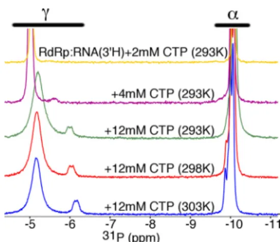
spectra of the RdRp䡠RNA䡠2⬘-dUTP and RdRp䡠RNA䡠CTP com-plexes (Figs. 3 and 4). We first performed the RdRp䡠RNA䡠CTP experiments using different concentrations of CTP and at dif-ferent temperatures. Here, we will focus first on the␥-P peak(s). As CTP is added, a second␥-P peak appears (␦⬃⫺5.8 ppm at 4 mM), and then both␥-P peaks begin to shift upfield as more CTP is added (␦⬃⫺5.4 and 6.2 ppm at 12 mMCTP and 293 K) (Fig. 3). As CTP is added, we also observe a broadening of the
␥-P peaks (Fig. 3). To better understand what is happening, it is important to keep in mind that the incorrect nucleotide CTP is
FIGURE 1.Structural dynamics and interactions in the poliovirus RNA-dependent RNA polymerase.A, overall structure of the poliovirus RdRp (PDB entry 3OL6) is shown with the conserved structural motifscolored(dark green, motif A;tan, motif B;dark cyan, motif C;blue, motif D;golden yellow, motif E;red, motif
F;light green, motif G). Also highlighted are motif F residues Lys-167 and Arg-174 (red), motif D residues Lys-359 and Thr-362 (blue), and locations of other
fidelity-altering substitutions (i.e.Gly-64 and His-273).B, conformational states of poliovirus RdRp based on X-ray crystal structures and MD simulations. The
boxed regioninA(left) is flipped 180º to improve clarity. The free state is in the absence of RNA and NTP (PDB entry 1RA6), the “binary” state is in the presence
of RNA (PDB entry 3OL6), and postincorporation complex represents the “ternary” state (PDB entry 3OL7). All of the residues shown, including residues in motifs B (i.e.Ser-288 and Asn-297), D (i.e.Lys-359), and F (i.e.Lys-167 and Arg-174), undergo structural rearrangements from the binary to ternary complex. These residues make important interactions with the incoming NTP; Ser-288 and Asn-297 are important for ribose sugar recognition, and Lys-167, Arg-174, and Lys-359 are all predicted to interact with the triphosphate. We have also previously proposed that Lys-359 acts as a general acid to protonate the pyrophos-phate leaving group. The “occluded” state is based on our previous MD simulations, which suggested that conserved residues that interact with the incoming NTP make interactions with each other, in the absence of NTP.C, NMR probes monitor rearrangements in the conserved structural motifs. The chemical shift positions of the⑀-13CH3Met-187 and Met-354 resonances are sensitive to structural rearrangements in motifs B and D (and other parts of the active site), where structural changes in these motifs are likely to be important for nucleotide selection. Residues are orientated similar toA(left).
bothMg2⫹ ionsandbasicaminoacidsidechainslikeLysand
Arg.Suchinteractionswouldalsotendtocreateamore elec-trophilic␣-P.Incontrast,the␥-Ppeakoftheprotein-bound nucleotidewasupfieldshiftedcomparedwiththe␥-PofMg2⫹ -UTPandhadachemicalshiftsimilartothatofthe␥-Poffree UTPintheabsenceofMg2⫹ (Fig.2C).Althoughthisfinding
mightsuggestthatthe␥-phosphatewasnolongerassociated with Mg2⫹, wedonotbelieve such ascenario islikely.Itis
less tightly associated with the RdRp than correct UTP, and as such, the spectra reflect an exchange between “free” (in solu-tion) and protein-bound CTP. When the time scale of this exchange is similar to that of the chemical shift differences between free and protein-bound CTP, then we observe chemi-cal shift changes for both the apparent free and protein-bound
␥-P peaks. In other words, the chemical shift of the free␥-P peak is “contaminated” with contributions from the chemical shift of the protein-bound ␥-P peak (and vice versa), so the apparent free ␥-P peak shifts toward the apparent protein-bound␥-P peak as more CTP is added. For variants with weaker association with CTP (e.g.K167R; see Fig. 4), the faster time scale of exchange can lead to the coalescence of the free and protein-bound peaks, leaving a single peak that represents a weighted population average of the underlying free and pro-tein-bound peaks (see Ref. 29). A similar broadening/shifting of the␣-P peaks does not occur because of the smaller chemical shift difference between the free and protein-bound␣-P peaks. This behavior is also not observed for the WT RdRp䡠RNA䡠UTP complex probably because UTP is held more tightly and
exchange on/off the enzyme is much slower. It should also be kept in mind that the31P NMR experiments were set up to
minimize the signal from free (d)NTP, including a short recycle delay, precluding estimates of the proportion of incorrect (d)NTP bound to the protein and free in solution. For the␥-P peaks for the RdRp䡠RNA䡠CTP complex, we assign the more intense (at␦⬃⫺5.4 ppm) and less intense (at␦⬃⫺6.2 ppm) peaks to the free and protein-bound CTP, respectively.
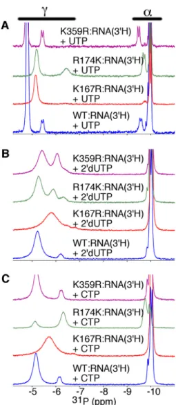
There were substantial chemical shift differences in the
31P NMR spectra of RdRp䡠RNA䡠UTP (Fig. 4A) and the
RdRp䡠RNA䡠2⬘-dUTP (Fig. 4B) and RdRp䡠RNA䡠CTP (Fig. 4C) complexes, strongly suggesting that the orientation and/or binding microenvironments of the correct and incorrect nucle-otides were substantially different between the two complexes. In particular, both the␣-P (␦ ⫺9.84 ppm for RdRp䡠RNA䡠2⬘ -dUTP and ⫺9.87 ppm for RdRp䡠RNA䡠CTP) and the ␥-P (␦ ⫺6.21 ppm for RdRp䡠RNA䡠2⬘-dUTP and ⫺6.16 ppm for RdRp䡠RNA䡠CTP) peaks of the incorrect nucleotide were not as downfield shifted as that of the correct nucleotide (Fig. 4). Besides exchange on/off the enzyme, conformational motions of the triphosphate of the incorrect nucleotide when bound to the RdRp may also have contributed to peak broadening; struc-tural studies of DNA polymerases suggest that incorrect NTP can fluctuate among an ensemble of conformations within the polymerase active site to prevent alignment and reaction with the primer 3⬘-OH (30 –35).
Motif D and F Substitutions Change the Binding Environment of the Nucleotide Triphosphate—To gain more insight into the microenvironment surrounding the triphosphate moiety of the incoming nucleotide and the roles of amino acid residues inter-acting directly with the triphosphate, we also generated com-plexes for the K167R, R174K, and K359R variants (Fig. 4). Arg-174, Lys-359, and Lys-167 are proposed to interact primarily with the␣-,-, and␥-phosphates, respectively (Fig. 1B). The
FIGURE 2.Formation of RdRp RNA and nucleotide complexes can be fol-lowed using31P NMR.A, the indicated 10-mer RNA can be used as a
sym-metrical substrate-template ligand. Incorporation of 3⬘-dATP terminates RNA synthesis, allowing the addition of a second nucleotide, UTP, to form the ternary RdRp䡠RNA(3⬘H)䡠UTP complex. Spin desalting columns can be used to remove excess free nucleotide.B, the triphosphates of the nucleotides can be detected using31P NMR. Not shown are the peaks for the phosphate back-bone of the RNA. The spectra for the RdRp complexes at 293 K (RdRp䡠RNA⫹3⬘ -dATP, RdRp䡠RNA(3⬘H), RdRp䡠RNA(3⬘H)⫹UTP, and RdRp䡠RNA(3⬘H)䡠UTP (desalted) shown ingolden yellow,magenta,green, andred, respectively) and 303 K (RdRp䡠RNA(3⬘H)䡠UTP (desalted);blue) are shown.C, these spectra are also compared with the spectra for free NTPs in the absence and presence of Mg2⫹, where temperatures are indicated on the plots. The assignments of the
␣-,-, and␥-phosphates are also shown. Spectra for protein complexes were collected with 250MRdRp, 500MRNA, 3.1 mM3⬘-dATP, and 4 mMUTP, where indicated, using buffer consisting of 10 mMHEPES, pH 8.0, 200 mM NaCl, 0.02% NaN3, 5 mMMgCl, and 10MZnCl. Spectra for free NTPs (i.e.in the absence of protein and RNA) were collected under the same buffer conditions using 2 mMNTP.
conservative amino acid changes should still allow these posi-tively charged residues to interact with the NTP triphosphate, albeit in an altered fashion that could be monitored by 31P
NMR.
Arg-174 is probably important for the alignment of the
␣-phosphate for reaction with the primer 3⬘-OH. Consistent with the role of Arg-174 in interacting with the␣-phosphate, the chemical shift for the protein-bound␣-P was upfield shifted in the R174K variant (␦⫺9.74 ppm) compared with WT RdRp (␦⫺9.36 ppm). The chemical shift positions of the␣-P and␥-P for the R174K䡠RNA䡠UTP and R174K䡠RNA䡠CTP complexes were remarkably similar (Fig. 4); only the relative intensities and widths of the peaks were different. The changes in peak intensities may just reflect the difference in the concentrations
of the nucleotides used in these experiments (i.e.[UTP]⫽4 mM; [CTP]⫽12 mM) and different exchange rates on/off the enzyme. Intriguingly, the R174K䡠RNA䡠2⬘-dUTP complex (Fig. 4B) gave rise to three peaks associated with ␥-P (␦ ⫺5.28, ⫺5.89, and ⫺6.32), suggesting that the ␥-phosphate for the “bound” 2⬘-dUTP might fluctuate between two or more conformations.
We have proposed that binding of correct, but not incorrect, nucleotide results in a structural rearrangement in the motif D loop to bring Lys-359 into the active site to interact with the
-phosphate (13). If so, we expected that the K359R substitu-tion would lead to changes in the31P NMR spectrum of the
correct RdRp䡠RNA䡠UTP complex but not the RdRp complexes bound with incorrect nucleotide. Consistent with this proposal, the K359R substitution led to small upfield chemical shift changes in the␣-P and␥-P peaks of the RdRp䡠RNA䡠UTP com-plex (␦ ⫺9.49 and⫺5.43 ppm; Fig. 4A) but did not lead to substantial chemical shift changes in the RdRp䡠RNA䡠CTP spec-trum (␦ ⫺9.87 and ⫺6.25 ppm; Fig. 4C) compared with the corresponding WT complexes. However, there was also a change in the␥-P peaks for the K359R䡠RNA䡠2⬘-dUTP spectrum (␦⫺5.40 and⫺6.03 ppm) compared with the WT complex (Fig. 4B). These changes may reflect different nucleotide exchange kinetics and/or changes to the binding microenvironment for 2⬘-dUTP once bound to the K359R variant.
The K167R substitution also led to differences in the spectra for the ternary complexes. In particular, there was one very broad ␥-P peak for the RdRp䡠RNA䡠2⬘-dUTP and RdRp䡠RNA䡠CTP complexes, which probably reflects changes in the nucleotide exchange kinetics. In other words, exchange on/off the enzyme is fast compared with the chemical shift dif-ferences between free and protein-bound NTP, such that the single␥-P peak represents a weighted average of the free and protein-bound states. This proposal is consistent with the higher apparent nucleotide dissociation constants for the K167R variant compared with WT RdRp (see Table 1). The protein-bound peak intensities for the K167R䡠RNA䡠UTP complex were also quite weak, which might be indicative of decreased UTP binding and/or additional exchange processes on the enzyme that would broaden out these signals. These results were consistent with Lys-167 making important inter-actions with the triphosphate, which might be important for nucleotide binding affinity.
The R174K Substitution Also Prevents Motif D Structural Rearrangements as Monitored by Protein NMR—The31P NMR
spectra for the K167R, R174K, and K359R variants indicated that these amino acid substitutions lead to changes in how the triphosphate interacts with the RdRp. We also collected NMR spectra of [methyl-13C]Met-labeled RdRp to gain insight into how these amino acid substitutions change RdRp structural dynamics. In particular, we have previously noted that Met-354 is a good reporter of any structural and/or dynamic changes in the motif D loop (see also Fig. 1C), which we propose are impor-tant for repositioning Lys-359 for catalysis. Chemical shift changes to Met-354 are generally accompanied by chemical shift changes to other informative resonances, such as Met-6, Met-74, and Met-225 (Fig. 5).
For the K167R variant, there were no substantial chemical shift differences compared with the corresponding WT com-plexes (Fig. 5,A–C), suggesting that the overall protein confor-mation was not substantially affected by this amino acid change. In contrast, binding of the correct UTP nucleotide to the R174K variant did not result in the same chemical shift changes as observed for WT RdRp (Fig. 5D). In fact, the chem-ical shift changes induced by binding of correct and incorrect nucleotides were similar for the R174K variant and similar to those changes induced by the binding of the incorrect nucleo-tide to WT RdRp (Fig. 5,EandF). These findings suggest that conformational changes in the motif D loop and other associ-ated structural changes were severely impaired in the R174K variant. Altogether, these findings are consistent with our pre-vious results that indicated that the␣- and-phosphates but not the␥-phosphate of the incoming nucleotide is required for the conformational changes in motif D and surrounding areas (13).
Motif F Substitutions Also Lead to Changes around Motif B— We have also previously used the Met-187 resonance as a probe to support molecular dynamics (MD) simulations suggesting that motif B and nearby regions fluctuate between conforma-tions competent for and occluded from NTP binding (see also Fig. 1C) (22). In the occluded conformation (Fig. 1B), Arg-174 is involved in a salt-bridging interaction with Asp-238, such that this interaction and others must be broken before the RdRp䡠RNA complex can bind NTP (22); Met-187 is a reporter of these interactions and rearrangements (22). For the WT pro-tein, there were distinct chemical shift changes when correct
UTP and incorrect CTP were added to the RdRp䡠RNA complex (Fig. 6A). The K167R, R174K, and K359R substitutions all led to changes to the Met-187 resonances, especially for the RdRp䡠RNA and RdRp䡠RNA䡠CTP complexes, suggesting that these substitutions have long range effects on the structure/ dynamics of motif B and surrounding regions (Fig. 6). In con-trast, the Met-187 resonances were very similar for the RdRp䡠RNA䡠UTP complexes (Fig. 6). The one exception was for the R174K variant, in which there were two additional reso-nances, where one of these overlapped that for the R174K RNA䡠CTP complex (Fig. 6C). Altogether, these results sug-gested that motif B and surrounding regions can attain a similar conformation when correct NTP binds for all variants, but the conformations in the absence of NTP or in the presence of incorrect NTP may differ.
Other changes to the Met-187 resonances were also informative. There were two resonances for the K167R RdRp䡠RNA䡠CTP complex, with one of those resonances more similar to what was observed for the K167R RdRp䡠RNA䡠UTP complex (Fig. 6B). There was no apparent Met-187 resonance for the R174K RdRp䡠RNA complex, suggesting conformational exchange on the intermediate NMR time scale (Fig. 6C). One potential explanation would be that the R174K substitution dis-rupts the interactions with Asp-238, such that the conforma-tional exchange kinetics between NTP binding-competent and occluded conformations was moved to the intermediate NMR time scale. Finally, the Met-187 resonances for the K359R ter-nary complexes had similar chemical shift positions, in contrast to what was observed for WT RdRp (Fig. 6D). A comparable TABLE 1
Comparison of the kinetic constants of nucleotide incorporation for variant and WT RdRp enzymes
Variant Metal NTP kpol Kd(app) kpol/Kd(app)
s⫺1
M M⫺1s⫺1 Correct NTP
WT Mg2⫹ ATP 59⫾1 36⫾2 1.6
ATP (D2O)
a 27⫾1 15⫾1 1.8
Mn2⫹ ATP 16⫾1 1.7⫾0.1 9.4
ATP (D2O)
a 5.3⫾0.1b NDc ND
K167M Mg2⫹ ATP 26⫾0.2 683⫾29 3.8⫻10⫺2
K167R Mg2⫹ ATP 56⫾1 27⫾1 2.1
ATP (D2O)
a 12⫾1 7.5⫾1.0 1.6
Mn2⫹ ATP 21⫾1 4.1⫾0.3 5.3
ATP (D2O)
a 3.1⫾0.1 ND ND
R174K Mg2⫹ ATP 4.2⫾0.2 19⫾4 0.22
ATP (D2O)
a 1.7⫾0.1 2.5⫾0.1 0.68
Mn2⫹ ATP 1.7⫾0.1 0.20⫾0.03 8.5
ATP (D2O)
a 0.40⫾0.01b ND ND
K359L Mg2⫹ ATP 0.62⫾0.01 272⫾15 2.3⫻10⫺3
R174K/K359L Mg2⫹ ATP 0.011⫾0.001 148⫾24 7.4⫻10⫺5
Incorrect sugar
WT Mg2⫹ 2⬘-dATP 0.89⫾0.01 134⫾4 6.6⫻10⫺3
Mn2⫹ 2⬘-dATP 7.4⫾0.1 6.1⫾0.1 1.2
K167R Mg2⫹ 2⬘-dATP 3.4⫾0.1 321⫾25 1.1⫻10⫺2
Mn2⫹ 2⬘-dATP 15⫾1 9.5⫾1.0 1.6
R174K Mg2⫹ 2⬘-dATP 2.4⫾0.1⫻10⫺4
26⫾6 9.2⫻10⫺7
Mn2⫹ 2⬘-dATP 0.5 7.6⫾0.5 6.6⫻10⫺2
Incorrect nucleobase
WT Mg2⫹ GTP 0.011⫾0.001 142⫾15 7.7⫻10⫺5
Mn2⫹ GTP 0.80⫾0.01 98⫾3 8.2⫻10⫺3
K167R Mg2⫹ GTP 3.7⫾0.1⫻10⫺2 223⫾20 1.7⫻10⫺4
Mn2⫹ GTP 1.9⫾0.1 168⫾19 1.1⫻10⫺2
R174K Mg2⫹ GTP 3.0⫾0.1⫻10⫺5d ND ND
Mn2⫹ GTP 0.11⫾0.01 57⫾5 1.9⫻10⫺3
a
Kinetic constants were determined in D2O solvent. b
kpolwas estimated using [ATP]⫽20M(i.e.⬎10⫻Kd(app)in H2O and Mn2⫹). c
ND, no data.
d
The R174K Substitution Reduces Catalytic Efficiency and Increases Nucleotide Selection Fidelity—Not surprisingly, the
31P and [methyl-13C]Met spectra suggested that conserved
amino acid substitutions at residues proposed to interact with the NTP triphosphate alter the nucleotide binding kinetics
FIGURE 5.Amino acid substitutions at triphosphate-interacting positions can change RdRp structural dynamics at motif D.A–E, comparison of [methyl -13C]Met HSQC spectra between WT (black), K167R (red), and R174K (green) RdRp bound with RNA lacking a 3⬘-OH and correct UTP nucleotide or incorrect 2⬘-dUTP or incorrect CTP nucleotide. The chemical shift signatures for R174K and WT RdRp bound with RNA and correct UTP nucleotide were quite different. More insight was gleaned from the finding that the spectra for the R174K variant bound with correct UTP (black) and incorrect CTP (green) nucleotides were nearly identical (F). This finding suggests that the R174K variant was impaired in its ability to form a “closed” conformation like WT RdRp. Spectra were collected at 293 K using 250MRdRp with 500MRNA and 4 mMUTP (12 mMUTP for the R174K variant), 8 mM2⬘-dUTP, or 12 mMCTP and a D2O-based buffer consisting of 10 mMHEPES, pH 8.0, 200 mMNaCl, 0.02% NaN3, 5 mMMgCl, and 10MZnCl.
and/or conformation. To gain more insight, we analyzed the abil-ity of RdRp variants to bind RNA and assemble into catalytically competent complexes and measured the maximum polymerase rate constant (kpol) and the apparent dissociation constant for the incoming nucleotide (Kd(app)). To compare RNA binding between
variant and WT RdRp, we monitored interactions with 6-car-boxyfluorescein (6-FAM)-labeled sym/sub-UA RNA using a fluo-rescence polarization assay (Table 2). This assay indicated that most substitutions at Lys-167, Arg-174, and Lys-359 had little effect on RNA binding affinity (Table 2). The sole exception was the R174L substitution, and the large decrease in RNA binding affinity precluded further assessment of this variant.
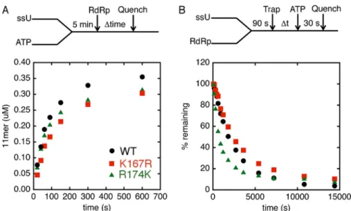
To ensure that the variants could assemble efficiently on the sym/sub-UA template and catalyze phosphodiester bond for-mation with the incoming nucleotide, we initiated the reaction with the addition of RdRp and followed RNA synthesis over various time points (Fig. 7A). The assay indicated that the vari-ants could assemble for catalysis in a similar time frame as WT RdRp. We also compared the stabilities of the RdRp䡠RNA com-plexes between variant and WT RdRps by incubating RdRp enzyme with labeled sym/sub-UA for 90 s to allow the
RdRp䡠RNA binary complexes to form before the addition of excess unlabeled sym/sub-UA to trap free and dissociating enzyme (Fig. 7B). ATP was then added at various times after the addition of the unlabeled sym/sub-UA, and the reaction was quenched 30 s later. This assay indicated that the R174K variant dissociated more quickly from the labeled RNA compared with WT RdRp (Table 2). Nonetheless, the RdRp䡠RNA complex for R174K RdRp was deemed sufficiently stable for our single nucleotide incorporation assays to measurekpolandKd(app)
val-ues. It is important to note that we also checked the RdRp cat-alytic activity under buffer conditions that we used for the31P
and [methyl-13C]Met NMR experiments, which did not reveal
any substantial differences compared with the buffer system we used for our kinetic assays (Fig. 8).
The K167R substitution had little effect on the kpol and Kd(app)values for correct nucleotide incorporation (i.e.ATP, in
the presence of Mg2⫹) but led to increases inkpol,Kd(app), and
catalytic efficiency (kpol/Kd(app)) for incorrect nucleotide
incor-poration compared with WT RdRp (Table 1). As mentioned above, the change inKd(app)for incorrect nucleotide may help to explain the broader peaks in the31P NMR spectrum for the
K167R䡠RNA䡠2⬘-dUTP and K167R䡠RNA䡠CTP complexes (Fig. 4), which suggests a change in the binding on/off rates. These results also indicate that the K167R variant is less able to dis-criminate against incorrect nucleotide and has lower fidelity compared with WT RdRp. The K167M variant also had a sub-stantial increase in the Kd(app) for correct nucleotide. The
higherKd(app)values precluded further kinetic assessment of incorrect nucleotide incorporation for this variant.
Perhaps not surprisingly, the R174K variant led to a 14-fold drop inkpolfor correct nucleotide incorporation (i.e.ATP, in the presence of Mg2⫹) compared with WT RdRp (Table 1). More remarkable was the finding that the change inkpolwas
even more substantial for incorrect nucleotide incorporation, either with incorrect sugar (i.e.2⬘-dATP) or incorrect nucleo-base (i.e. GTP). Specifically, there were 3,700- and 370-fold drops inkpolfor 2⬘-dATP and GTP incorporation, respectively,
compared with WT RdRp (kpolfor GTP was estimated based on
using 1000MGTP (i.e.almost 10-fold greater than theKd(app) for WT RdRp; Table 1). Thesekpolvalues also indicated that the
R174K variant was more faithful (i.e. kpol(correct)/kpol(incorrect)
values were 17,500 and 140,000 for 2⬘-dATP and GTP, respec-tively) than WT RdRp (kpol(correct)/kpol(incorrect)values were 70
and 5400 for 2⬘-dATP and GTP, respectively).
Metal and Solvent Effects Help to Gauge the Effects of Amino Acid Substitutions on the Prechemistry Conformational Change and Chemical Steps—To gain more insight into the effects of these amino acid substitutions, we also assayed the effects of
FIGURE 6.Amino acid substitutions at triphosphate-interacting positions change RdRp structural dynamics around motif B.It was shown previously that the Met-187 resonance is a reporter of conformational changes in motif B, including those changes associated with RNA and NTP binding (22). Met-187 resonances from [methyl-13C]Met HSQC spectra are shown for RdRp bound to sym/sub-UA lacking a 3⬘-OH (black), bound to RNA lacking a 3⬘-OH and correct UTP (blue), and bound to RNA lacking a 3⬘-OH and incorrect CTP (green) for WT (A), K167R (B), R174K (C), and K359R (D) complexes. Spectra were collected at 293 K using 250MRdRp with 500MRNA and 4 mMUTP (12 mMUTP for the R174K variant), 8 mM2⬘-dUTP, or 12 mMCTP and a D2O-based buffer consisting of 10 mMHEPES, pH 8.0, 200 mMNaCl, 0.02% NaN3, 5 mM MgCl, and 10MZnCl.
TABLE 2
Interactions between variant and WT RdRps and sym/sub-UA RNA
RdRp Kd kd
nM ⫻10⫺4s⫺1
WT 575⫾68 4.0⫾0.2
K167M 254⫾28 1.4⫾0.6
K167R 767⫾203 3.5⫾0.1
R174K 603⫾35 11⫾1
R174L 1951⫾304 5.7⫾0.8
K359L 397⫾28 1.4⫾0.1
more rate-limited by the chemical step in the presence of Mn2⫹, presumably because the correct nucleotide more readily
induces the prechemistry conformational change with this metal (37). However, the chemistry step is less discriminatory in the presence of Mn2⫹, and the prechemistry step largely
governs polymerase fidelity (37). The relative contribution of the chemistry step can also be assayed by performing the assays in D2O, because the solvent deuterium kinetic isotope effect FIGURE 7.Formation and stability of RdRp䡠RNA complexes is similar between WT RdRp and motif F variants.A, RdRp䡠RNA䡠NTP assembly assay. Reactions were initiated by adding RdRp (1M) into sym/sub-UA (0.5Mduplex) and ATP (500M), which were incubated at 30 °C for 5 min. At the indicated times, reactions were quenched by adding 25 mMEDTA. The results for the RdRp䡠RNA䡠NTP assembly assay are shown for WT (black circles), K167R (red squares), and R174K (green triangles).B, RdRp䡠RNA dissociation assay. RdRp and RNA were incubated at 30 °C for 90 s, at which time trap (100Munlabeled RNA) was added to the reaction buffer. After the indicated times, the reaction buffer was mixed with ATP (500M) and then quenched after 30 s by adding EDTA (25 mM). The results for the RdRp䡠RNA dissociation assay are shown for WT (black circles), K167R (red squares), and R174K (green triangles).
FIGURE 8.UMP incorporation into sym/sub-UA is not affected by the composition of the buffers used for NMR experiments.A, reaction scheme. Buffer 1 was preincubated with 500MATP and 1Msym/sub-UA for 5 min, and then the reaction was initiated by the addition of 5MWT RdRp to incorporate AMP into sym/sub-UA. After 5 min, the AMP incorporated RdRp䡠RNA complexes were added into 500MUTP and buffers 1, 2, and 3, respectively, via a 1:10 ratio so that the final concentrations of RdRp, RNA, and ATP were 0.5, 0.1, and 50M, respectively. The reactions were then quenched at the indicated times by the addition of EDTA to a final concentration of 50 mM.B, products from reactions described inAwere resolved by electrophoresis on a denaturing 23% polyacrylamide gel.C, comparison of UMP incorporation kinetics using buffers 1, 2, and 3. Buffer 1 contained 50 mMHEPES, pH 7.5, 10 mM-mercaptoethanol, 5 mMMgCl2, and 60MZnCl2; buffer 2 contained 10 mMHEPES, pH 8.0, 200 mMNaCl, 5 mMMgCl2, 10MZnCl2, and 0.02% NaN3in 100% D2O; and buffer 3 contained the same components as buffer 2 except that the final concentration of D2O was 10%. All reactions were performed at 30 °C.
changingthe metal ion and the solvent. In the presenceof Mg2⫹,theincorporationofcorrectnucleotidecatalyzedbythe
(SDKIE;i.e. kpol(H2O)/kpol(D2O)) reports on proton transfer steps occurring during phosphodiester bond formation (11).
The SDKIEs for the K167R and R174K variants were higher than WT RdRp in the presence of Mg2⫹(4.67⫾0.40, 2.47⫾
0.05, and 2.19⫾0.03 for K167R, R174K, and WT RdRp, respec-tively) or Mn2⫹(6.77⫾0.40, 4.25⫾0.02, and 3.02⫾0.03 for
K167R, R174K, and WT RdRp, respectively) (Table 1). These results suggest that the K167R and R174K substitutions both decreased the rate of the chemistry step. Nucleotide discrimi-nation for the K167R and R174K variants was more similar to that of WT RdRp in Mn2⫹(i.e. kpol(ATP)/kpol(2⬘-dATP)values are
1.4, 3.4, and 2.2 for K167R, R174K, and WT RdRp respectively; kpol(ATP)/kpol(GTP)values are 11, 15, and 20) than Mg2⫹. These
results suggest that the differences in RdRp fidelity in the pres-ence of Mg2⫹are probably due to the K167R and R174K sub-stitutions primarily affecting the chemical step for incorrect nucleotide incorporation, such that most of these effects disap-pear with Mn2⫹.
Kinetic Experiments Indicate That the R174K Variant Exchanges Nucleotides More Readily—The NMR experiments suggested that the NTP and protein structural changes for R174K variant were substantially different from that observed for WT enzyme. To gain more insight, AMP-CPP (or buffer as control) was preincubated with RdRp䡠RNA binary com-plexes for 5 min to trap the “closed” state by forming RdRp䡠RNA䡠AMP-CPP ternary complexes, and then the reac-tion was initiated by the addireac-tion of an excess amount of ATP (Fig. 9). The reaction occurs only when AMP-CPP is exchanged with ATP. The rate for nucleotide incorporation was deter-mined with and without AMP-CPP. The delay in nucleotide incorporation induced by AMP-CPP is a measure of the reverse rate of the prechemistry conformational change. For WT and K167R RdRps, the rates were delayed by 40 –50-fold (Fig. 9). However, in R174K RdRp, the rate was delayed by only 3-fold (Fig. 9). These results suggested that the R174K substitution destabilized the “closed” state of the ternary complex, consis-tent with the NMR experiments (Figs. 4 – 6). The slower rate of catalysis together with the enhanced ability to exchange nucle-otides probably provide additional opportunities for the R174K variant to release incorrect nucleotide, leading to a higher fidel-ity enzyme.
The Functional Roles of Arg-174 and Lys-359 Are Largely Independent—It was shown previously that the K359R and K359L substitutions lead to decreases inkpoland increases in
polymerase fidelity (5, 12, 13). Because both Arg-174 and Lys-359 (13) appear to be important for the conformational changes in motif D observed by NMR (Fig. 5), we wanted to understand whether the roles of these residues were related or independent from one another. To address this question, we compared the kinetics of the variants with single amino acid substitutions (i.e. R174K and K359L) with the variant with both amino acid sub-stitutions (i.e.R174K/K359L). RdRp with a nonpolar residue at position 359 (i.e.K359M) does not undergo the same confor-mational changes as WT enzyme (13), similar to what is observed for the R174K variant (Fig. 5). The R174K/K359L var-iant demonstrated decreased affinity to RNA (Table 2) and was much more impaired in its ability to incorporate correct ATP
nucleotide compared with either of the two single variants (Table 1).
To test whether the amino acid substitutions were thermo-dynamically coupled and hence whether the function of the residues were interdependent, we compared the free energy changes induced in the catalytic efficiency (i.e.⌬⌬G⫽ ⫺RTln ((kpol/Kd(app))variant/(kpol/Kd(app),app)WT) by the single and double amino acid substitutions, analogous to the classic dou-ble mutant cycle experiments performed by Fersht (38), Mild-vanet al.(39), and Serranoet al.(40). The effects of the R174K (⌬⌬G⫽1.2 kcal/mol) and K359L (⌬⌬G⫽4.0 kcal/mol) substi-tutions were nearly additive (assuming⬃20% error) compared with the R174K/K359L double amino acid substitution (⌬⌬G⫽ 6.0 kcal/mol), suggesting that these residues are very weakly thermodynamically coupled and that their roles are largely inde-pendent from one another. If the functions of these residues were interrelated, we would have expected a non-additive response. The differences in the NMR spectra for the R174K and K359R variants also suggest that these amino acid substi-tutions have substantially different effects on the nucleotide and protein structural dynamics.
Fidelity Variants Substantially Alter Triphosphate Binding and Reorientation—Our NMR and kinetic studies indicated that amino acid substitutions at residues that interact with the NTP triphosphate lead to changes in catalytic efficiency and polymerase fidelity. We were interested in testing whether other amino acid substitutions that change polymerase fidelity
FIGURE 9.The R174K substitution destabilizes the “closed” conforma-tion.A, the minimal kinetic mechanism for the dissociation of AMP-CPP and incorporation of AMP. Reactions contained 50 mMHEPES, pH 7.5, 10 mM
-mercaptoethanol, 5 mMMnCl2, and 60MZnCl2. Enzyme was preincubated with sym/sub-UA to form binary complexes for 90 s. Then 200MAMP-CPP or buffer was added and incubated for an extra 5 min to form RdRp䡠RNA䡠 AMP-CPP ternary complexes. Reactions were initiated by the addition of an excess amount of ATP. AMP incorporation was then monitored by fluorescent changes using a stopped flow apparatus. Final concentrations of ATP, sym/ sub-UA, and RdRp were 1 mM, 1M(WT and K167R) or 2M(R174K), and 1M, respectively. B–D, the comparison of AMP incorporation in the presence (blue) and absence (red) of AMP-CPP for WT (B), K167R (C), and R174K RdRp (D).
FIGURE 10.Fidelity variants of RdRp can change the conformational dynam-ics of motif D.A–C,close-upsof [methyl-13C]Met HSQC spectra showing the Met-354 resonances for WT, H273R, and T362I RdRp bound with RNA lacking a 3⬘-OH and correct UTP nucleotide (black) or incorrect 2⬘-dUTP (blue) or incorrect CTP (red) nucleotide. Spectra were collected at 293 K using 250MRdRp with 500M RNA and 4 mMUTP, 8 mM2⬘-dUTP, or 12 mMCTP and a D2O-based buffer consist-ing of 10 mMHEPES, pH 8.0, 200 mMNaCl, 0.02% NaN3, 5 mMMgCl, and 10MZnCl.
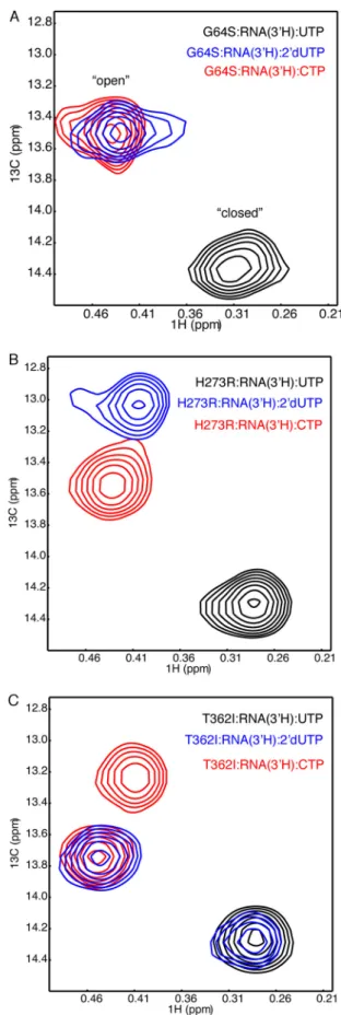
andattenuatethevirusalsoleadtochangestothe triphosphate-bindingenvironment.Inparticular,theG64SandH273R sub-stitutionsarerelativelyremotefromtheactivesite(⫻20Åaway fromthecatalyticcenter)butgeneratehigherandlowerfidelity polymerases,respectively(41–43).Althoughtheseaminoacid changesarerelativelyremotefromtheactivesite,interaction pathwayssuggesthow thesesubstitutionsmightaffectRdRp catalysis and fidelity. For example, Gly-64 is involved in a hydrogenbondingnetworkinvolvingAla-239andLeu-241in motifAandGly-285inmotifB.His-273makeshydrogenbond interactionswithThr-156ina-strandthatmakesinteractions withthe-strandcontainingArg-174.MD simulationshave alsoshownhowtheG64SandH273Rsubstitutionsaffect struc-turaldynamicsaroundtheactivesiteandmoreglobally(22,44). AnotherintriguingfidelityvariantisT362IRdRp(18).Wewere initiallyinterestedinthisaminoacidsubstitutionbecauseitis encodedbytheSabinIvaccinestrainandislocatedinmotifD, nearLys-359(45).OurstudiesindicatedthattheT362I substi-tutionleadstoalowerfidelitypolymerase,probablybecauseit canaccessamoreactiveconformationeveninthepresenceof incorrectnucleotide(18).TheT362Isubstitutionbyitself(i.e. intheabsenceofotherSabinImutations)alsoleadstosome attenuationoftheencodingPVstrain(18).Inthepresenceof otherSabinIsubstitutions,theseeffectsarelargelymitigated (46).Nonetheless,theT362Ivariantcanserveasanimportant tool in understanding how the conformational dynamics of motifDare(un)relatedtotherealignmentofthetriphosphate. WeassessedproteinstructuralchangesintheG64S,H273R, andT362Ivariantsbyusing[methyl-13C]MetNMR.Wewere
especiallyinterestedintrackingtheMet-354resonance,asit responds tostructural changesin motif Dand surrounding areas(Fig.1).TheG64Ssubstitutioninducedonlyverysmall chemicalshiftdifferencesinthe[methyl-13C]Metspectrafor
theRdRp䡠RNA䡠UTP,RdRp䡠RNA䡠2⬘-dUTP,andRdRp䡠RNA䡠CTP complexes (Fig.10A)compared withWT RdRp (Fig.5). In contrast,theH273R䡠RNA䡠2⬘-dUTP,T362I䡠RNA䡠2⬘-dUTP,and T362I䡠RNA䡠CTP complexes all ledtospectralchanges com-paredwiththesameWTandG64Scomplexes(Fig.10,Band C).WehadpreviouslynotedthattheT362Ivariantgivesriseto two setsof resonances fortheRdRp䡠RNA䡠2⬘-dUTP complex (18);onesetofresonances overlapswhatisobservedforthe WT䡠RNA䡠UTPcomplex,andtheothersetofresonances over-laps what is observed forthe WT䡠RNA䡠CTP complex. This finding prompted us to suggest that the T362I variant can accessamoreactiveconformationeveninthepresenceofthe incorrect 2⬘-dUTP (18),suggestingwhy itis alowerfidelity variant.TheT362I䡠RNA䡠CTPcomplexalsoappearedto fluctu-ateintotwo(ormore)conformations,consideringthepresence oftwoMet-354resonances.
the RdRp䡠RNA䡠UTP complex was similar to that for WT enzyme, but the Met-187 resonance for the RdRp䡠RNA䡠CTP complex was at a chemical shift position different from that for WT RdRp (22).
To test the effects of the G64S, H273R, and T362I substitu-tions on the binding and microenvironment of the NTP triphosphate, we collected31P NMR spectra of their correct RdRp䡠RNA䡠UTP and incorrect RdRp䡠RNA䡠2⬘-dUTP and RdRp䡠RNA䡠CTP complexes (Fig. 11). It should be kept in mind that any changes in the31P NMR spectra of the G64S, H273R,
and T362I variants must be due to indirect effects, because these residues do not make direct interactions with RNA or NTP. Nonetheless, there were substantial changes in the31P
NMR spectra for these variants compared with WT RdRp,
␣-P peaks between the RdRp䡠RNA䡠CTP and RdRp䡠RNA䡠UTP complexes (␦⫺9.46 ppm) compared with the same complexes for WT enzyme. This result might suggest that the ␣ -phos-phates for these complexes were in more comparable chemical environments for the H273R variant compared with WT RdRp. If true, this finding would help to explain why the H273R vari-ant has a lower ability to discriminate between correct and incorrect nucleotides. This result is also consistent with the proposal that the H273R substitution stabilizes the NTP-bind-ing competent conformation, helpNTP-bind-ing to bypass this fidelity checkpoint (22).
In contrast to what is observed for the G64S and H273R variants, the T362I substitution did not lead to additional␥-P peaks for the RdRp䡠RNA䡠2⬘-dUTP complex. In fact, the 31P
spectrum was very similar to that for WT RdRp (Fig. 11), which was in stark contrast to the corresponding [methyl-13C]Met
spectra (Fig. 8). The T362I substitution also induced a chemical shift change in the␥-P peak (␦⫺5.90 ppm) compared with what was observed for the WT enzyme (Fig. 11C), although this might just reflect a change in the CTP exchange kinetics on/off the enzyme (i.e.the␥-P peaks are less broad and more intense for the T362I variant). The T362I substitution also induced changes in the RdRp䡠RNA䡠UTP spectra, which was a bit sur-prising considering that the corresponding [methyl-13C]Met
spectra were very similar to WT enzyme. These findings
sug-FIGURE 11.Lower and higher fidelity RdRp variants change the orienta-tion and/or microenvironment of the triphosphate for the incoming nucleotide.Comparisons of RdRp䡠RNA complexes bound with correct nucle-otide (i.e.UTP) (A), bound with incorrect nucleotide with incorrect sugar (i.e. 2⬘-dUTP) (B), and bound with incorrect nucleotide with incorrect nucleobase (i.e.CTP) (C) for WT (black), G64S (red), H273R (green), and T362I (blue) RdRp. The G64S substitution leads to a higher fidelity polymerase (41), and the H273R (22) and T362I (18) substitutions lead to lower fidelity polymerases. Spectra were collected at 303 K with 250MRdRp, 500MRNA, 3.1 mM 3⬘-dATP, and 4 mMUTP, 8 mM2⬘-dUTP, or 12 mMCTP and a buffer consisting of 10 mMHEPES, pH 8.0, 200 mMNaCl, 0.02% NaN3, 5 mMMgCl, and 10M ZnCl.
especiallyfortheG64SandH273Rvariants(Fig.11).In partic-ular,theH273Rsubstitutioninducedchemicalshiftchangesfor the␣-Pand␥-Ppeaksin theRdRp䡠RNA䡠UTPcomplex (Fig. 11A),suggestingthatcorrectnucleotidemaybindinadifferent manner,and/orthesurroundingmicroenvironmenthasbeen alteredintheH273Rvariant.Theseeffectsmightbeduetothe seriesofinteractionsconnectingHis-273tothe-strand con-taining Arg-174, as suggested above, and/or changes in the structuraldynamicsaroundtheactivesite,asobserved previ-ouslyinMDsimulations(22).
changes in motifs B and D (Fig. 1C) and surrounding areas. Our studies provide insight into not only which fidelity checkpoints are altered by these amino acid substitutions, but provide infor-mation about the connections between different fidelity check-points through comparisons of fidelity variants (Fig. 12).
In this report, we have developed a31P NMR assay to probe the binding environment of the NTP triphosphate (Fig. 2). Crystal structures of nucleic acid polymerase superfamily members have indicated that there is a reorientation of the triphosphate of the incoming nucleotide following initial bind-ing to properly align the␣-phosphate for nucleophilic attack by the primer 3⬘-OH (10, 48) (Fig. 12). Having a mispaired primer terminus and/or binding incorrect nucleotide does not result in the same structural rearrangement (32–34, 49). As such, the reorientation of the triphosphate probably reflects an impor-tant fidelity checkpoint (10, 37, 50 –52). Our31P NMR studies
on PV RdRp are consistent with these proposals, where the binding of correct and incorrect NTP led to different31P NMR
signatures (Figs. 3 and 4). In the interpretation of these experi-ments, it should be kept in mind that the spectra can represent a mixture of states. If the triphosphate is fluctuating between different conformations on a time scale fast relative to the chemical shift differences between these states, we would
FIGURE 12.Nucleotide selection checkpoints altered by PV RdRp fidelity variants.Conformational changes important for nucleotide incorporation include a reorientation of the triphosphate of the incoming nucleotide for direct in-line attack by the RNA 3⬘-OH, a conformational change in structural motif D to reposition conserved residue Lys-359, and conformational changes in structural motif B to allow for interactions with the 2⬘-OH of the incoming nucleotide. We have developed diagnostic NMR probes for each of these rearrangements; our31P NMR studies provide insight into the environment surrounding the triphosphate of the incoming nucleotide, and our [methyl-13C]Met NMR studies provide probes for structural changes in motifs B (i.e.Met-187) and D (i.e. Met-354). Our results indicate that the fidelity-altering amino acid substitutions differentially affect these checkpoints, suggesting that the checkpoints are not strictly dependent on each other.
gestthattheT362Isubstitutioncanhavedifferingeffectsonthe NTPtriphosphateandmotifB/Dstructuraldynamics.
Discussion
Enzymes within the nucleic acid polymerase superfamily appeartoshareacommonkineticmechanism,includinga con-formationalchangeintoamoreactiveformbeforecatalyzing phosphodiester bond formation (1). There has been much debateoverthestructuraldetailsofthisconformationalchange (47).Itislikelythattheprechemistryconformationalchange identified in thesekinetic mechanisms actually represents a numberofconformationalevents.InRdRps,thereare struc-turalrearrangementsin motifsA andBtointeractwith the NTPribosehydroxyls,arealignmentoftheNTPtriphosphate forproperorientation,andarepositioningofthemotifD ly-sine(Lys-359inPVRdRp)toactasageneralacidtoprotonate the pyrophosphate leaving group. We have now developed diagnosticNMRexperimentstoprobeeachofthesestructural changes,allowingustoinvestigatethenucleotide incorpora-tioncycleforWTandfidelity-alteringvariants.Specifically,the
31PNMRexperimentsthatwehavedevelopedhereprobethe
observe only a single set of peaks for protein-bound NTP that represents a weighted average of these different states. Chang-ing residues proposed to interact with the NTP triphosphate led to changes in rates and fidelity of nucleotide addition (Table 1). Interestingly, modification of Arg-174 and Lys-359, pro-posed to interact with the␣- and-phosphates, respectively, increased fidelity, whereas modification of Lys-167, proposed to interact with the␥-phosphate, decreased fidelity. The SDKIE and metal effects suggested that these modifications primarily affected the chemistry step itself.
We have also suggested that conformational changes to reposition Lys-359 for catalysis, analogous to the conforma-tional change in the O/P helix in the A/B family DNA polymer-ases (32, 34, 53– 61), represent an important fidelity check-point. The Met-354 resonance has been diagnostic of conformational changes in motif D (13, 21) (Figs. 1Cand 5). The chemical shift changes experienced by Met-354 are abso-lutely dependent on the protonation state of Lys-359 (13); only under conditions in which the side-chain amine of Lys-359 is protonated do these structural changes occur. We do not observe similar chemical shift changes upon binding incorrect nucleotide. Consistent with this proposal, the K359R substitu-tion changed the31P NMR spectrum relative to WT for the
correct RdRp䡠RNA䡠UTP complex but not for the incorrect RdRp䡠RNA䡠CTP complex (Fig. 4). This result suggested that this residue was only brought near the NTP triphosphate when correct NTP was present. Unfortunately, PV RdRp crystal structures have failed to capture Lys-359 in a position to pro-tonate the pyrophosphate leaving group. We note here that the
31P and1H-13C spectra (Figs. 4 and 5) and the AMP-CPP trap
assay (Fig. 9) indicated that the R174K variant was impaired in its ability to fluctuate into a closed conformation. These results might indicate that R174K is capable of catalyzing phosphodi-ester bond formation through an open-like conformation (i.e. when Lys-359 is not in a position to donate a proton to the
-phosphate). Likewise, catalysis in the RdRp crystals may not require the repositioning of Lys-359. It is known that Lys-359 is not absolutely required for catalysis, considering that amino acid substitutions at this position only decrease the catalytic rate by up to 50-fold (12).
Structural rearrangements in motifs A/B upon interacting with the NTP ribose are also well known to be important for RdRp fidelity (62). The Met-187 resonance serves as a diagnos-tic probe for structure/dynamic changes in motif B and sur-rounding areas (Fig. 1C). It is intriguing that most of the fidelity variants resulted in some change to the Met-187 resonances (Fig. 6) (22). The pervasiveness of these changes to Met-187 and its surrounding environment are consistent with motif A/B-ribose interactions being one of the first steps in NTP binding and selection, necessary to trigger subsequent conformational changes.
One question regarding RdRp fidelity is whether these fidel-ity checkpoints, involving structural changes in motifs A/B induced by interacting with the NTP ribose, rearrangement of the NTP triphosphate, and repositioning of the general acid Lys-359, are interdependent. Our NMR and kinetic analyses suggest that fidelity-altering substitutions have differing, potentially independent effects on these checkpoints. For
␥-phosphates (e.g.Lys-167 and Arg-174 in motif F) are segre-gated from the positively charged residue that interacts with the
-phosphate and acts as the general acid (i.e.Lys-359 on motif D). Our findings suggest that the binding and alignment of the triphosphate necessary for the bond formation between the
␣-phosphate and the primer 3⬘-O group are effectively sepa-rated in time and space from the repositioning of the general acid whose actions facilitate bond breakage between the␣- and
-phosphates. The separation of these fidelity checkpoints probably allows independent verification of the incoming NTP, which might be especially important for polymerases that lack proofreading domains.
Experimental Procedures
Materials—[␥-32P]ATP (⬎7000 Ci/mmol) was from VWR-MP Biomedical; nucleoside 5⬘-triphosphates and 2⬘ -deoxy-nucleoside 5⬘-triphosphates (all nucleotides were ultrapure solutions) were from GE Healthcare; 3⬘-deoxyadenosine 5⬘ -triphosphatewasfromTrilinkBiotechnologies.AllRNAoligo-nucleotides were from Dharmacon Research, Inc. (Boulder, CO). [methyl-13C]Methionine was from Cambridge Isotope
Laboratories. All other reagents were of the highest grade avail-able from Sigma or Fisher.
Site-directed Mutagenesis, Protein Overexpression, and Puri-fication of PV RdRp—All mutants were generated using the QuikChange method (Stratagene) and appropriate primers. Mutations were confirmed by DNA sequencing (Nucleic Acid Facility, Pennsylvania State University). It should be noted that WT and all variant RdRps also have L446D and R455D substi-tutions to prevent RdRp self-association. Overexpression and example, fidelity variants may change interactions with the NTP triphosphate or lead to structure/dynamic changes in motif D,but notnecessarilyboth (Fig.12).Inthesecases,it appearsthatchangesintheNTPtriphosphate binding/confor-mationandstructure/dynamicsinmotifDarenotstrictly cou-pled. For instance, there were additional ␥-P peaks for the G64S䡠RNA䡠2⬘-dUTP and H273R䡠RNA䡠2⬘-dUTP complexes (Fig.11),butthesewerenotaccompaniedbyadditionalpeaksin the [methyl-13C]Met spectra (Fig. 10). Likewise, there were
additionalresonancesinthe[methyl-13C]Metspectra,butnot
the31Pspectra,fortheT362I䡠RNA䡠2⬘-dUTPandT362I䡠RNA䡠
CTP complexes.Theseresults were alsoconsistentwith the findingthataminoacidsubstitutionsatArg-174andLys-359 were not thermodynamically coupled, according to double mutant cycle analysis (Table 1). Likewise, the behavior (i.e. chemical shift, number ofresonances) of theMet-187 reso-nance(s)(Fig.6)(22)wasnotcoupledtowhatwasobservedfor theMet-354resonance(Figs.5and10)orthe31Ptriphosphate
spectra(Figs.4and11).
Author Contributions—X. Y., X. L., J. J. A., C. E. C., and D. D. B. designed the study and wrote the paper. X. Y. collected and analyzed the NMR data. X. L. performed and analyzed the kinetic studies. D. M. M. helped in the purification and NMR characterization of the proteins. I. M. M. helped to design Figs. 1 and 12. All authors ana-lyzed the results and approved the final version of the manuscript.
References
1. Trakselis, M. A., and Murakami, K. S. (2014) Introduction to nucleic acid polymerases: families, themes and mechanisms.Nucleic Acids Mol. Biol.
30,1–15
2. Pfeiffer, J. K., and Kirkegaard, K. (2005) Increased fidelity reduces poliovi-rus fitness and virulence under selective pressure in mice.PLoS Pathog.1,
e11
3. Vignuzzi, M., Stone, J. K., Arnold, J. J., Cameron, C. E., and Andino, R. (2006) Quasispecies diversity determines pathogenesis through coopera-tive interactions in a viral population.Nature439,344 –348
4. Vignuzzi, M., Wendt, E., and Andino, R. (2008) Engineering attenuated virus vaccines by controlling replication fidelity.Nat. Med.14,154 –161 5. Weeks, S. A., Lee, C. A., Zhao, Y., Smidansky, E. D., August, A., Arnold, J. J.,
and Cameron, C. E. (2012) A polymerase mechanism-based strategy for viral attenuation and vaccine development. J. Biol. Chem.287,
31618 –31622
6. Gna¨dig, N. F., Beaucourt, S., Campagnola, G., Borderı´a, A. V., Sanz-Ramos, M., Gong, P., Blanc, H., Peersen, O. B., and Vignuzzi, M. (2012) coxsackievirus B3 mutator strains are attenuatedin vivo.Proc. Natl. Acad. Sci. U.S.A.109,E2294 –E2303
7. Graci, J. D., and Cameron, C. E. (2008) Therapeutically targeting RNA viruses via lethal mutagenesis.Future Virol.3,553–566
8. Gong, P., Kortus, M. G., Nix, J. C., Davis, R. E., and Peersen, O. B. (2013) Structures of coxsackievirus, rhinovirus, and poliovirus polymerase elon-gation complexes solved by engineering RNA mediated crystal contacts.
PLoS One8,e60272
9. Gong, P., and Peersen, O. B. (2010) Structural basis for active site closure by the poliovirus RNA-dependent RNA polymerase.Proc. Natl. Acad. Sci. U.S.A.107,22505–22510
10. Shu, B., and Gong, P. (2016) Structural basis of viral RNA-dependent RNA polymerase catalysis and translocation.Proc. Natl. Acad. Sci. U.S.A.113,
E4005–E4014
11. Castro, C., Smidansky, E., Maksimchuk, K. R., Arnold, J. J., Korneeva, V. S., Go¨tte, M., Konigsberg, W., and Cameron, C. E. (2007) Two proton trans-fers in the transition state for nucleotidyl transfer catalyzed by RNA- and DNA-dependent RNA and DNA polymerases. Proc. Natl. Acad. Sci. U.S.A.104,4267– 4272
12. Castro, C., Smidansky, E. D., Arnold, J. J., Maksimchuk, K. R., Moustafa, I., Uchida, A., Go¨tte, M., Konigsberg, W., and Cameron, C. E. (2009) Nucleic acid polymerases use a general acid for nucleotidyl transfer.Nat. Struct. Mol. Biol.16,212–218
13. Yang, X., Smidansky, E. D., Maksimchuk, K. R., Lum, D., Welch, J. L., Arnold, J. J., Cameron, C. E., and Boehr, D. D. (2012) Motif D of viral RNA-dependent RNA polymerases determines efficiency and fidelity of nucleotide addition.Structure20,1519 –1527
14. Castro, C., Arnold, J. J., and Cameron, C. E. (2005) Incorporation fidelity of the viral RNA-dependent RNA polymerase: a kinetic, thermodynamic and structural perspective.Virus Res.107,141–149
15. Gohara, D. W., Arnold, J. J., and Cameron, C. E. (2004) Poliovirus RNA-dependent RNA polymerase (3Dpol): kinetic, thermodynamic, and structural analysis of ribonucleotide selection.Biochemistry43,
5149 –5158
16. Gohara, D. W., Crotty, S., Arnold, J. J., Yoder, J. D., Andino, R., and Cam-eron, C. E. (2000) Poliovirus RNA-dependent RNA polymerase (3Dpol): structural, biochemical, and biological analysis of conserved structural motifs A and B.J. Biol. Chem.275,25523–25532
17. Korneeva, V. S., and Cameron, C. E. (2007) Structure-function relation-ships of the viral RNA-dependent RNA polymerase: fidelity, replication speed, and initiation mechanism determined by a residue in the ribose-binding pocket.J. Biol. Chem.282,16135–16145
18. Liu, X., Yang, X., Lee, C. A., Moustafa, I. M., Smidansky, E. D., Lum, D., Arnold, J. J., Cameron, C. E., and Boehr, D. D. (2013) Vaccine-derived mutation in motif D of poliovirus RNA-dependent RNA polymer-ase lowers nucleotide incorporation fidelity. J. Biol. Chem. 288,
32753–32765
19. McDonald, S., Block, A., Beaucourt, S., Moratorio, G., Vignuzzi, M., and Peersen, O. B. (2016) Design of a genetically stable high fidelity coxsacki-evirus B3 polymerase that attenuates virus growthin vivo.J. Biol. Chem.
291,13999 –14011
20. Curti, E., and Jaeger, J. (2013) Residues Arg283, Arg285, and Ile287 in the nucleotide binding pocket of bovine viral diarrhea virus NS5B RNA poly-merase affect catalysis and fidelity.J. Virol.87,199 –207
21. Yang, X., Welch, J. L., Arnold, J. J., and Boehr, D. D. (2010) Long-range interaction networks in the function and fidelity of poliovirus RNA-de-purificationofPVRdRpfollowedproceduresdescribed
previ-ously(21,23,63,64).
NMRSamplePreparationandSpectroscopy—NMRsamples ofthedifferentcomplexes(freeprotein,boundtoRNA,bound toRNAandNTP,andsoon)werepreparedasdescribed pre-viously(13,21).TheNMRbufferconsistsof10mM HEPES,pH 8.0,200mM NaCl,0.02%NaN3, 5 mM MgCl,and10M ZnCl.
For⑀-13CH
3 Met-labeledprotein,sampleswerein100%D2O,
butsamplesgeneratedfor31PNMRcontained90%H 2Oand
10%D2Oforthelocksignal.Datacollectionforthe⑀ -13CH
3
Met-labeledsamplesfollowedpreviousprocedures(13,21). The31PNMRspectrawerecollectedusingaBrukerAvance
III600-MHzNMRspectrometerwitha31P-selectiveprobeat
spectrometerfrequencies of 600.07 MHz for1H and 242.91
MHzfor31P.Theexperimentswereperformedwithfully
cali-brated90ºpulsesandusedarecycledelayof 1 s to suppress signal from free-floating RNA and NTP. Protons were decoupledduringdataacquisitionusingtheWaltz16sequence. The31Pchemicalshiftsareexpressedwithreferencetoan85% phosphoricacidexternalstandard.
RNABindingAssays—Fluorescencepolarizationassayswere conductedtodeterminesym/sub-UAbindingaffinityofvariant andWTRdRp.Reactionscontained50mM HEPES,pH7.5,10 mM 2-mercaptoethanol,5mM MgCl2, 60M ZnCl2,and10mM NaCl.Reactionswereconductedatroomtemperature.Enzyme wasaddedimmediatelyfromice,andminipolarizationwas mea-suredafter10minfromtheadditiontoreachthebinding equi-librium.The sym/sub-UA was modifiedwith 6-FAM atthe 5⬘-endoftheoligonucleotide.The concentrationof 6-FAM-sym/sub-UAwas1nM.Thevolumeofenzymeaddedintothe reactionwasnomorethanone-tenthofthetotalassayvolume. EnzymeKinetic Assays—Enzyme assays, including active-site titration assays, RdRp䡠RNA䡠NTP assembly assays, and RdRp䡠RNAdissociationassays, wereconducted as described previously(18,23).Thereactionscontained50mM HEPES,pH 7.5,10mM 2-mercaptoethanol,5mM MgCl2,and60M ZnCl2.