CELLULAR & MOLECULAR BIOLOGY LETTERS http://www.cmbl.org.pl
Received: 05 October 2007 Volume 13 (2008) pp 570-584 Revised form accepted: 26 March 2008 DOI:10.2478/s11658-008-0023-8 Published online: 07 June 2008 © 2008 by the University of Wrocław, Poland
* Author for correspondence; e-mail: [email protected], tel.: +81-6-6645-3722, fax: +81-6-6645-3721
Abbreviations used: Gbp45 – GTP-binding protein with a molecular weight of 45 kD; SSH – subtraction suppression hybridization; GST – glutathione-S-transferase protein
Research article
THE IDENTIFICATION AND CHARACTERIZATION OF A NEW GTP-BINDING PROTEIN (Gbp45) INVOLVED IN CELL PROLIFERATION
AND DEATH RELATED TO MITOCHONDRIAL FUNCTION
YUKIMI KIRA1 and MANABU NISHIKAWA2*
Departments of 1Central Laboratory and 2Biochemistry, Osaka City University Medical School, Osaka 545-8585, Japan
Abstract: We describe the identification and characterization of a GTP-binding
protein with a molecular weight of 45 kD (Gbp45). Gbp45 cDNA was found to overlap with a hypothetical human protein, PTD004, the sequence of which was previously deposited in the databases. The gene for PTD004 was recently found to be one of the ATPases, hOLA1 (human Obg-like ATPase 1). The Gbp45 gene encodes a protein of 396 amino acid residues. Immunocytochemical analysis and examination with GFP-tagged protein revealed that Gbp45 is primarily located in the cytosolic compartment. Immunoblot analysis showed that the Gbp45 protein is strongly expressed in the neuronal tissues and pancreas. T43N and T56N mutations resulted in a loss of Gbp45’s ability to bind to GTP and a loss of GTPase activity. In cultured cells, the transfection of wild-type Gbp45 accelerated cell proliferation, though T43N and T56N mutations induced cell death. Down-regulating Gbp45 expression decreased the cell proliferation rate and increased the rate of cell death induced by the inhibition of mitochondrial electron transport. These findings indicate that Gbp45 plays important roles in cell proliferation and death related to mitochondrial function.
Key words: Gbp45, GTPase, Small GTP-binding protein, Mutant, Cell growth,
INTRODUCTION
Small momomeric GTP-binding proteins have been found to play important roles in the regulation of basic cellular processes in all organisms [1]. Approximately one percent of the human genome encodes proteins that either regulate or are regulated by direct interaction with this type of protein [2]. Through a series of complex biochemical networks, these highly conserved molecular switches control some of the most fundamental cellular processes that are common to all eukaryotes, including morphogenesis, polarity, movement, and cell division [2]. All small GTPases work as binary switches existing in GTP-bound or GDP-bound conformations [1]. The GTP-bound state functions as the “on”-state based on its interaction with downstream effectors or target proteins. GTP binding induces a conformational change mainly in two regions, switch 1 and switch 2, which relay the signal by interacting with and modifying the activity of effector proteins. These target proteins function as the molecular links between a given GTP-binding protein and the regulated cellular process. Any event that decreases oxidative phosphorylation, issuing either from genetic damage or from the cell environment, can provide a proliferative advantage to transformed or tumor cells [3]. The mitochondria of rapidly growing tumors tend to be fewer in number and have smaller numbers of cristae than those of slowly growing tumors [4-6].
We report on the results of a cDNA subtraction screen performed with the aim of identifying GTP-binding proteins in human liver cancer cells treated with a mitochondrial respiratory inhibitor, and on the characterization of a human ORF, which was found to encode a novel GTP-binding protein. We also report on the identification and characterization of a newly identified GTP-binding protein, Gbp45, which does not exhibit sequence similarity with other GTP-binding proteins, although it contains GTP-GTP-binding motifs. An immuno-histochemical analysis was performed in mouse tissues to determine its distribution in the body. A cytochemical analysis was also carried out to determine the intracellular location of this protein. The GTP-binding and GTPase activities were examined in mutants of this protein. The expression of this protein was also enhanced or reduced in cultured cells in order to examine its characteristics. We also discuss the possible functions of this protein in cells.
MATERIALS AND METHODS
Materials
Constructing and sequencing the subtraction suppression hybridization (SSH) cDNA libraries
SSH libraries were generated using the reagents and protocols provided by BD Biosciences. To find the genes connected with mitochondrial electron transport, we selected HepG2 cells, because of their relatively high resistance to mitochondrial respiratory inhibition. In the SSH library, the RNA from HepG2 treated for 24 h with 0.1 µM antimycin A, a specific mitochondrial electron transport inhibitor, was used as a “tester” and the RNA from untreated HepG2 as a “driver”. A reverse transcriptase-polymerase chain reaction analysis of the SSH products showed that the level of the housekeeping gene glyceraldehyde- 3-phosphate dehydrogenase decreased more than 1000-fold in the SSH library compared with unsubtracted cDNA, suggesting that the subtraction procedure was effective. Clones from each SSH library were sequenced with M13 primers using an ABI BigDye Terminator v2.0 Cycle Sequencing kit (Applied Biosystems) and ABI 3700 DNA Analyzers (Applied Biosystems), according to the manufacturers’ protocols.
Annotation of the sequencing results
The sequences generated through deep sequencing of clones from the SSH library were submitted to BLAST searches of various online databases, including the NCBI, GenBank, EMBL, DDBJ, and PDB, to determine the identity of the clones. The sequences extended in silico were used to search for corresponding qualifiers.
cDNA cloning
According to the EST sequence of the selected clone, 30-mers and 27-mers were synthesized (5’-GGA ATT CCT TCT CTC GCT GCC GCT GGG ACC-3’ and 5’-TCC CCC GGG GGG GGG GGG GGT ACA CAG-3’) for PCR (94ºC, 30 s; 65ºC, 30 s; and 72ºC, 2 min, for 35 cycles) using 5 µl of a human liver cDNA library as a template. The 1153-base PCR product was cloned using a TA-cloning kit (BD Biosciences). Approximately 1,000 plaques were screened. Single positive clones were isolated after two more rounds of rescreening. The size of the inserts was determined by PCR using two primers from the pT-Adv vector, and the inserts were subcloned. DNA sequencing was performed on an Applied Biosystems automated DNA sequencer.
Animal experiments
Antibodies
Polyclonal anti-Gbp45 serum to Gbp45 was generated by immunizing rabbits with a peptide corresponding to 14 residues (WTIRKGTKAPQAAG) of the protein coupled via an N-terminal cysteine. The antiserum was subsequently affinity-purified against the antigenic peptide.
Gel electrophoresis and Western blotting
All the samples for Western blotting were first separated on 12% SDS-polyacrylamide minigels prior to transfer onto a polyvinylidene difluoride membrane. The blots were probed with the affinity-purified anti-Gbp45 antibody or anti-β-actin antibody at a dilution of 1:1000 as the primary antibody, followed by the addition of a horseradish peroxidase-coupled goat antirabbit IgG secondary antibody (PIERCE) used at a dilution of 1:1000. The amount of bound antibody was detected with the ECL Western Blotting Detection System (GE Healthcare). The expression of the Gbp45 protein was standardized with reference to that of β-actin.
Construction of the GFP-tagged Gbp45-encoding vector
pEGFP-Gbp45 encodes a fusion protein consisting of the GFP tag at the N-terminus and the amino acids of the human Gbp45 protein. To form this plasmid, a fragment encoding the EcoRI and SmaI restriction sites and amino acids was amplified by PCR as described above in cDNA cloning. pEGFP-Gbp45 was synthesized by ligating the EcoRI/SmaI-treated PCR products into the EcoRI/SmaI sites of the pEGFP-c1 vector. GFP-tagged mutant T43NGbp45
and T56NGbp45-encoding vectors were also constructed with Stratagene’s
QuikChange II site-directed mutagenesis kit, according to the manufacturer’s instructions.
Fluorescence imaging and vector transfection
HEK-293 cells were grown on coverslips, stained with anti-Gbp45 antibody followed by FITC-conjugated anti-rabbit IgG antibody, and used for fluorescence imaging. In another experiment, pEGFP-Gbp45 was transiently transfected into HEK-293 cells with LipofectamineTM 2000 (Invitrogen) according to the manufacturer’s instructions. The cells were then used for fluorescence imaging. Conventional fluorescence images were obtained with a Zeiss Axiovert 100 microscope equipped with a standard fluorescein filter and a Zeiss Plan-Neofluoar oil immersion lens.
Construction of the glutathione-S-transferase protein-tagged (GST-tagged) wild-type Gbp45 and mutant Gbp45-encoding vectors
the EcoRI/SmaI sites of the pGEX-4T-3 vector. GST-tagged mutant T43NGbp45 and T56NGbp45-encoding vectors were also constructed.
Construction of the hairpin siRNA expression vector
Gene silencing was performed via a plasmid-based siRNA method [7]. The sequence of the human Gbp45 mRNA from bp 169 to 187 was selected to construct the hairpin siRNA expression vector. The selected sequences were compared with the human genome database and EST database to ensure that only the Gbp45 gene was targeted. To design the hairpin siRNA insert, we specified a 19-nt sequence derived from the transcript, separated by a 9-nt spacer from the reverse complement of the same 19-nt sequence. The hairpin siRNA sequences were synthesized as two complementary DNA oligonucleotides, annealed and ligated into the BamI/HindIII sites of the pRNA-U6.1/Neo vector (Genscript, Piscataway, NJ) to form the vector for silencing the Gbp45 gene, pRNA-U6.1/Neo-Gbp45. The resulting transcript was predicted to form a 19-bp stem-loop structure.
Protein purification
GST-tagged wild-type Gbp45 and mutant T43NGbp45 and T56NGbp45 proteins
were purified as described below. The proteins were expressed as a fusion protein with GST in E. coli DE21. They were purified on a glutathione-Sepharose 4B affinity chromatography column by elution with 10 mM glutathione, and subsequently released from GST by cleavage with thrombin. The proteins were separated from GST by filtration through a Microcon Ultracel-YM 5000 membrane (Millipore).
GTP®S binding assay
The degree of binding of [35S]GTP
®S to Gbp45 and its mutants T43NGbp45 and
T56NGbp45 was assessed with the modified rapid-filtration technique described
previously [8]. 120 ng of protein was diluted in 40 µl of 1 µM [35S]GTP
®
S-containing (3.0 × 10³ cpm/pmol) reaction buffer consisting of 20 mM Tris-HCl
(pH 8.0), 5 mM MgCl2, 1 mM EDTA, 1 mM DTT, and 3 mM dimyristoyl
phosphatidylcholine. The incubations were carried out at 30ºC for 2 h, and the samples were added to 2 ml of ice-cold stop solution (20 mM Tris-HCl, pH 8.0, 25 mM MgCl2, and 100 mM NaCl2) and then applied to 25-mm nitrocellulose
filters (Schleicher & Shunell, BA-85). The filterswere rapidly washed (under suction) with four successive 2-mlvolumes of stopping solution, oven-dried, dissolved, and counted in scintillationfluid.
The GTPase activity assay
The GTPase activity was assayed by measuring the radioactivity of [32P]Pi released from [g-32P]GTP [8]. Purified Gbp45 and its mutants T43NGbp45 and
T56NGbp45 were incubated for 30 min at 30ºC in a reaction mixture (100 µl)
containing 1 µM [g-32P]GTP (3.0 x 10³ cpm/pmol), 20 mM Tris-HCl at pH 7.5,
After the incubation, a 50-µl aliquot was added to 0.75 ml of an ice-cold stopping solution containing 5% (w/v) charcoal and 50 mM NaH2PO4. The mixture was
centrifuged at 1,000 x g for 10 min at room temperature. The amount of [32P]Pi
released from [®-32P]GTP was then estimated by counting the radioactivity of 0.5
ml of the clear supernatant.
Transfection of Gbp45 and its mutant vectors and cell death assays
The pEGFP-Gbp45, pEGFP-T43NGbp45 and pEGFP-T56NGbp45 vectors were
transiently transfected into HEK-293 cells with LipofectamineTM 2000. After 24 h,
the cells were used for fluorescence imaging. Conventional fluorescence images were obtained with a Zeiss Axiovert 100 microscope equipped with a standard fluorescein filter and a Zeiss Plan-Neofluoar oil immersion lens. The degree of cell mortality was also determined using the trypan blue exclusion assay, performed as follows. Cells were suspended by gentle pipetting, and 50 µl of 0.4% trypan blue solution was mixed with 200 µl of the cell suspension (final concentration 0.08%) at room temperature. The stained cells were counted within 3 min of mixing with the trypan blue solution. 100 cells were counted for each trypan blue exclusion assay. The cell mortality was then determined as the percentage of trypan blue-stained cells, representing the population of dead cells in the whole cell population, including both adherent and floating cells, at the termination of the experiments.
Establishing a cell line with stably reduced expression of Gbp45
HEK-293 cells were transfected with pRNA-U6.1/Neo-Gbp45 and cultured in DMEM medium supplemented with 10% FBS, antibiotics, and 0.1 mg/ml of G418 sulfate. After a 2-week culture, G418-resistant colonies were collected, and single cell clones (HEK-293-siRNA-Gbp45) were obtained by limited dilution. For the experimental controls, the empty vector (pRNA-U6.1/Neo) was also transfected into HEK-293 cells, followed by G418 selection to obtain HEK-293-siRNA.
Statistical analysis
All the experiments were repeated at least three times with independent treatments, each of which yielded essentially the same results. All the death and cell-viability experiments were performed with n = 7. All the values in the figures are the means ± SD. The statistical examination was performed by the analysis of variance followed by the post hoc test, with findings of p < 0.05 considered significant.
RESULTS
Identification and sequence characterization
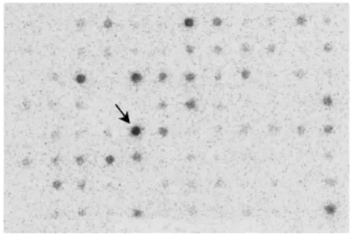
Fig. 1. Subtraction suppression hybridization. The spots expressed in antimycin A-treated cells are presented as described in the text. One of the strongly expressed spots (arrow) was cloned.
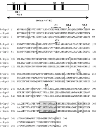
We cloned the cDNA based on the database using PCR and sequenced it completely. The cloned cDNA has 1759 bp and encodes a 396-amino acid polypeptide (Fig. 2). The cDNA was found to overlap with a human protein, PTD004, whose sequence was previously deposited in the databases (GenBank accession no. AF078859). When homology protein searches were performed against databases for other species, two conceptual translation products in mouse (NP_080218.1) and rat (NP_001029099.1) were found with extremely high identity scores (Fig. 3). The protein sequence identities with Xenopus tropicalis (NP_001008044.1), Drosophila melanogaster (NP_572580.1) and Arabidopsis thaliana (NP_174346.1) were respctively 86%, 74% and 62%. Its theoretical pI is 7.64, and its predicted molecular weight is 44.7 kDa (as predicted by the Compute pI/Mw tool). The predicted functional domains of G1, G2-1, G2-2, G3, G4, and G5 are suggested in Fig. 3. There are no other remarkable domains in the protein.
The expression of the Gbp45 protein in various tissues
The tissue distribution of the Gbp45 protein in male and female mice is summarized in Fig. 4. Gbp45 was found to have a relatively high expression in the neuronal tissues and pancreas, but only a very low expression was detected in other tissues.
Fig. 4. The expression of Gbp45 protein in the mouse tissues. Western blotting analysis was performed on male and female mice as described in the text.
Subcellular localization of Gbp45
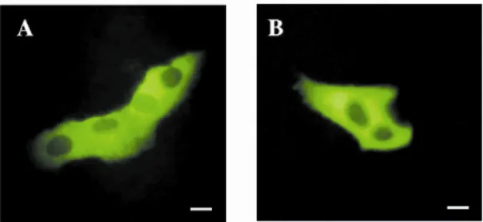
The SOSUI membrane protein predictor (http://bp.nuap.nagoya-u.ac.jp/sosui/) predicted Gbp45 to be a soluble protein, while the PSORT protein-sorting signal and localization site predictor (http://psort.nibb.ac.jp/) suggested that it is a cytoplasmic protein. To determine the subcellular location of the Gbp45 protein, HEK-293 cells were transfected with the pEGFP-Gbp45 vector, and observed with confocal microscopy. GFP-tagged protein fluorescence was detected in the cytosolic compartment (Fig. 5A). To determine the location of the endogenous Gbp45, HEK-293 cells were analyzed by immunofluorescence staining of Gbp45. Again, immunofluorescence was mostly detected in the cytosolic compartment by a peptide competition-inhibitable mechanism (Fig. 5B).
The GTP-binding activity and GTPase activity of Gbp45
Thr43 and Thr56 were predicted as functional domains included in the G2 box of
Gbp45. To determine the importance of Thr43 (G2-1) and Thr56 (G2-2), the
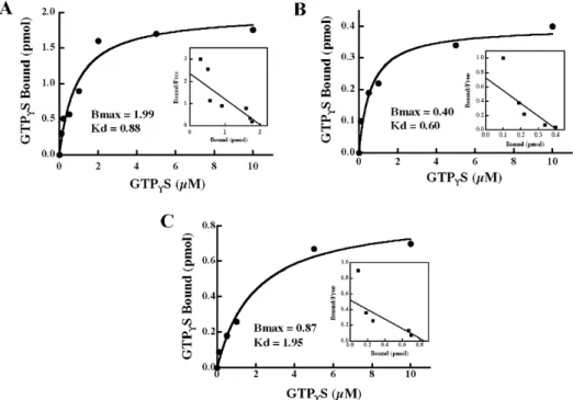
GTP-binding activities of Gbp45 (Fig. 6A) and its mutants T43NGbp45 (Fig. 6B) and T56NGbp45 (Fig. 6C) were examined. The Scatchard plot analysis revealed that
Gbp45, T43NGbp45 and T56NGbp45 respectively bound maximally 0.74, 0.15 and
0.32 mol of this nucleotide per mol proteins, with respective Kdvalues of 0.88, 0.60 and 1.95µM. The time and dose dependencies of the GTPase activity of these proteins was also examined (Fig. 7). The respective specific activities of Gbp45 and its mutants T43NGbp45 and T56NGbp45 were calculated to be 0.22,
0.13 and 0.17 mol Pi/mol of protein/min.
Fig. 6. The GTP-binding activity of Gbp45 and its mutants. The GTP -binding activities of wild-type Gbp45 and its mutants T43NGbp45 and T56NGbp45 were examined as described in the text. A – Dose-dependent effect. 120 ng of Gbp45, T43NGbp45 and T56NGbp45 was incubated with various doses of [35S]GTP
®S for 30 min at 30ºC under the standard
conditions. B – Scatchard plots. The results shown are representative of three independent experiments.
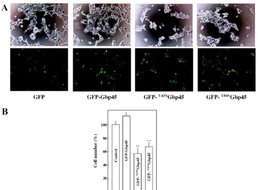
The biological activities of Gbp45
To explore the biological activities of Gbp45, Gbp45,
pEGFP-T43NGbp45 and pEGFP-T56NGbp45 vectors were transiently transfected into
Fig. 7. The GTPase activity of Gbp45 and its mutants. This is a dose-dependent effect: various doses of wild-type Gbp45 (open circles) and its mutants T43NGbp45 (closed circles) and T56NGbp45 (open squares) were incubated with 1 µM [g-32P]GTP for 30 min at 30ºC under the standard conditions (A). Time course: the GTPase activity of 120 ng of wild-type Gbp45 (open circles) and its mutants T43NGbp45 (closed circles) and T56NGbp45 (open squares) was assayed with 1 µM [g-32P]GTP for various periods of time at 30ºC (B). The results shown are representative of three independent experiments.
the same (data not shown). The rate of proliferation of GFP-Gbp45-expressing cells was about 1.2-fold that of empty vector-transfected cells (Fig. 8B). When transfected with pEGFP-T43NGbp45 and pEGFP-T56NGbp45 vectors, about 60%
and 70% of cells, respectively, were viable.
By transfecting pRNA-U6.1/Neo-Gbp45, a hairpin siRNA expression vector targeting Gbp45 mRNA, into HEK-293 cells, we could obtain cells (HEK-293-siRNA-Gbp45) in which Gbp45 was stably silenced. However, a control vector, pRNA-U6.1/Neo, exhibited no silencing response in HEK-293-siRNA (Fig. 9A). The growth rate of HEK-293-siRNA-Gbp45 was lower than that of the cells transfected with the empty vector (Fig. 9B). When treated with the mitochondrial electron transport inhibitors Rotenone or Antimycin A, HEK-293-siRNA-Gbp45 exhibited a decreased resistance to the cytotoxic activity of these agents (Fig. 9C).
Fig. 9. The effect of silencing Gbp45 on cell growth and mitochondrially mediated cell death. A – HEK-293 cells tranfected with empty vector pRNA-U6.1/Neo (HEK-293-siRNA) or pRNA-U6.1/Neo-Gbp45 (HEK-293-siRNA-Gbp45) were analyzed by Western blotting using anti-Gbp45 antibody. Lane 1: HEK-293-siRNA; lane 2: HEK-293-siRNA-Gbp45. B – At the indicated times, the number of cells was counted and plotted. Open squares: HEK-293-siRNA; closed squares: HEK-293-siRNA-Gbp45. The values are the means ± SD (n = 7). *P < 0.01 vs. HEK-293-siRNA group on day 3, **P < 0.01 vs. HEK-293-siRNA group on day 4. C – The cells were placed for 24 h in a medium containing Rotenone (1 µM) or Antimycin A (0.1 µM). Cell mortality was determined via the trypan blue exclusion assay. Open bars, HEK-293-siRNA; closed bars, HEK-293-siRNA-Gbp45. The values are the means ± SD. #P < 0.01 vs. HEK-293-siRNA group treated with Rotenone, ##P < 0.01 vs. HEK-293-siRNA group treated with Antimycin A.
DISCUSSION
a homologue of yeast-44.2 kD protein (Zhang et al., 1998, direct submission). Recently, the gene for PTD004 was found to be one of the ATPases, hOLA1 (human Obg-like ATPase 1) [9]. The protein was characterized and found to bind and hydrolyze ATP. Although this protein contains GTP-binding motifs, it does not share sequence similarity with other GTP-binding proteins, and has thus been classified as a member of a novel group, the GTP1/Obg family, the functions of which remain uncertain. Only suggestions exist concerning the functions of the members of this family. For example, Sikora-Borgula et al. [10] recently reported that homologues of CgtA, the common GTP-binding protein of V. harveyi, play roles in the coupling of DNA replication to cell growth and cell division. As homologues of the V. harveyi CgtA protein found in different organisms exhibit high levels of similarity to this protein, it might be speculated that this type of role is also played by other members of the GTP1/Obg subfamily, including GTP-binding protein PTD004.
Using the protein sequence of Gbp45 to search the NCBI/EMBL protein database revealed that proteins homologous to it exist in many eukaryotic species from A. thaliana to humans. Although these were hypothetical proteins (without experimental evidence for their actual existence), this high degree of evolutionary conservation suggests that Gbp45 may have important biological functions. This was confirmed by our findings that cell proliferation was accelerated by overexpression of Gbp45, and that cell viability was decreased by the expression of mutants of the protein.
The amounts of Gbp45 in the cerebrum, cerebellum, spinal cord and pancreas were significantly larger than those in the heart, lung, liver and spleen. This protein is thus expressed relatively strongly in the tissue of the nervous system. The subcellular localizations of Gbp45 predicted by PSORT were the cytosol (probability 56.5%), mitochondria (17.4%), and nucleus (13%). Indeed, the results of immunocytochemical analysis and examination with GFP-tagged protein suggested that Gbp45 is probably located primarily in the cytosolic compartment.
Thr43 (G2-1) and Thr56 (G2-2) were predicted as functional domains included in
the G2 box of the GTP-binding protein. The GTP-binding activity and GTPase activity of the mutants T43NGbp45 and T56NGbp45 was significantly decreased
compared with that of the wild-type. Thr43 and Thr56 thus play critical roles in
the biological activities of Gbp45. It should be noted that the rate of proliferation of Gbp45-overexpressing cells was slightly increased. On the other hand, expression of T43NGbp45 and T56NGbp45 markedly decreased the number of
viable cells. In the experiment using hairpin siRNA expression vectors, silencing Gbp45 reduced the cell proliferation rate. These findings revealed that Gbp45 plays an essential role in cell growth. Furthermore, T43N and T56N mutants possibly affect the cellular status not only with their dominant negative activity but also their cytotoxic effect. Further study on the details is needed.
O2- by mitochondrial electron transport inhibition [11, 12]. Hydrogen peroxide,
which is itself a dangerous oxidant due to its production of hydroxyl radicals via events such as the Fenton reaction, is generated both in the spontaneous dismutation of O2- or in the catalytic dismutation of it by superoxide dismutase.
In this study, the cells with stably reduced expression of Gbp45 exhibited a decreased resistance to the cytotoxic activity of the mitochondrial electron transport inhibitors Rotenone or Antimycin A. This indicated that Gbp45 plays important roles related to mitochondrial electron transport inhibition, which causes ATP depletion and uncontrolled ROS production. It should be noted that Gbp45 expression had no effect on oligomycin-induced cell death (oligomycin is an inhibitor of ATP production; data not shown). The molecular mechanisms based on these phenomena are the subject of our current investigations.
In summary, this study identified a novel and highly conserved GTP-binding protein, Gbp45, which may be representative of a new group of proteins. Investigation of Gbp45 will likely reveal new pathways of signaling in the cell cycle and cell death.
REFERENCES
1. Takai, Y., Sasaki, T. and Matozaki, T. Small GTP-binding proteins.
Physiol. Rev. 81 (2001) 153-208.
2. Jaffe, A.B. and Hall, A. Rho GTPases: biochemistry and biology. Annu.
Rev. Cell Dev. Biol. 21 (2005) 247-269.
3. Mathupala, S.P., Rempel, A. and Pedersen, P.L. Aberrant glycolytic metabolism of cancer cells: a remarkable coordination of genetic, transcriptional, post-translational, and mutational events that lead to a critical role for type II hexokinase. J. Bioenerg. Biomembr. 29 (1997) 339-343.
4. Pedersen, P.L. Tumor mitochondria and the bioenergetics of cancer cells.
Prog. Exp. Tumor Res. 22 (1978) 190-274.
5. Weinhouse, S. Oxidative metabolism of neoplastic tissues. Adv. Cancer Res. 3 (1955) 269-325.
6. Papa, S., Scacco, S., Schliebs, M., Trappe, J. and Seibel, P. Mitochondrial diseases and aging. Mol. Aspects Med. 17 (1996) 513-563.
7. Sui, G., Soohoo, C., Affar el, B., Gay, F., Shi, Y., Forrester, W.C. and Shi, Y. A DNA vector-based RNAi technology to suppress gene expression in mammalian cells. Proc. Natl. Acad. Sci. USA 99 (2002) 5515-5520.
9. Koller-Eichhorn, R., Marquardt, T., Gail, R., Wittinghofer, A., Kostrewa, D., Kutay, U. and Kambach, C. Human OLA1 defines an ATPase subfamily in the Obg family of GTP-binding proteins. J. Biol. Chem. 282 (2007) 19928-19937.
10. Sikora-Borgula, A., Slominska, M., Trzonkowski, P., Zielke, R., Mysliwski, A., Wegrzyn, G. and Czyz, A. A role for the common GTP-binding protein in coupling of chromosome replication to cell growth and cell division.
Biochem. Biophys. Res. Commun. 292 (2002) 333-338.
11. Chance, B., Sies, H. and Boveris, A. Hydroperoxide metabolism in mammalian organs. Physiol. Rev. 59 (1979) 527-605.