APPLICATION OF CHLORANIL AND FLUORANIL
π-ACCEPTORS FOR THE SPECTROPHOTOMETRIC
DETERMINATION OF MESALAMINE IN PHARMACEUTICALS
Theia'a N. Al-Sabha,
[a]Mohammed S.
Al-Enizzi
[b]and Omar A. Al-Taee
[a]Keywords: spectrophotometry, mesalamine, aqueous solution, p-chloranil, p-fluoranil
Two simple, sensitive and accurate spectrophotometric methods for the determination of mesalamine are described. The methods are based on charge-transfer complex formation reactions of the drug with two π-electron acceptors, p-chloranil and p-fluoranil, in aqueous medium. The coloured complexes having maximum absorbance at 346 nm and 347 nm by using p-chloranil and p-fluoranil reagents respectively. Under the optimized experimental conditions, the calibration curves showed a linear relationship over the concentration ranges of 1–35 and 1-30 μg ml−1 with molar absorptivity values 4.60×10³and 5.67×10³L mol-1 cm-1. using above reagents respectively. The limits of detection
and quantitation were found 0.376 and 1.25 μg ml−1 using p-chloranil, respectively, and 0.333 and 1.111 μg ml−1 using p-fluoranil,
respectively. The complexes were found to be formed in ratio of 1:1 species in both methods, with stability constants of 6.9104 and
5.1105 L mol-1 for mesalamine complexes with p-chloranil and p-fluoranil respectively. The methods were applied successfully to the
assay of mesalamine in pharmaceutical formulations and was agreed well with its certified value.
* Corresponding Authors
E-Mail: [email protected]
[a] Chemistry department, College of Education, Mosul University, Mosul, Iraq
[b] Chemistry department, Education College for Girls, Mosul University, Mosul, Iraq
Introduction
Mesalamine also known as mesalazine, chemically known as 5-aminosalicylic acid (Scheme1) is used for its local effects in the treatment of inflammatory bowel disease, including ulcerative colitis and Crohn’s disease.1,2
Mesalamine has been shown to be a potent scavenger of reactive oxygen species that play a significant role in the pathogenesis of inflammatory bowel disease, inhibition of natural killer cell activity, inhibition of antibody synthesis, inhibition of cyclo-oxygenase and lipoxygenase pathways and impairment of neutrophil function.3,4
Scheme 1. Chemical structure of mesalamine
Different analytical methods have been described for determination of mesalamine such as potentiometry5
HPLC,6-8 differential pulse voltammetry9 and fluorimetry.10
These methods are often time-consuming, expensive, lack selectivity and cumbersome. Spectrophotometry continues to be very popular, because of its simplicity, versatility and low cost. Several spectrophotometric methods using different reagents have been reported for determination of mesalamine. Diazotization coupling method using resorcinol11 and N-(1-naphthyl)ethylenediamine
dihydro-chloride12 as coupling agents. Oxidative coupling reaction
using orcinol, resorcinol and cresol in the presence of hydrogen peroxide and horseradish peroxidase13 and
3-methyl-2-benzothiazolinone hydrazone in the presence of Fe III.14 Schiff base formation reactions using vanillin,15
p-dimethylaminobenzaldeyde and 1,2-naphthoquinone-4-sulphonate,16 redox reactions using Folin Ciocalteu,15 ferric
chloride in the presence of 2,2'–bipyridyl or potassium ferricyanide17 and ion-pairing spectrophotometric methods
using bromocresol green and bromocresol purple.18
However, the most of these methods involve complicated procedures, which require several manipulation steps,11,12
low sensitivity,14,15 using of organic medium,16 indirect
determination procedure17 and an extraction step needed.18 It
is well known that p-benzoquinones such as tetrachloro-1,4-benzoquinone (p-chloranil) and 2,3,5,6-tetrafluoro-1,4-benzoquinone (p-fluoranil) as -electron acceptors often form highly colored charge transfer complexes with various donors which provides the possibility of determination of drugs by spectrophotometric methods. Previously, we reported charge transfer complex formation reactions for determination of mesalamine using o-chloranil,19 tetracyanorthylene (TCNE) and
2,3-dichloro-5,6-dicyano-1,4-benzoquinone (DDQ) acceptors.20 The aim
of the present study is to extend the application of charge transfer complex formation reactions by using of other -acceptors such as p-chloranil and p-fluoranil for the spectrophotometric determination of mesalamine in bulk and pharmaceutical formulations.
Experimentals
Apparatus
All absorption measurements were made on a Shimadzu UV-210A double - beam spectrophotometer supplied with a digital printer DP80Z and matched 1-cm optical silica cells. Heating of solutions was carried out on a water bath of frost instruments, LTD. The reading of pHs made on a PW 9420 C7H7NO3
pH meter supplied with an electrode type CE 10-12 pH. Weighing was carried out on a balance type of Mettler H 54 AR.
Materials and reagents
Mesalamine and its pharmaceutical formulations (tablet and capsule) were kindly provided by state company for Drug Industries and Medical Appliance-(SDI) Sammara-Iraq. p-Chloranil and p-fluoranil and other chemicals were obtained from Fluka and BDH companies. All solvents were analytical reagent grade and water was distilled.
Working standard solution of mesalamine: 250 μgml-1
mesalamine solution was prepared by dissolving of 0.025 g of its pure form in 5 ml ethanol and diluted to 100 ml with distilled water in a volumetric flask.
Reagent solution: 1×10⁻³ M p-chloranil (p-CA) and 2×10⁻³
M p-fluoranil (p-FA) solutions were prepared by dissolving 0.123 g and 0.018 g, respectively, in absolute ethanol and diluted to 50 ml in calibrated flasks with the same solvent.
Borate buffer solution (pH 9): 0.05 M sodium tetraborate was prepared in distilled water and adjusted to pH 9 by pH meter.
Analytical Procedure
Aliquots of the working solution of mesalamine were transferred into two series sets of 25 ml calibrated flasks. In the first set, 0.5 ml of pH9 and 2 ml of 1×10⁻³ M p-CA
solutions were added, in the second set, 1ml of pH9 and 2 ml of 2×10⁻³ M p-FA solutions were added. The solutions
were diluted to the mark with distilled water and left for 20 min at 40ºC and 50ºC using p-CA and p-FA respectively. The absorbance, for the first set, was measured at 346 nm and 347 nm for the second set against their respective reagent blank.
Analytical procedure for pharmaceutical formulations
Ten mesacol tablets or capsule contents (each tablet or capsule containing 400 mg mesalamine) were accurately weighed and pulverized. A portion of the fine and homogenized powder equivalent to 400 mg mesalamine was accurately weighed and dissolved in least amount of ethanol and diluted to 100 ml with distilled water, mixing well and filtered with Whatmann filter paper no.1. The filtrate was diluted to the 100 with distilled water in a volumetric flask obtaining 4000 μg mL-1. a suitable volume was diluted, and
the above procedure was followed.
Results and discussion
The interaction of mesalamine, as n-donor, with p-chloranil and p-fluoranil, as π-acceptors, in the presence of borate buffer solution of pH9 yielded intense purple coloured species in aqueous medium having new bands with maximum absorption at 346 nm and 347 nm respectively where the corresponding blank gave low absorbance at these
wavelengths (Fig. 1). These new bands may be attributed to the formation of radical cation for mesalamine and radical anions for both acceptors of charge transfer complexes.
Figure 1. Absorption spectra of 10 μg mL-1mesalamine with p
-FA (a) and p-CA (b) against their reagent blank (c) and (d) respectively.
Optimization of reaction conditions
The influence of different parameters on the color development was studied to determine optimum conditions for the assay procedures.
Effect of pH and buffer solution
The effect of pH on the absorbance of complex solutions containing mesalamine (10µg mL-1) and p-CA (1×10⁻³ M)
or p-FA (2×10⁻³ M) were studied. It was found that
complexes are formed in basic medium in the presence of NaOH with maximum absorbance at pH 9.0, (Fig.2). The effect of buffer solutions such as, borate, carbonate and phosphate buffers with pH 9.0 were examined.
Figure 2. Effect of pH on the absorption intensity of 10 µg mL-1
mesalamine complexes with p-CA (purple ) and p-FA (red ).
Figure 3. Effect of borate buffer solution (pH 9) amount on the intensity of 10 µg mL-1 mesalamine complexes with p-CA (purple )
and p-FA (red )
Effect of reagents concentration
The effect of changing the p-CA and p-FA concentrations on the absorbance of solution containing a fixed amount of mesalamine (10µg mL-1) were studied. It was observed that
the absorbance increases with increasing the reagent concentrations and reached maximum on using 2.0 ml of 1×10⁻³ M p-CA and 2×10⁻³ M p-FA (i.e. 0.8×10⁻⁴ M and
1.6×10⁻⁴ M in final solution respectively), (Figure 4).
Therefore, these volumes of these concentrations were used in the subsequent work.
Figure 4. Effect of p-CA (1×10⁻³M) (▪) and p-FA (2×10⁻³M) (♦)
volumes on the absorption of 10 μg mL-1 mesalamine
Effect of surfactant
Effect of various surfactants including sodium dodecyl sulphate (SDS), cetylperydinum chloride (CPC), cetyltrimethylammonium bromide (CTAB), Tween-80 and Triton x-100 were tested. It was found that these surfactants decreased the absorbance of solutions.
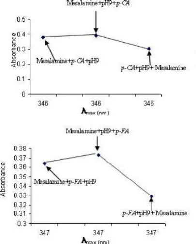
Effect of order of addition
The order of addition of reactants on the color development was examined. Maximum sensitivity was achieved when mesalamine and pH9 were added before adding the p-CA or p-FA reagents (Fig. 5). Hence, the method was performed in the order: Mesalamine + pH 9 + p-CA: or Mesalamine + pH 9 + p-FA
Figure 5. Effect of order of reactants on the absorption of 10 μg mL-1 mesalamine
Effect of temperature and reaction time
The reaction time was determined by following the color development at room temperature and in thermostatically controlled water-bath at different temperatures up to 60○C. The absorbance was measured at 5 and 10 minutes intervals against reagent blank treated similarly. It was observed that the complexes were formed at room temperature after addition of p-CA or p-FA immediately and the absorbance were increased with increasing the temperature and reached at maximum after 20 min and remain constant for 50 min at 40ºC and 50ºC for both complexes using p-CA and p-FA reagents respectively, (Fig. 6). However; after these temperatures and time stability the absorbance was decreased indicating the dissociation of the complexes. Therefore; 20 min developing time at 40ºC and 50ºC as optimum temperatures were selected using p-CA and p-FA reagents respectively in the subsequent experiments.
Figure 6. Effect of temperature and reaction time on the absorbance of 10 μg mL-1mesalamine in the presence of p-CA and
Method validation
In order to investigate the range in which the colored complex adhere to Beer's law, the absorbance of the complexes were measured at 346 nm and 347 nm using p-CA and p-FA reagents respectively, after developing the color by following the recommended procedure for a series of solutions containing increasing amounts of mesalamine. The Beer's law limits and molar absorptivity values were evaluated and given in Table 1, which are indicated that the method is sensitive. The linearity was represented by the regression equations and the corresponding correlation coefficients for the studied determined drug by the proposed method represents excellent linearity. The relative standard deviation (RSD) and accuracy (average recovery %) for the analysis of five replicates of each three different concentrations for pure mesalamine indicated that the method is precise and accurate. Limit of detection (LOD) and limit of quantitation (LOQ) are determined by taking the ratio of standard deviation of the blank with respect to water and the slope of calibration curve by applying the following equations:
whereas:
σB= Standard deviation for six determinations of blank S= slope of calibration graph for each isomer
LOQ is approximately 3.3 times LOD. Naturally, the LOQ slightly crosses the lower limit of Beer's law range
Table 1. Summary of optical characteristics and statistical data for the proposed method
*Average of five determinations.**Y=aX+b, where X is the concentration of drug in μg mL-1.
Interference
The extent of interference by some excipients which often accompany pharmaceutical preparations were studied by measuring the absorbance of solutions containing fixed amount of mesalamine and various amounts of diverse species, which is present the pharmaceutical formulations of mesalamine, in a final volume of 25 ml. It was found that the studied excipients up to 25 fold excess did not interfere seriously (Table 2). However; an error of 5.0 % in the absorbance readings was considered tolerable.
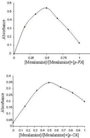
Analytical applications
The proposed method was successfully applied to determine mesalamine in pharmaceutical tablets and capsules preparations. The obtained results were compared statistically by a Student's t-test for accuracy at the 95 % confidence level with three degrees of freedom. As cited in Table 3, the results showed that the experimental t-test was less than the theoretical values (t=4.303), indicating that the method is accurate.
Comparison of the proposed method with other spectrophoto-metric methods
The proposed spectrophotometric method for determination of mesalamine have been compared favorably with other spectrophotometric methods depending on charge transfer complex formation reactions using different -acceptor reagents such as o-chloranil, tetracynoethylene (TCNE) and 2,3-dicloro-5,6-dicyno-p-benzoquinone (DDQ). As seen in Table 4, p-chloranil reagent is more sensitive than o-chloranil reagent but less than TCNE and DDQ reagents, where as the linearity range of p-fluoranil method is more than other methods. However; other parameters seem to be the same with some different in λmax, pH and temperature.
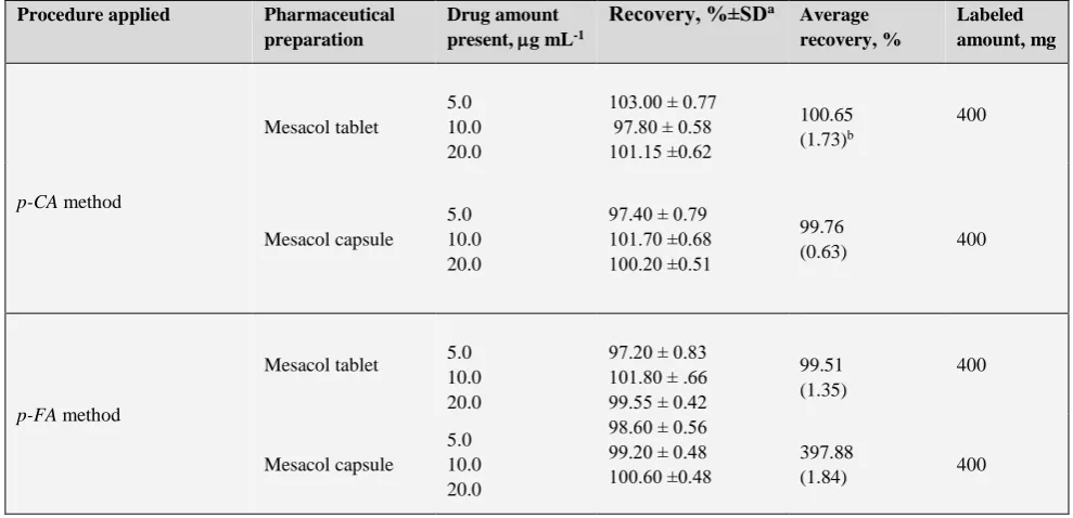
Stoichiometry and stability constant
The molar ratio of the n-π charge transfer complex formed between the mesalamine with p-CA and p-FA reagents were investigated by applying the continuous variation (Job's) method.21 Experiments were performed by mixing different
volumes of equimolar solutions of drug and reagents [1.63×10⁻³M] and keeping total volume at 6 ml at final
dilution of 25 ml. Under the optimum conditions described above, the plot of absorbance vs molar ratio between drug and reagents were prepared, and results indicated that complexes were formed in the ratio of 1:1 (Figures 7). This finding supports that the n-π CT complexes are formed through amino group present in mesalamine. The stability constant (Kst) of the complexes were calculated using the
following equation:
where
Kst is the stability constant, L mol
-1
, the dissociation degree,
C the concentration of the product which is equal to the concentration of mesalamine,
Am the absorbance of the complexes at optimum amount of p-CA or p-FA reagent and
As the absorbance of the complexes at stoichiometric amounts of p-CA or p-FA reagent according to the 1:1 ratios under the optimum conditions reaction.
It was found 6.9×10⁴and 5.1×10⁵L.mol-1 using p-CA and
p-FA reagents respectively, indicating good stability.
Parameter p-CA p-FA
Beer's law limits, μg mL-1 1-35 1-30
Molar absorptivity, L mol-1 cm-1 4.60×10³ 5.67×10³
LOD, μg mL-1 0.376 0.333
LOQ, μg ml-1 1.250 1.111
Average recovery, %* 99.16 98.80 Correlation coefficient 0.996 0.997 Regression equation, Y**
Slope, a 0.0301 0.0371
Intercept, b 0.1058 0.0993
RSD a ≤ 1.53 ≤ 2.07
3.3 B 10 B
LOD LOQ
S S
st 2
m
m
1
s K
C
A A
A
Table 2. Effect of excipients for assay of mesalamine
Table 3. Assay of mesalamine in pharmaceutical preparations using theproposed method and comparison with the official method
Labeled amount, mg Average
recovery, %
Recovery, %±SDa
Drug amount present, g mL-1
Pharmaceutical preparation Procedure applied 400 100.65 (1.73)b
103.00 ± 0.77 97.80 ± 0.58 101.15 ±0.62 5.0
10.0 20.0 Mesacol tablet
p-CA method
400 99.76
(0.63) 97.40 ± 0.79
101.70 ±0.68 100.20 ±0.51 5.0 10.0 20.0 Mesacol capsule 400 99.51 (1.35) 97.20 ± 0.83
101.80 ± .66 99.55 ± 0.42 5.0
10.0 20.0 Mesacol tablet
p-FA method
400 397.88
(1.84) 98.60 ± 0.56
99.20 ± 0.48 100.60 ±0.48 5.0
10.0 20.0 Mesacol capsule
aAverage of three determinations. bFigures in parenthesis are the calculated values for t.
Table 4. Comparison of spectrophotometric methods for mesalamine determination
Excipient Recovery % of 10 µg mL-1 of mesalamine per µg mL-1 foreign added
p-CA p-FA
20 50 100 250 20 50 100 250
Lactose 102.00 104.28 104.94 103.71 96.19 95.16 98.42 100.47 Sodium chloride 95.98 96.40 98.20 98.53 96.74 95.95 100.19 98.98 Arabic Gum 98.53 96.87 101.72 101.85 100.47 101.77 100.47 102.51 Starch 103.38 101.39 98.07 96.07 98.98 96.16 98.23 101.95 Sucrose 105.61 105.00 103.85 103.28 97.95 96.74 99.72 95.23 Glucose 95.64 95.50 95.65 97.00 97.21 99.44 99.81 102.73
Literature method Present method
Analytical parameters
DDQ19
TCNE19
o-Chloranil 18
p-Fluoranil p-Chloranil 420 346 571.5 347 346 λmax, nm
9 3 9.8 9 9 pH 40 R.T 25 50 40 Temp., °C 50 10 5 20 20 Development time, min
>60 30
45 50
50 Stability period, min
0.4-10 0.8-12
1.25-30 1-35
1-30 Beer’s law, μg mL-1
5.9586×10⁴
2.9785×10⁴
3.4×10³
5.67×10³
4.60×10³
Molar absorptivity, L mol-1cm-1
0.893 1.018
0.143 0.376
0.333 LOD, μg mL-1
2.976 3.393
0.4773 1.250
1.111 LOQ, μg mL-1
Figure 7. Continuous variations plots for complexes of mesalamine (1.63×10⁻³M)with 1.63×10⁻³Mof each p-CA and
p-FA under the optimum reaction conditions.
Proposed chemical reactions
The charge transfer complex forming reactions occur when π-acceptors react with the basic nitrogenous compounds which act as n-donors. Charge-transfer complex formation is characterized by electronic transition(s) to an excited state in which there is a partial transfer of electronic charge from the donor to the acceptor moiety, followed by the formation of a radical anion. Complete electron transfer from the donor to the acceptor moiety took place with the formation of intensely colored radical ions.22
Therefore, aliphatic and aromatic amines, nitrogenous base acting as n-donor, is made to react with p-CA and p-FA as π-acceptors to produce a colored charge transfer complexes in aqueous medium according to the scheme 2.
Conclusion
Determination of mesalamine is based on n-π charge transfer complex formation reactions using p-CA and p-FA acceptors. The proposed method is found to be simple. It has the advantage of being accurate, does not require the removal of excipients, temperature control and solvent extraction step. The statistical parameters and recovery study data clearly indicate the reproducibility and accuracy of the method. It was applied successfully to pharmaceutical preparations.
Scheme 2. Proposed mechanism of charge transfer complex formation reactions for determination of mesalamine by p-CA and p-FA. HOOC
O H
NH2
Mesalamine
pH=9
HOOC
O H
NH2 (V)
HOOC
N H2
(V)
OH O
O
Cl Cl
Cl Cl
O
O F
F F
F
HOOC
O H
NH2 + (III)
HOOC
OH
N H2
+ (III)
O-(III)
O(III)
Cl Cl
Cl Cl
O-(III)
O(III) F
F F
F n - CT intermediates
colored species O
O
Cl Cl
Cl Cl
O
O F
F F
References
1Cai, Q. X., Zhu, K. J., Chen, D. and Gao, L. P., Eur. J. Pharm.
Biopharm., 2003, 55, 203.
2Gotti, R., Pomponio, R., Bertucci, C. and Cavrini, V., J.
Chromatogr. A,2001, 916, 175.
3Geier, D. L. and Miner, P. B., Am. J. Med., 1992, 93, 208. 4Palumbo, G., Carlucci, G. and Mazzeo, P., J. Pharm. Biomed.
Anal. Oxford, 1995, 14, 175.
5British Pharmacopoeia Commission, British Pharmacopeia,
London, Stationery Office, 2003, 2, 1257, [CD-ROOM].
6United States Pharmacopeia 24. Ed. Rockville, United States
Pharmacopeial Convention, 2000, [CD-ROOM].
7Pastorini, E., Locatelli, M., Simoni, P., Roda, G., Roda, E. and
Roda, A., J. Chromatogr.B Anal. Technol. Biomed. Life Sci., 2008, 872, 99.
8Nigović, B. and Simunić, B., J. Pharm. Biomed. Anal., 2003, 31,
169.
9Darak, V., Karadi, A. B., Appal, R. S., Arshad, M. D. and Ganure
A. L. , Pharma Sci.Monitor, 2012, 3, 74.
10Zadeh, H. A. and Kohansal, S., J. Braz. Chem. Soc., 2012, 23,
473.
11Reddy, M. P., Prabhavahi, K., Reddy, N. R. and Reddy, P. R.,
Global J. Pharmacol., 2011, 5, 101.
12Patel, K. M., Patel, C. N., Panigrahi, B., Parikh, A. S. andn
Patzel, H. N., J. Young Pharm.,2010, 2, 284.
13Shakeela, S., Ram, B. S. and Sekaran, C. B., Chiang Mai J. Sci.,
2011, 38, 551.
14Narala, S. R. and Saraswathi, K., Int. J. Pharm. Sci. Res., 2011, 2,
366.
15Chandra, B. S., Bhogela, S. S., Shaik, M., Vadlamudi, C. S.,
Chappa, M. andMaddirala, N. S., Quim. Nova, 2011, 34, 1068.
16Sama, N. S., Gurupadayya, B. M. and Kumar, A., J. Pharm. Res.,
2011, 4, 39.
17Narala, S. and Saraswathi, K., Int. J. Res. Pharm. Biomed. Sci.,
2010, 1, 10.
18Darak,V., Karadi, A. B., Arshad, M. D. and Raju, S., Appl. Res.
J. Pharm. Techn., 2011, 4, 962,.
19Al-Enizzi, S. M., Al-Sabha, T. N. and Al-Ghabsha, T. S., Jordan.
J.Chem., 2012, 7, 87.
20Al-Sabha, T. N. and Al-Taee, A. O., J. Educ. Sci., 2008, 21, 49.
21Hargis, L. G., Analytical Chemistry, Principles and Techniques,
Prentice- Hall Inc., Jersey, 1988, 424.
22Alzoman, N. Z., Sultan, M. A., Maher, H. M., Alshehri, M. M.,
Wani, T. A. and Darwish, I. A., Molecules, 2013, 18, 7711.