Open Access
Review
Talking to chromatin: post-translational modulation of polycomb
group function
Hanneke EC Niessen
1, Jeroen A Demmers
2and Jan Willem Voncken*
1Address: 1Molecular Genetics, GROW School for Oncology and Developmental Biology, Maastricht University, Maastricht, The Netherlands and 2Proteomics Center, Erasmus MC University Medical Center, Rotterdam, The Netherlands
Email: Hanneke EC Niessen - [email protected]; Jeroen A Demmers - [email protected]; Jan Willem Voncken* - [email protected]
* Corresponding author
Abstract
Polycomb Group proteins are important epigenetic regulators of gene expression. Epigenetic control by polycomb Group proteins involves intrinsic as well as associated enzymatic activities. Polycomb target genes change with cellular context, lineage commitment and differentiation status, revealing dynamic regulation of polycomb function. It is currently unclear how this dynamic modulation is controlled and how signaling affects polycomb-mediated epigenetic processes at the molecular level. Experimental evidence on regulation of polycomb function by post-translational mechanisms is steadily emerging: Polycomb Group proteins are targeted for ubiquitylation, sumoylation and phosphorylation. In addition, specific Polycomb Group proteins modify other (chromatin) associated proteins via similar post-translational modifications. Such modifications affect protein function by affecting protein stability, protein-protein interactions and enzymatic activities. Here, we review current insights in covalent modification of Polycomb Group proteins in the context of protein function and present a tentative view of integrated signaling to chromatin in the context of phosphorylation. Clearly, the available literature reveals just the tip of the iceberg, and exact molecular mechanisms in, and the biological relevance of post-translational regulation of polycomb function await further elucidation. Our understanding of causes and consequences of post-translational modification of polycomb proteins will gain significantly from in vivo validation experiments. Impaired polycomb function has important repercussions for stem cell function, development and disease. Ultimately, increased understanding of signaling to chromatin and the mechanisms involved in epigenetic remodeling will contribute to the development of therapeutic interventions in cell fate decisions in development and disease.
Introduction
Polycomb group (PcG) proteins preserve transcriptionally silenced states through epigenetic marking of target genes in higher eukaryotes. Currently, at least two biochemically and functionally distinct polycomb repressive complexes (PRC) are recognized, PRC2 and PRC1, which contribute to establishment and maintenance of gene repression
pro-files (Figure 1a) [1-3]. As such, PcG function equips the cell with a transcriptional memory throughout develop-ment and differentiation. It is becoming increasingly clear that PcG-chromatin association is subject to dynamic reg-ulation; PcG complex composition and chromatin associ-ation change throughout eukaryotic development [4,5]. In a constantly changing environment (for example, dur-Published: 1 September 2009
Epigenetics & Chromatin 2009, 2:10 doi:10.1186/1756-8935-2-10
Received: 13 May 2009 Accepted: 1 September 2009
This article is available from: http://www.epigeneticsandchromatin.com/content/2/1/10
© 2009 Niessen et al; licensee BioMed Central Ltd.
ing differentiation), cells respond to a plethora of extracel-lular and intrinsic cues. Mechanistic insight in epigenetic regulation in response to signaling is essential to under-stand how cell fate, function and physiology are control-led. It is, however, still largely unknown how cells 'talk' to chromatin to facilitate appropriate cellular responses. Physiological adaptation of cells is initially mediated by mostly transient and reversible covalent post-translational modifications (PTMs) at specific amino acid residues including ubiquitylation, sumoylation, and phosphoryla-tion (see Appendix). Through altered protein-protein interaction, subcellular localization, enzyme activity and protein stability, PTMs ultimately also affect gene
expres-sion. We review current established PTMs on PcG proteins and the effect they have on interaction and enzymatic activity. In addition, we extracted PcG-specific PTMs from published analyses and used these to predict upstream kinase pathways signaling to Polycomb. The final section presents a tentative integrated view on signaling to chro-matin in the context of PcG PTM.
Ubiquitylation: Polycomb-mediated
ubiquitylation
Ubiquitylation plays a central role in PRC-mediated silencing. Histone 2A (H2A) is one of the most abundant ubiquitylated nuclear proteins, and H2AK119Ub1 is
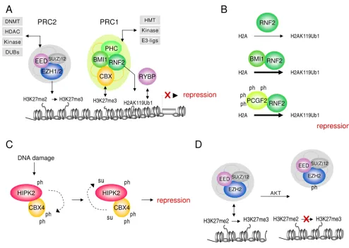
Examples of post-translational modulation of Polycomb group and associated proteins
Figure 1
Examples of post-translational modulation of Polycomb group and associated proteins. (a) Simplified model of Polycomb Group (PcG)-mediated repression. The histone methyltransferase EZH2 trimethylates histone H3 at lysine 27, this mark is recognized by chromobox homolog (CBX) proteins via the chromodomain. RNF2/RING1 homologs are E3 ubiquitin ligases for H2A; RYBP binds H2AK119Ub1. Combined, these activities induce/maintain transcriptional repression. Gray boxes depict Polycomb Repressive Complex (PRC)-associated, epigenetically relevant enzymatic activities. (b) RNF2 E3 ligase activity is significantly enhanced in the presence of BMI1 or phosphorylated PCGF2. (c) DNA damage-induced phosphorylation of HIPK2 leads to phosphorylation of CBX4. Phosphorylation of CBX4 at T495 in turn enhances the HIPK2 sumoylation. (d)
AKT-induced phosphorylation of EZH2 on S21 impairs its binding to histone H3, thereby inhibiting H3K27 trimethylation. me = methylation, ph = phosphorylation, su = sumoylation; ub = ubiquitylation.
RYBP EZH1/2
SU(Z)12 EED
PHC
RNF2 BMI1
CBX
repression
H3K27me2 H3K27me3
H2AK119Ub1 H3K27me3
PRC2 PRC1
EZH2
SU(Z)12
EED
H3K27me2 H3K27me3
EZH2
SU(Z)12
EED
H3K27me2 H3K27me3
AKT
A
D
B
C
RNF2 BMI1
RNF2
RNF2 PCGF2
H2AK119Ub1 H2A
H2AK119Ub1 H2A
H2AK119Ub1 H2A
CBX4 HIPK2 DNA damage
CBX4 HIPK2
repression
DNMT
HDAC
Kinase
HMT
Kinase
E3-ligs
DUBs
ph ph ph
ph
su
su ph
ph
ph ph
ph
repression
required for PcG-mediated gene repression [6]. Published data shows that the PRC1 protein ring finger protein 2 (RNF2, also known as RING1B and RING2) ubiquitylates H2A, as loss of RNF2 dramatically decreases global H2Aub levels and derepresses PcG-controlled genes. The RING domain of the really interesting new gene protein 1 (RING1, also known as RING1A) substitutes for that of RNF2 in vitro [7]. Consistently, H2AK119Ub1 is main-tained in RING1 or RNF2 single null cells, but not in dou-ble knockout cells, supporting functionally redundant roles for these proteins in certain biological contexts [8]. RNF2-mediated H2A ubiquitylation is important in X chromosome inactivation [8,9]. The exact role of RNF2 and H2AK119Ub1 in X-inactivation is currently under debate, however, as recent studies suggest that H2AK119Ub1 may not be sufficient for X-inactivation and conversely, Xist-initiated silencing occurs in the absence of RNF2 and H2AK119Ub1 [10,11]. Addition-ally, histone variant H2A.Z may be a target for RNF2-mediated ubiquitylation, as knockdown of RNF2 reduces H2Aub and H2A.Zub levels in vitro [12].
Whereas in Drosophila melanogaster multiple E3 ligase complexes contribute to H2A ubiquitylation, the PRC1 protein RNF2 is currently considered the major E3 ligase for H2AK119 (Figure 1b) [13,14]. Although within the PcG core complex, containing RING1, RNF2, BMI1 and polyhomeotic homolog 2 (PHC2), most in vitro H2A-E3 ligase activity is attributed to RNF2 [8], other PcG RING-type E3 ligases control H2A-directed ubiquitylation and affect HOX gene silencing: BMI1 (Polycomb Group ring finger 4 (PCGF4)) and homologues PCGF2 (MEL18) and PCGF1 (NSPc1) enhance H2A ubiquitin (Ub) E3 ligase activity when complexed to RNF2 (Figure 1b) [15-18]. In addition, PcG ubiquitin E3 ligase activity is enhanced within the molecular context of an intact PRC1 complex: fully reconstituted complexes containing RNF2, RING1, BMI1 and chromobox homolog 8 (CBX8) show highest activity compared to RNF2 alone or subcomplete PRC1 complexes [15]. Crystal structure analyses of interacting RING domains of mammalian BMI1 and RNF2 reveals extensive contacts between the RNF2 and BMI1 RING domains: the N-terminal 'arm' of RNF2 embraces the BMI1 RING domain [7,19]. RING protein partnerships occur frequently in cell biology [20]; RING domain pro-teins function as adapters, bringing together E2 conju-gases and their substrates (see Appendix) [21]. Based on structural analogy to a breast cancer type 1 susceptibility protein (BRCA1)-BRCA1-associated RING domain 1 (BARD1) complex, it was suggested that RNF2 contains an E2 binding site, whereas BMI1 is involved in substrate binding [19]. E2 ubiquitin conjugating enzymes (Ubc) UbcH5 subtypes a, b, c and UbcH6 promote H2A Ub con-jugation, although these Ubcs do not bind the RING RNF2/BMI1 complex [7]. Which E2 conjugases contribute
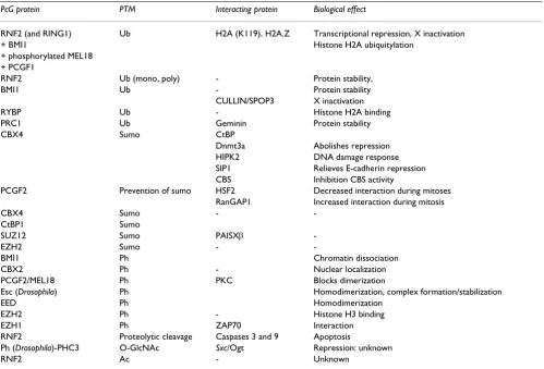
to H2A ubiquitylation in vivo is currently not known. A summary of PcG-related PTMs and their functional rele-vance is provided in Table 1.
Ubiquitylation of PcG proteins
The above relatively simple picture is complicated by additional levels of PTM. Both RNF2 and BMI1 are ubiq-uitin-conjugated proteins and differential autoubiquityla-tion of RNF2 is required for H2AK119Ub1 [7,22]. Polyubiquitylation of RNF2 requires Lys6, Lys27 and Lys48 linkage on the same ubiquitin molecule and RNF2 autoubiquitylation is promoted by Ubc5 in vitro [22]. Some of these K residues are involved in epigenetic silenc-ing, as the ability of RNF2 to promote H2AK119Ub1 relates to the availability of UbK6 and UbK27, not UbK48 [22]. Although inhibition of Ub-dependent degradation with proteasome inhibitors increases RNF2 levels, autou-biquitylation mutant RNF2(I53S) proteins are still effi-ciently degraded, suggesting the involvement of other E3 ligase(s) and/or Ub site(s). BMI1 inhibits ubiquitylation of RNF2 and coexpression of RNF2 and BMI1 blocks its degradation in a RING domain-dependent manner [22]. Unlike RNF2, BMI1 lacks autoubiquitylation activity. However, like RNF2, it is stabilized by proteasome inhibi-tion. As for RNF2, the identity of the ubiquitin E3 ligase responsible for proteasomal degradation of BMI1 is not known. Whereas RNF2K112ub appears dispensable for H2A E3 ligase activity [7], mutant RNF2 with an intact RING domain, however missing most ubiquitylation sites (K92-198R) still binds BMI1, but lacks E3 ligase activity [22]. Thus, seemingly at odds with each other: although RNF2 autoubiquitylation is required for H2A ubiquityla-tion, it is inhibited by BMI1, yet overall, BMI1 promotes RNF2 H2A-E3 ligase activity and blocks its proteolytic degradation [22]. A possible explanation for this discrep-ancy may involve number, length and linkage type of ubiquitin chains, aside from molecular context.
A candidate E3 ubiquitin ligase for BMI1 is the CULLIN3/ Speckle-type POZ protein (SPOP) complex: in vitro and in vivo analyses confirmed that CULLIN3 and SPOP are required for BMI1 ubiquitylation in cells. As RNAi-medi-ated knockdown of CULLIN3 or SPOP does not affect BMI1 protein levels, CULLIN3/SPOP-mediated ubiquit-ylation of BMI1 most likely has no bearing on protein sta-bility [23]. Interestingly, a human BMI1 polymorphism resulting in a C18Y substitution increases ubiquitylation and proteasomal degradation [24]. Whether or not this has any effect on human health is currently not clear.
interacts with RING1 and RNF2, it prevents formation of Polycomb bodies in osteosarcoma cells, suggesting a dominant negative role in PcG recruitment [25]. Thus, RYBP may play a role in engagement of specific transcrip-tion factors, and hence directranscrip-tion of PcG complexes to spe-cific target genes [17,26].
Combined, the above data shows that PRC proteins are engaged in numerous ubiquitin-dependent regulatory mechanisms, and that ubiquitylation is important for PcG-mediated silencing at multiple levels.
Sumoylation: Polycomb-mediated sumoylation
and Polycomb sumoylation
Although mechanistically not completely understood, one of the potential functional outcomes of PcG protein sumoylation is induction of transcriptional repression [27]. In support of a biologically relevant role in the con-text of transcriptional repression, sumoylation appears conserved throughout evolution. A genome-wide RNA interference screen in Drosophila cells identified proteins that, when absent, relieve small ubiquitin-like modifier (sumo)-dependent inactivation of the transcription factor
Sp3 [28]. Among interactors identified were the PcG pro-tein Sfmbt, the zinc finger propro-tein MEP-1 and dMi-2, an ATP-dependent chromatin remodeler which shows genetic interaction with PcG [29]; all three proteins bind Sp3-sumo in vitro and all are recruited to promoters in a Sp3-sumoylation-dependent manner [28]. Additionally, in Caenorhabditis elegans a link was established between sumoylation and PcG proteins: the PcG-like protein SOP-2 interacts with UBC9 via its conserved sterile α motif/self association motif (SAM) domain [30]. SOP-2 sumoyla-tion is required for in vivo localizasumoyla-tion to nuclear bodies and repression of HOX genes [30].
The identification of CBX4 as a sumo E3 ligase forged the first link between PcG function and sumoylation. C-ter-minus binding protein (CtBP; an interaction partner of RING1 and other PcG proteins [31]) is sumoylated [32,33]. CBX4 interacts with E2 Ubc9 and CtBP and sequesters both proteins to PcG bodies. Multiple biologi-cal CBX4 targets have been identified which begin to link PcG-mediated sumoylation to relevant biological proc-esses [32,34-38]. Among these targets is Dnmt3a (de novo DNA methyltransferase). Despite its highly conserved Table 1: Post-translational modifications (PTMs) in polycomb group (PcG) biology
PcG protein PTM Interacting protein Biological effect
RNF2 (and RING1) Ub H2A (K119), H2A.Z Transcriptional repression, X inactivation
+ BMI1 Histone H2A ubiquitylation
+ phosphorylated MEL18 + PCGF1
RNF2 Ub (mono, poly) - Protein stability,
BMI1 Ub - Protein stability
CULLIN/SPOP3 X inactivation
RYBP Ub - Histone H2A binding
PRC1 Ub Geminin Protein stability
CBX4 Sumo CtBP
Dnmt3a Abolishes repression
HIPK2 DNA damage response
SIP1 Relieves E-cadherin repression
CBS Inhibition CBS activity
PCGF2 Prevention of sumo HSF2 Decreased interaction during mitoses
RanGAP1 Increased interaction during mitosis
CBX4 Sumo -
-CtBP1 Sumo
SUZ12 Sumo PAISXβ
-EZH2 Sumo -
-BMI1 Ph Chromatin dissociation
CBX2 Ph - Nuclear localization
PCGF2/MEL18 Ph PKC Blocks dimerization
Esc (Drosophila) Ph Homodimerization, complex formation/stabilization
EED Ph Homodimerization
EZH2 Ph - Histone H3 binding
EZH1 Ph ZAP70 Interaction
RNF2 Proteolytic cleavage Caspases 3 and 9 Apoptosis
Ph (Drosophila)-PHC3 O-GlcNAc Sxc/Ogt Repression: unknown
RNF2 Ac - Unknown
nature among the PcG proteins CBX2, CBX6, CBX7 and CBX8, only the C-terminal COOH box of CBX4 interacts with Dnmt3a in a yeast two-hybrid setting. Dnmt3a is polysumoylated; sumoylation terminates the interaction of Dnmt3a with histone deacetylase (HDAC)1/2 and completely abolishes its repressive ability in vitro, suggest-ing a role for PTMs in dynamic epigenetic regulation of gene expression [35,36]. DNA damage-induced homeo-domain interacting protein kinase 2 (HIPK2) phosphor-ylates and activates CBX4 E3 sumo activity, whereas CBX4-mediated sumoylation of HIPK2 in turn enhances its ability to repress genes in response to DNA damage (Figure 1c) [38]. This autoregulatory feedback loop is likely relevant in the context of cellular DNA damage responses. Sumoylation of SMAD interacting protein 1 (SIP1) interferes with CtBP interaction and relieves repres-sion of E-cadherin, an important regulator of epithelial mesenchymal transition (EMT) during development and tumorigenesis [37]. Furthermore CBX4 targets cystathio-nine β-synthase (CBS), an enzyme involved in homo-cysteine to homo-cysteine conversion [34]. Sumoylation of CBS inhibited its enzymatic activity [34]. As homocysteine to cysteine conversion is an important step in the synthesis of S-adenosylmethionine (SAM), a major methyl donor reagent for essential methylation reactions [39], this may have obvious implications for local and global epigenetic regulation.
In the context of PRC1 function, the PcG protein PCGF2, appears to oppose sumoylation. PCGF2 binds, both in vitro and in vivo, heat shock factor 2 (HSF2), which is pre-dominantly sumoylated during mitosis [40]. PCGF2 dis-sociates from HSF2 during mitosis and, conversely, recombinant PCGF2 inhibits in vitro sumoylation of HSF2. Thus PCGF2 may act as an anti-sumo regulator [40]. PCGF2 also inhibits sumoylation of Ran GTPase activating protein (RanGAP)1, which is independent of its RING domain [41]. Intriguingly, in contrast to HSF2, the interaction between PCGF2 and RanGAP1 increases dur-ing mitosis, suggestdur-ing a sumo-dependent switch of inter-action partners of PCGF2 [41].
PRC2 complex function is associated with sumoylation as well: suppressor of zeste 12 (SUZ12) and enhancer of zeste homolog 2 (EZH2) (Figure 1a) are both sumoylated; the E3 ligase for SUZ12 appears to be PAISXβ, not CBX4. The exact biological role of SUZ12 sumoylation is not clear, as wild type or non-sumoylatable SUZ12 3KR mutants both show similar H3K27me3 in a SUZ12
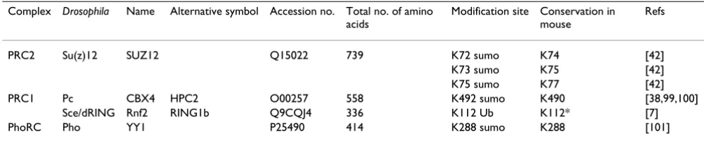
-/-background, and 3KR colocalization with EZH2 and embryonic ectoderm development (EED) is not affected [42]. An overview of experimentally confirmed site-spe-cific sumoylation and ubiquitylation sites on PcG pro-teins is presented in Table 2. Although a total of 33 sumoylation sites are predicted on human PcG proteins, both their occurrence and biological relevance await vali-dation in vitro and in vivo (Additional file 1).
In summary, sumoylation, like ubiquitylation, emerges as a PTM relevant for regulation of gene expression. In con-trast to ubiquitylation, PcG-mediated sumoylation has not directly been linked to histone modifications yet. Instead, currently available data suggest a relevant role for sumoylation in dynamic interaction with non-PcG pro-teins in the context of cell physiology.
Phosphorylation of PcG proteins
Although thus far no PcG proteins have been identified as kinases, phosphorylation is a common PTM on PcG pro-teins. Recent observations have shown that signaling, which triggers downstream phosphorylation events, affects subcellular localization, protein interactions within complexes, enzymatic activity and chromatin asso-ciation of several PcG proteins and other factors. The first report of PcG protein phosphorylation suggested a mean-ingful regulatory role for PcG phosphorylation, as phos-phorylated BMI1 dissociates from chromatin at late S-phase, when chromatin is assembled de novo [43]. Addi-tionally, MEL18 and BMI1-like RING finger protein (MBLR; PCGF6) is predominantly phosphorylated in G2/ M [44]. In vitro kinase assays suggested PCGF6 as a sub-strate for CDK7 [44]. Interestingly, Trithorax orthologs are also phosphorylated in a cell cycle-dependent manner [45], suggesting phosphorylation as a common
mecha-Table 2: Site-specific polycomb sumoylation and ubiquitylation sites
Complex Drosophila Name Alternative symbol Accession no. Total no. of amino acids
Modification site Conservation in mouse
Refs
PRC2 Su(z)12 SUZ12 Q15022 739 K72 sumo K74 [42]
K73 sumo K75 [42]
K75 sumo K77 [42]
PRC1 Pc CBX4 HPC2 O00257 558 K492 sumo K490 [38,99,100]
Sce/dRING Rnf2 RING1b Q9CQJ4 336 K112 Ub K112* [7]
PhoRC Pho YY1 P25490 414 K288 sumo K288 [101]
nism to temporarily relocate chromatin-associated pro-teins. Phosphorylation may affect individual PcG proteins in other biologically relevant ways. CBX2/M33 phospho-rylation affects nuclear localization: high mobility (unmodified) CBX2 resides in the cytoplasm in mouse liv-ers, whereas low mobility isoforms localize to the nucleus [46]. Dimerization of PCGF2, is blocked in the presence of protein kinase C (PKC) [47]. Additionally, NSPc1/ PCGF1 is functionally targeted by phosphorylation: a syn-thetic phosphomutant GAL4-DBD-NSPc1 no longer tran-scriptionally represses a GAL4-LUC reporter [48].
Phosphorylated Drosophila PRC2 protein Extra sex combs (Esc) is preferentially found in a 600 kDa complex with PcG protein enhancer of zeste (E(z)) and Esc phosphoryla-tion is required for formaphosphoryla-tion and stability of a larger Esc/ E(z) complex containing PCL and RPD3 [49,50]. Both Esc and EED (its mammalian ortholog) are phosphorylated in a domain responsible for Esc/EED homodimerization [50]. Casein kinase (CK)1 and CK2 may be the responsi-ble kinases, as they phosphorylate Esc and EED in vitro and promote Esc/EED homodimerization. In addition, phosphorylation appears to regulate EZH2, a PRC2 his-tone methyl transferase (HMT) which trimethylates lysine 27 on histone 3 (H3K27me3) [51]: AKT (protein kinase B)-induced EZH2 Ser21ph suppresses its methyltrans-ferase activity by impeding EZH2 binding to histone 3 (Figure 1d) [52]. In addition, phosphorylation affects self-recruitment of EZH2 to its own mark, which may have bearing on maintenance of repression throughout cell divisions [53,54]. Additionally, the PcG EZH2 homo-logue EZH1 is phosphorylated: the tyrosine kinase p56lck
is required for the phospho-dependent association between EZH1 and zeta-associated protein 70 (ZAP-70) in activated T cells [55]; whether phosphorylated residues within EZH1 and ZAP-70 are directly involved in binding awaits experimental validation.
Classification of PcG phosphorylation sites
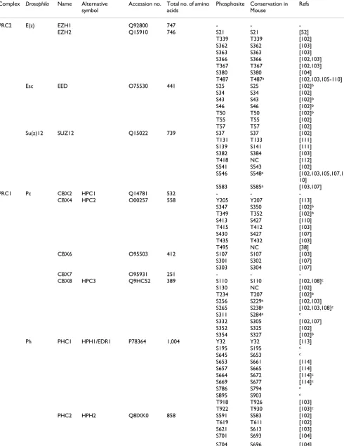
Most current published information on PcG phosphoryla-tion neither unequivocally identifies responsible kinases nor targeted amino acid residues, and the majority of existing in vitro data will have to be confirmed and func-tionally tested in vivo. Over recent years several large-scale phosphoproteomic analyses have been published. We have extracted PcG-related data from these and our own studies; this reveals numerous PcG phosphorylation sites: to date 118 human (Table 3) and 31 mouse (Additional file 2) PcG phosphorylation sites have been published; so far 14 of these phosphorylation events are shared by both species (Table 4). Amino acids surrounding phosphoryla-tion sites direct kinase binding and hence define kinase-substrate specificity. Classification of currently known PcG phosphorylation sites into predicted phosphosite cat-egories shows that most sites group in the proline-directedor acidic class (Table 5; Figure 2a-c). Kinases that recog-nize/bind the proline-directed residues belong to the mitogen-activated protein (MAP) kinase (MAPK) family; acidic motifs are phosphorylated by CK2; kinases target-ing basic motifs belong to the AKT/PKB and PKA types [56]. Consistent with our analysis, published PCGF2/6 (CK1/2) and EZH2 phosphorylation sites (AKT/PKB) clas-sify in the acidic and basic target group, respectively.
Although it is not known at present whether PcG phos-phorylation directly affects chromatin association, their correlation (that is, signaling induced phosphorylation and chromatin dissociation) suggests cells use phosphor-ylation cascades to relay environment cues to chromatin, where reprogramming of gene activity includes epigenetic remodeling events [57]. Consistent with a functional link between MAPK signaling and PcG function, we recently showed that PRC1/chromatin association is disrupted downstream of MAPK activation [57]. Mitogen-activated protein kinase-activated protein kinase (MK3; MAPKAPK3, 3pK) associates with chromatin and occupies PcG target genes. Indeed, phosphopeptide analysis sug-gests that MK3 directly targets PcG proteins (HN and JWV, unpublished results). Although the involved PTMs have not been fully mapped, phosphorylation of PcG proteins affects specific protein interactions and, hence, PRC1 complex composition. Several observations underscore this hypothesis: immunofluorescence studies showed dif-ferential subnuclear localization of PHC1 compared to other PRC1 proteins downstream of signaling-induced phosphorylation [57]. Secondly, PHC1/2 proteins are not stably associated to the PRC1 core complex, but instead show increased heterotypical interaction (HN and JWV, unpublished results) [15,58]. Thirdly, PHC and CBX pro-teins show distinct chromatin enrichment/occupation profiles in response to physiological stimuli in chromatin immunoprecipitation studies; whereas CBX8 is mostly found on genes where H3K27me3 is also present, PHC1 enrichment profiles seem to be more global as are those for the PHC1 interacting kinase MK3 (HN and JWV, unpublished results).
Table 3: Human polycomb phosphorylation sites
Complex Drosophila Name Alternative symbol
Accession no. Total no. of amino acids
Phosphosite Conservation in Mouse
Refs
PRC2 E(z) EZH1 Q92800 747 - -
-EZH2 Q15910 746 S21 S21 [52]
T339 T339 [102]
S362 S362 [103]
S363 S363 [103]
S366 S366 [102,103]
T367 T367 [102,103]
S380 S380 [104]
T487 T487a [102,103,105-110]
Esc EED O75530 441 S25 S25 [102]b
S34 S34 [102]
S43 S43 [102]b
S46 S46 [102]b
T50 T50 [102]b
T55 T55 [102]
T57 T57 [102]
Su(z)12 SUZ12 Q15022 739 S37 S37 [102]
T131 T133 [111]
S139 S141 [111]
S382 S384 [103]
T418 NC [112]
S541 S543 [102]
S546 S548a [102,103,105,107,1
10]
S583 S585a [103,107]
PRC1 Pc CBX2 HPC1 Q14781 532 - -
-CBX4 HPC2 O00257 558 Y205 Y207 [113]
S347 S350 [102]b
T349 T352 [102]b
S413 S427 [110]
T415 T412 [103]
S430 S427 [107]
T435 T432 [103]
T495 NC [38]
CBX6 O95503 412 S107 S107 [103]
S301 S302 [107]
S303 S304 [107]
CBX7 O95931 251 - -
-CBX8 HPC3 Q9HC52 389 S110 S110 [102,108]c
S130 NC [102]
T234 T207 [102]b
S256 S229a [102,103]
S265 S238a [102,103,108]c
S311 S284a c
S332 S305 [102,107]
S352 S325 [102]
S354 S327 [102]b
Ph PHC1 HPH1/EDR1 P78364 1,004 Y32 Y32 [113]
S195 S195 c
S645 S653 c
S653 S661 [114]
S657 S665 [114]
S664 S672 [114]c
S669 S677 [114]c
S786 S794 c
S895 S903 c
T918 T926 [103]
T922 T930 [103]c
PHC2 HPH2 Q8IXK0 858 S591 S583 [102]
T619 T611 [102]
S621 S613 [103]
S701 S693 [104]
S733 S725 [115]c
S745 S737 [105]
PHC3 HPH3/EDR3 Q8NDX5 983 S229 S227 [115]
S263 S261 [105,110]
S315 S312 [102]c
T609 T607a [102,103,105,107,1
09,110]c
T614 T612a [102,107]b
S616 S614a [102,103,105,107,1
09,110]c
S724 S722 [102]b
S761 S759 [102]
S762 S760 [102]
S862 S860 c
Sce/dRING RING1 RING1A/RNF1 Q06587 406 S38 S38 [102,103,107]c
S187 S187 [102,103]
S188 S188 [102,103]
S190 S190 [102,103]
S229 S229 c
RNF2 RING1B/RING2 Q99496 336 S41 S41 [69]
S168 S168 [110]
Psc BMI1 PCGF4 P35226 326 S251 S249a [110]
S253 S251a [110]
S255 S253a [110]
PCGF2 RNF110/MEL18 P35227 344 Y197 Y197 [113]
Y205 Y205 [113]
Y206 Y206 [113]
T344 gap [102]
PhoRC Pho YY1 P25490 414 S118 S120a [109]
S184 S184 [104]
S247 S247a [102,103,105,108,1
09]
Y251 Y251 [102]
T348 T348 [102,116]
T378 T378 [102,103]
Other Psc PCGF1 NSPc1 Q9BSM1 259 - -
-PCGF6 MBLR/RNF134 Q9BYE7 350 S30d S34 [44]
Scm SCMH1 Q96GD3 660 S649 S653 [102,114]
SCML1 Q9UN30 208 S15 NA [102]b
S17 NA [102]
S117 NA [108]
SCML2 Q9UQR0 700 S255 NA [102]b
S256 NA [102]
S258 NA [102]
S267 NA [102]
S299 NA [102]
S300 NA [102]
T305 NA [102]
S495 NA [102]b
S499 NA [102,103,105,106]
T503 NA [102]
S511 NA [102,103,105,106]
S570 NA [115]
S576 NA [102]
T581 NA [102]b
S583 NA [102]
S590 NA [102,103]
S594 NA [102,103]
S606 NA [102]
RYBP Q8N488 228 S127 S127 [102]
S130 S130 [102]
S203 S203 [102]b
T215 T215 [102]b
a Sites that were detected as phosphorylation sites in mouse cells as well; b these putative phosphorylation sites do not reach a sufficiently high
probability based on prediction algorithms used; c marks unpublished results (HN, JD, JWV); d site was referred to as S32 in original article. NA = not available; NC = non-conserved
function in release of methyl-binding interfaces (Figure 3). Of note, CK2 was recently identified as an interactor of RNF2 and CBX8 [62,63]. It is however currently unclear whether and how CK2 regulates PcG function.
In summary, it is clear that phosphorylation is a relevant part of PcG biology. Further characterization and func-tional validation of these phosphorylation sites will be pivotal for our understanding of the cellular conditions under which PcG phosphorylation takes place, the regula-tory pathways involved and how cells use these pathways to modulate chromatin structure to allow biologically appropriate responses.
Other PTMs in PcG biology
N-Acetylglucosamine (GlcNAc)ylation
Recently, the Drosophila PcG gene super sex combs (sxc) was reported to encode the enzyme O-linked O-GlcNAc trans-ferase (Ogt) [64]. PRC1 protein polyhomeotic (Ph) is Glc-NAcylated by sxc/Ogt in vivo. PRC1-mediated repression is dependent on functional sxc/Ogt, possibly via GlcNAcyla-tion of Ph [64]. Interestingly, the process appears con-served across species, as the mammalian ortholog PHC3 is O-GlcNAcylated as well [65]. Analogous to PcG, GlcNA-cylation of the histone methyltransferase MLL5, a Tritho-rax-like protein, generally assumed to counteract PcG function, regulates methylation of H3K4 [66]. These find-ings begin to show that GlcNAcylation of chromatin fac-tors, such as PcG proteins, is relevant for epigenetic regulation and provide important leads for future study.
Proteolytic cleavage
Other than ubiquitin-26S proteasome-dependent protein degradation, as holds for PcG proteins RNF2 and BMI1 [22], proteolytic cleavage may also occur via activated cas-pases [67]. Caspase substrates include, among others, structural proteins, such as actin, vimentin and nuclear lamins and proteins involved in transcription and transla-tion, such as nuclear factor (NF)κB and translation initia-tion factors [67]. Recently, RNF2 was identified as a direct substrate of caspases 3 and 9 in apoptotic cells [68]. The exact function of RNF2 cleavage during apoptosis remains illusive, but it is possibly a prerequisite for nuclear con-densation and DNA fragmentation to occur. Whether PcG protein cleavage is relevant outside the context of apopto-sis is currently unclear.
Acetylation
Currently acetylation of only one PcG protein has been reported: RNF2 is acetylated at S2, which is accompanied by a N-terminal methionine excision [69]. The signifi-cance of this modification in relation to PcG function is not known.
Signaling to chromatin: post-translational
crosstalk to Polycomb
It is evident that PcG proteins are subject to numerous dif-ferent PTMs (among others, ubiquitylation, sumoylation, phosphorylation and GlcNAcylation) and, besides, har-bor intrinsic enzymatic modifying activities themselves (HMT, ubiquitin and sumo E3 ligase, O-GlcNAc trans-ferase). How these PTMs relate to each other is currently far from clear, however, several studies begin to unveil an intricate interplay between different PTMs, their effect at the molecular level and their biological relevance (Figure 1). Complex formation between PCGFs and RNF2 regu-lates H2A ubiquitin E3 ligase activity [16]. PCGF2 phos-phorylation (Additional file 2) is required for substrate recognition in a relevant chromatin context, as dephos-phorylated RNF2/PCGF2 complexes no longer efficiently ubiquitylate nucleosomes [16]. Similar regulation likely holds true for its close relatives BMI1 and PCGF1. Phos-phorylated EZH2 has reduced methyltransferase activity toward histone 3, as a result of reduced substrate binding [52]. Phosphorylation and sumoylation-mediated Table 4: Summary of polycomb group (PcG) phosphorylation sites
Residue Human Mouse Human + mouse non-overlapping Human + mouse overlapping
Serine 87 (74%) 26 (83%) 101 (75%) 12 (86%)
Threonine 25 (20%) 5 (16%) 28 (21%) 2 (14%)
Tyrosine 6 (6%) - 6 (4%)
-Total 118 (100%) 31 (100%) 135 (100%) 14 (100%)
Classification of PcG phosphorylation sites based on common phosphoamino acids.
Table 5: Classification of polycomb group (PcG) S/T phosphorylation sites
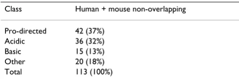
Class Human + mouse non-overlapping
Pro-directed 42 (37%)
Acidic 36 (32%)
Basic 15 (13%)
Other 20 (18%)
Total 113 (100%)
autoregulatory feedback between CBX4 and HIPK2 has obvious relevance for DNA damage responses [38]. Recently the PRC1 complex was identified as the E3 ubiq-uitin ligase for geminin, an inhibitor of replication licens-ing factor Cdt1. PHC1 (Rae28) deficiency in mice impairs ubiquitin/S26-proteasome-mediated degradation of gem-inin and has direct consequences for cell cycle progression in the haematopoietic lineage [70].
In keeping with differential chromatin association of Ph as compared to other PRC1 proteins, GlcNAcylated Ph does not copurify with other PRC1 components in larval extracts, whereas it does in embryonic nuclear extracts [64]. Phosphorylation and GlcNAcylation mostly affect the same amino acids (S/T), hence an intricate interplay between these modifications may be predicted, either via competitive occupancy at the same site or alternative occupancy at adjacent sites [71]. Taken together these observations support differential regulation within the PRC1 complex upon activation of signaling cascades (Fig-ure 4), implicitly suggesting differential roles for and/or regulation of PHC proteins and other members of the PRC1 complex in response to cellular signaling.
Integration of signaling at the chromatin level
Signaling-induced PTMs clearly affect non-histone and histone proteins concurrently, suggesting that signaling-induced phosphorylation evokes an integrated response at the level of chromatin-associated proteins and nucleo-somes. Histone H3Ser phosphorylation occurs down-stream of signaling induced by arsenite (Akt1, extracellular signal-related kinase (ERK2) and RSK2), ultraviolet B (ERK, p38 MAPK, c-Jun N-terminal kinase (JNK1) and MSK1), TPA (MSK2 and MSK1) and anisomy-cin (MSK2 and Mitogen and stress-activated protein kinase-1 (MSK1)) [72-76]. Even though MSK1 phosphor-ylates histone H3S10 and S28 within the same peptide in vitro, this apparently does not happen in vivo, where sub-nuclear localization of histone H3S10ph and H3S28ph are distinct [77,78]. The spatial distribution of these H3 PTMs is likely controlled by context-dependent kinase recruitment (by, for example, HP1 or PcG complexes).Classification of S/T phosphorylation sites into general kinase recognition sequence categories
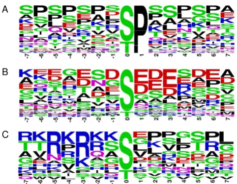
Figure 2
Classification of S/T phosphorylation sites into gen-eral kinase recognition sequence categories. At present, not all consensus phosphorylation motifs are known for all kinases. A number of general phosphosite classes have been annotated based on amino acid sequences surrounding S/T phosphorylation sites: proline-directed, acidic, basic and otherwise [97]. (a-c) Sequence logos for Polycomb Group (PcG) phosphorylation motifs of proline-directed ((a), n = 42) acidic ((b), n = 36) and basic ((c), n = 15) categories where the phosphorylated residue (S/T) is centered were generated with Weblogo [98]. Only serine and threonine phosphorylation sites were taken into account when a full 15-mer sequence was available. To avoid sequence bias only non-overlapping human and mouse PcG phosphorylation sites were used. Centered 15-mer sequences were assigned to a motif class sequentially by following a binary decision tree as follows: P at +1 (Pro-directed), 5 or more E/D at +1 to +6 (acidic), R/K at -3 (basic), D/E at +1/+2 or +3 (acidic), 2 or more R/K at -6 to -1 (basic), otherwise. Colors corre-spond to the chemical properties of the amino acids: hydro-phobic (black), basic (blue), acidic (red) and polar (green). (a)
Although phosphosite numbers are low and thus no solid deductions are warranted, it is apparent for the proline-directed class (classified by a P in the +1 position), that pro-lines are not limited to +1, but are abundant in various - and + positions. (b) In the acidic motif, characterized by the presence of the amino acids glutamic acid (E) and aspartic acid (D) in the + residues, E/Ds are present in the - positions.
(c) In basic motifs, arginine (R) is predominantly found in -3 positions, in addition to -5.
A
C B
Hypothetical mechanism for phosphorylation-induced disso-ciation of Polycomb Repressive Complex (PRC)1 from chro-matin
Figure 3
Hypothetical mechanism for phosphorylation-induced dissociation of Polycomb Repressive Com-plex (PRC)1 from chromatin. The chromodomain of chromobox homolog (CBX) proteins interacts with H3K27me3. Chromatin dissociation may be the result of ARKS motif methyl-phos switching, by phosphorylation of H3S28 (upper panel). Phosphorylation of conserved resi-due(s) within the chromodomain of CBX may contribute to chromatin dissociation (lower panel).
CBX
H3
-XXXARKSXXX-Me3
H3 -XXXARKSXXX-CBX
ph Me3
H3S28ph H3
-XXXARKSXXX-Me3
CBX ph
CBX
28 27
ph ph
Although the correlation between PcG phosphorylation and chromatin dissociation is clear, both cell cycle dependent and cell cycle independent underlying molec-ular mechanisms remain largely obscure [43,44,57]. Comparison of PcG modules to a related histone binding protein family, HP1 (associated with constitutive hetero-chromatin and regions of facultative heterohetero-chromatin) may begin to reveal molecular mechanisms [79]. HP1 and PcG CBX proteins share a chromodomain, which binds specific histone 3 trimethyl marks. Both chromodomain protein classes are released from chromatin during mito-sis. HP1 dissociates from chromatin in M-phase, despite unchanged H3K9me3 levels. Aurora kinase B-induced phosphorylation of H3S10 peptides adjacent to the K9me3 mark strongly reduces HP1 binding in vitro and
releases HP1 from chromatin in vivo [80]. H3S10 phos-phorylation was put forward as part of a 'binary switch' mechanism of the 'methyl/phos' type: phosphorylation adjacent to a methyl mark leads to induced loss of methyl-based binding of a factor or complex [59,81]. Dynamic 'methyl/phos' switching modules also provide a tentative molecular basis for heritable transcriptional memories. Such a switching mechanism may not be limited to the K9/S10 region of H3, but may be more common: both K9 and K27 in histone 3 reside in nearly identical amino acid contexts (ARKS); hence 'methyl/phos' switching may con-trol CBX binding to H3 as well (Figure 3). Indeed, H3S28 is also phosphorylated in M-phase [82,83]. Relevantly, we reported a close correlation between signaling-induced PRC1 dissociation and H3S28ph, whereas this was not
Integrated hypothetical model for post-translational modification (PTM)-dependent regulation of Polycomb Repressive Com-plex (PRC)1-mediated repression
Figure 4
Integrated hypothetical model for post-translational modification (PTM)-dependent regulation of Polycomb Repressive Complex (PRC)1-mediated repression. Three independent chromatin states in the context of PRC1 func-tion are recognized: (a) the repressed state, which requires ubiquitylation and sumoylation of PRC1 compounds. Detectable baseline phosphorylation may indicate a prerequisite for PRC function and/or differences in PRC activity at local targets throughout the genome. Signaling to chromatin alters PTM states and chromatin association, and eventually releases PRC silencing (b,c). Observations from our and other groups suggest differential regulation of polyhomeotic homolog (PHC) pro-teins versus RNF2, BMI1 and chromobox homolog (CBX) propro-teins; this may involve N-acetylglucosamine (GlcNAc) modifica-tion in mammals as well. (c) Whether or not full expression of a Polycomb Group (PcG) target gene requires complete removal or relocation of PHC is currently not known. Likewise, whether ubiquitin-mediated proteolysis of BMI1 and RNF2 is needed to release gene repression is purely speculative. ph = phosphorylation; su = sumoylation; ub = ubiquitylation. Black tri-angles = H3K27me3; open circles = H3S28ph; unmarked circular/oval structures represent general transcription factors and/or unknown proteins.
PHC
ph
ph expressed state
gene target
C
repressed state/poised?
derepressed state/poised?
PHC
gene target
X
RNF2
CBX
BMI1
ph
B
phSCMh
GlcNAc
GlcNAc
RNF2
CBX BMI1
ph
ph
ph
ubub ubub
ub ub
ub ub
SCMh
X
ph
gene target RNF2
CBX PHC
BMI1
A
phsu ub
ub
seen in relation to H3S10ph [57], suggesting similar molecular strategies between related proteins. Hence, the larger family of MSK, ERK and RSK kinases integrate regu-lation of gene expression at several functional levels by targeting transcription factors, chromatin binding com-plexes and nucleosomal components.
Although clearly numerous PTMs affect PcG biology, molecular mechanisms in signaling to chromatin, down-stream modulation of epigenetic marking and the estab-lishment and transfer of heritable epigenetic states remain largely elusive. Specific consensus/motif based kinase-substrate interactions most likely define and direct signal-ing-induced remodeling and/or other epigenetically rele-vant events at the chromatin level. In contrast, context-dependent variation in sumoylation sites is less well defined and the role of select consensus motifs in ubiquit-ylation is largely unknown. Although speculative, this may suggest that once signaling is triggered, a hierarchical sequence of PTMs is initiated, the target specificity (that is, networks, pathways, complexes) of which is defined by phosphorylation events, whereas downstream effects (among others, altered protein interactions, activity or sta-bility) are 'merely' consequential, and controlled by numerous other PTMs. Indeed ubiquitylation, sumoyla-tion, GlyNAcylation and phosphorylation are probably functionally linked in PcG biology, as important cross talk between these PTMs exists in many ways in other systems [84]. In this context important open issues are for instance how exactly PcG PTMs functionally relate to each other, that is, whether PTMs act separately, processively or com-binatorially. Signaling-induced PTMs are generally revers-ible, proteolytic cleavage excluded, hence many chromatin-associated epigenetic regulators are presuma-bly rapidly converted to their initial post-translational sta-tus upon signal recession. However, at the receiving end, stable, heritable covalent histone modifications appear somehow exempt from such regulation. Notions such as these should provide important basis for future hypothe-sis-driven research.
Conclusion
It is evident that chromatin-associated protein complexes, like PcG proteins, are targets for cell signaling. These sign-aling events lead to PTMs that may affect chromatin bind-ing, complex composition and catalytic activity. We and others have found that multiple kinases target PcG pro-teins. In addition, PcG proteins are subject to ubiquityla-tion, sumoylation and additional PTMs. Studies reviewed in this manuscript have only just begun to unravel the complexity and multiple layers of regulation of PcG func-tion. PcG-mediated transcriptional silencing already appears a much more complex process than the anti-quated view that PRCs physically obstruct transcription factor binding to DNA, and ongoing studies refine
posi-tioning of PcG function in the proper cellular context. Current conservative estimates predict the existence of anywhere between 60 to 100 or more mammalian PcG(-related) proteins, each likely with multiple phosphoryla-tion and/or ubiquitylaphosphoryla-tion, sumoylaphosphoryla-tion and potentially numerous other PTM sites, including acetylation and methylation [85,86]. In stark contrast with this, at the moment only two PcG phosphospecific antisera exist [44,52]. Clearly there is a need for additional experimen-tal tools and approaches.
Ultimately PTMs are aimed at concerted regulation of a number of chromatin-based processes in which PcG pro-teins play a role, including dynamic transcriptional regu-lation, long-term silencing, DNA replication and DNA damage responses, to ensure proper regulation of cell fate and survival. Increased insight into mechanisms employed by cells to target chromatin and chromatin-associated factors, including PcG, and their physiological consequences at the chromatin level will be important for further development and application of epigenetic strate-gies in for instance regenerative medicine. As many of the processes targeting and involving PcG function are etio-logically linked to disease (for example, overexpression in cancer, bypass of replicative senescence [62,87-89]), a bet-ter fundamental understanding of gene-environment interactions at the molecular level will eventually contrib-ute to the development of therapeutic and preventive strategies relevant for Western world-type diseases, including obesity, diabetes and cancer.
Competing interests
The authors declare that they have no competing interests.
Authors' contributions
HN wrote the initial draft and JD and JWV edited the uscript. All authors have read and approved the final man-uscript.
Appendix: Most common PTMs in polycomb
Ubiquitylation
then conjugated to the E2, that, assisted by an E3 ligase, transfers ubiquitin to a lysine residue onto the substrate protein [90]. The diversity and combination of these enzymes ultimately determines substrate specificity and biological responses. Two classes of E3 ligases are recog-nized: the HECT domain E3s and the RING domain E3s; RING-type E3 ligases are thought to function as bridging factors between the E2 and substrate [91]. Ub protein sub-strates are deubiquitylated by deubiquitylases (DUBs). The large number of different E2 and E3 enzymes and DUBs identified to date and their involvement in a great variety of processes underscores the complexity of regula-tion by ubiquitylaregula-tion [91].
Sumoylation
Sumo proteins are approximately 10 kDa in size [92]. Like ubiquitylation, sumoylation involves three distinct enzy-matic activities: an E1 activating enzyme (AOS1/UBA2 heterodimer) an E2 conjugating enzyme (UBC9) and an E3 ligase. Currently three classes of sumo E3 ligases have been distinguished: SP-RING motif E3 ligases, such as protein inhibitor of activated STAT (PIAS) proteins, a sec-ond class containing RanBP2, and a third class with no apparent homology to either other class comprising CBX4 (HPC2). In contrast to ubiquitylation, sumoylation takes place at sumo acceptor sites: ΨKXE (Ψ and X represent hydrophobic and any amino acids).
Phosphorylation
Phosphorylation is probably the most widespread and best studied PTM in cellular signaling. Protein kinases cat-alyze the transfer of γ-phosphate from ATP to a substrate protein, thereby generating ADP [93]. Phosphorylation is reversible: phosphatases remove the attached phosphate moiety. An estimated 30% of all proteins are phosphor-ylated on at least one residue. Phosphate is most com-monly linked to Ser (S), Thr (T) or Tyr (Y)), but in addition occurs on His (H), Lys (K), Arg (R), Asp/Glu (D/ E), and Cys (C) [84].
Additional material
Acknowledgements
Collection of PcG annotations was based on Uniprot [94] and Phosphosite [95]. Cross-species sequence conservation was verified by aligning human and murine homologous sequences in Geneious software (Biomatters, Auckland New Zealand), using default settings. PTM-related protein nomenclature was based on published consensus [96]. The authors thank M Vidal (Madrid, Spain) and R Shiri-Sverdlov for critically reading the man-uscript. The authors received financial support from the European Molec-ular Biology Organization (Germany) 186.00-06; the Dutch Science Organization (ZonMW-NWO): Research Support grant 908-02-040 to JWV and VIDI grant 016.046.362; the National Rheuma Foundation (Reu-mafonds) grant LLP14, a grant from the transnational University Limburg (tUL 0810) to JWV and a Netherlands Genome Initiative (NGI) fellowship 050-72-422 to HN.
References
1. Lewis EB: A gene complex controlling segmentation in Dro-sophila. Nature 1978, 276:565-570.
2. Orlando V: Polycomb, epigenomes, and control of cell iden-tity. Cell 2003, 112:599-606.
3. van Lohuizen M: Functional analysis of mouse polycomb group genes. Cell Mol Life Sci 1998, 54:71-79.
4. Bracken AP, Dietrich N, Pasini D, Hansen KH, Helin K: Genome-wide mapping of polycomb target genes unravels their roles in cell fate transitions. Genes Dev 2006, 20:1123-1136.
5. Otte AP, Kwaks THJ: Gene repression by polycomb group pro-tein complexes: a distinct complex for every occasion? Curr Opin Genet Dev 2003, 13:448-454.
6. Jason LJ, Moore SC, Lewis JD, Lindsey G, Ausio J: Histone ubiquiti-nation: a tagging tail unfolds? Bioessays 2002, 24:166-174. 7. Buchwald G, Stoop P van der, Weichenrieder O, Perrakis A, van
Lohuizen M, Sixma TK: Structure and E3-ligase activity of the Ring-Ring complex of polycomb proteins Bmi1 and Ring1b. EMBO J 2006, 25:2465-2474.
8. de Napoles M, Mermoud JE, Wakao R, Tang YA, Endoh M, Appanah R, Nesterova TB, Silva J, Otte AP, Vidal M, Koseki H, Brockdorff N: Polycomb group proteins Ring1A/B link ubiquitylation of his-tone H2A to heritable gene silencing and X inactivation. Dev Cell 2004, 7:663-676.
9. Fang J, Chen T, Chadwick B, Li E, Zhang Y: Ring1b-mediated H2A ubiquitination associates with inactive X chromosomes and is involved in initiation of X inactivation. J Biol Chem 2004, 279:52812-52815.
10. Leeb M, Wutz A: Ring1B is crucial for the regulation of devel-opmental control genes and PRC1 proteins but not X inacti-vation in embryonic cells. J Cell Biol 2007, 178:219-229. 11. Schoeftner S, Sengupta AK, Kubicek S, Mechtler K, Spahn L, Koseki
H, Jenuwein T, Wutz A: Recruitment of PRC1 function at the initiation of X inactivation independent of PRC2 and silenc-ing. EMBO J 2006.
12. Sarcinella E, Zuzarte PC, Lau PN, Draker R, Cheung P: Monoubiq-uitylation of H2A.Z distinguishes its association with euchro-matin or facultative heterochroeuchro-matin. Mol Cell Biol 2007, 27:6457-6468.
13. Wang H, Wang L, Erdjument-Bromage H, Vidal M, Tempst P, Jones RS, Zhang Y: Role of histone H2A ubiquitination in polycomb silencing. Nature 2004, 431:873-878.
14. Lagarou A, Mohd-Sarip A, Moshkin YM, Chalkley GE, Bezstarosti K, Demmers JA, Verrijzer CP: dKDM2 couples histone H2A ubiq-uitylation to histone H3 demethylation during polycomb group silencing. Genes Dev 2008, 22:2799-2810.
15. Cao R, Tsukada Y, Zhang Y: Role of Bmi-1 and Ring1A in H2A ubiquitylation and Hox gene silencing. Mol Cell 2005, 20:845-854.
16. Elderkin S, Maertens GN, Endoh M, Mallery DL, Morrice N, Koseki H, Peters G, Brockdorff N, Hiom K: A phosphorylated form of Mel-18 targets the Ring1B histone H2A ubiquitin ligase to chromatin. Mol Cell 2007, 28:107-120.
17. Gearhart MD, Corcoran CM, Wamstad JA, Bardwell VJ: Polycomb group and SCF ubiquitin ligases are found in a novel BCOR complex that is recruited to BCL6 targets. Mol Cell Biol 2006, 26:6880-6889.
Additional file 1
Prediction of human polycomb sumoylation sites. A table containing predicted sumoylation sites on human PcG proteins.
Click here for file
[http://www.biomedcentral.com/content/supplementary/1756-8935-2-10-S1.pdf]
Additional file 2
Mouse polycomb phosphorylation sites. A table containing published mouse polycomb phosphorylation sites.
Click here for file
18. Wu X, Gong Y, Yue J, Qiang B, Yuan J, Peng X: Cooperation between EZH2, NSPc1-mediated histone H2A ubiquitina-tion and Dnmt1 in HOX gene silencing. Nucleic Acids Res 2008, 36:3590-3599.
19. Li Z, Cao R, Wang M, Myers MP, Zhang Y, Xu RM: Structure of a Bmi-1-Ring1B polycomb group ubiquitin ligase complex. J Biol Chem 2006, 281:20643-20649.
20. Kentsis A, Borden KL: Construction of macromolecular assem-blages in eukaryotic processes and their role in human dis-ease: linking RINGs together. Curr Protein Pept Sci 2000, 1:49-73. 21. Laney JD, Hochstrasser M: Substrate targeting in the ubiquitin
system. Cell 1999, 97:427-430.
22. Ben-Saadon R, Zaaroor D, Ziv T, Ciechanover A: The polycomb protein Ring1B generates self atypical mixed ubiquitin chains required for its in vitro histone H2A ligase activity. Mol Cell 2006, 24:701-711.
23. Hernandez-Munoz I, Lund AH, Stoop P van der, Boutsma E, Muijrers I, Verhoeven E, Nusinow DA, Panning B, Marahrens Y, van Lohuizen M: Stable X chromosome inactivation involves the PRC1 polycomb complex and requires histone MACROH2A1 and the CULLIN3/SPOP ubiquitin E3 ligase. Proc Natl Acad Sci USA
2005, 102:7635-7640.
24. Zhang J, Sarge KD: Identification of a polymorphism in the RING finger of human Bmi-1 that causes its degradation by the ubiquitin-proteasome system. FEBS Lett 2009, 583:960-964. 25. Arrigoni R, Alam SL, Wamstad JA, Bardwell VJ, Sundquist WI, Sch-reiber-Agus N: The polycomb-associated protein Rybp is a ubiquitin binding protein. FEBS Lett 2006, 580:6233-6241. 26. Trimarchi JM, Fairchild B, Wen J, Lees JA: The E2F6 transcription
factor is a component of the mammalian Bmi1- containing polycomb complex. Proc Natl Acad Sci USA 2001, 98:1519-1524. 27. Gill G: Something about SUMO inhibits transcription. Curr
Opin Genet Dev 2005, 15:536-541.
28. Stielow B, Sapetschnig A, Kruger I, Kunert N, Brehm A, Boutros M, Suske G: Identification of SUMO-dependent chromatin-asso-ciated transcriptional repression components by a genome-wide RNAi screen. Mol Cell 2008, 29:742-754.
29. Kehle J, Beuchle D, Treuheit S, Christen B, Kennison JA, Bienz M, Muller J: dMi-2, a hunchback-interacting protein that func-tions in polycomb repression. Science 1998, 282:1897-1900. 30. Zhang H, Smolen GA, Palmer R, Christoforou A, Heuvel S van den,
Haber DA: SUMO modification is required for in vivo Hox gene regulation by the Caenorhabditis elegans polycomb group protein SOP-2. Nat Genet 2004, 36:507-511.
31. Sewalt RG, Gunster MJ, Vlag J van der, Satijn DP, Otte AP: C-Termi-nal binding protein is a transcriptioC-Termi-nal repressor that inter-acts with a specific class of vertebrate polycomb proteins. Mol Cell Biol 1999, 19:777-787 [http://mcb.asm.org/cgi/content/full/19/ 1/777?view=long&pmid=9858600].
32. Kagey MH, Melhuish TA, Wotton D: The polycomb protein Pc2 is a SUMO E3. Cell 2003, 113:127-137.
33. Wotton D, Merrill JC: Pc2 and SUMOylation. Biochem Soc Trans
2007, 35:1401-1404.
34. Agrawal N, Banerjee R: Human polycomb 2 protein is a SUMO E3 ligase and alleviates substrate-induced inhibition of cys-tathionine β-synthase sumoylation. PLoS ONE 2008, 3:e4032. 35. Li B, Zhou J, Liu P, Hu J, Jin H, Shimono Y, Takahashi M, Xu G:
Poly-comb protein Cbx4 promotes SUMO modification of de novo DNA methyltransferase Dnmt3a. Biochem J 2007, 405:369-378.
36. Ling Y, Sankpal UT, Robertson AK, McNally JG, Karpova T, Robert-son KD: Modification of de novo DNA methyltransferase 3a (Dnmt3a) by SUMO-1 modulates its interaction with histone deacetylases (HDACs) and its capacity to repress transcrip-tion. Nucl Acids Res 2004, 32:598-610.
37. Long J, Zuo D, Park M: Pc2-mediated sumoylation of Smad-interacting protein 1 attenuates transcriptional repression of E-cadherin. J Biol Chem 2005, 280:35477-35489.
38. Roscic A, Moller A, Calzado MA, Renner F, Wimmer VC, Gresko E, Ludi KS, Schmitz ML: Phosphorylation-dependent control of Pc2 SUMO E3 ligase activity by its substrate protein HIPK2. Mol Cell 2006, 24:77-89.
39. Fontecave M, Atta M, Mulliez E: S-adenosylmethionine: nothing goes to waste. Trends Biochem Sci 2004, 29:243-249.
40. Zhang J, Goodson ML, Hong Y, Sarge KD: MEL-18 interacts with HSF2 and the SUMO E2 UBC9 to inhibit HSF2 sumoylation. J Biol Chem 2008, 283:7464-7469.
41. Zhang J, Sarge KD: Mel-18 interacts with RanGAP1 and inhibits its sumoylation. Biochem Biophys Res Commun 2008, 375:252-255. 42. Riising EM, Boggio R, Chiocca S, Helin K, Pasini D: The polycomb repressive complex 2 is a potential target of SUMO modifi-cations. PLoS ONE 2008, 3:e2704.
43. Voncken JW, Schweizer D, Aagaard L, Sattler L, Jantsch MF, van Lohu-izen M: Chromatin-association of the polycomb group pro-tein BMI1 is cell cycle-regulated and correlates with its phosphorylation status. J Cell Sci 1999, 112:4627-4639 [http:// jcs.biologists.org/cgi/content/abstract/112/24/4627].
44. Akasaka T, Takahashi N, Suzuki M, Koseki H, Bodmer R, Koga H: MBLR, a new RING finger protein resembling mammalian polycomb gene products, is regulated by cell cycle-depend-ent phosphorylation. Genes Cells 2002, 7:835-850.
45. Muchardt C, Reyes JC, Bourachot B, Leguoy E, Yaniv M: The hbrm and BRG-1 proteins, components of the human SNF/SWI complex, are phosphorylated and excluded from the con-densed chromosomes during mitosis. EMBO J 1996, 15:3394-3402.
46. Noguchi K, Shiurba R, Higashinakagawa T: Nuclear translocation of mouse polycomb m33 protein in regenerating liver. Bio-chem Biophys Res Comm 2002, 291:508-515.
47. Fujisaki S, Ninomiya Y, Ishihara H, Miyazaki M, Kanno R, Asahara T, Kanno M: Dimerization of the polycomb-group protein Mel-18 is regulated by PKC phosphorylation. Biochem Biophys Res Commun 2003, 300:135-140.
48. Gong Y, Wang X, Liu J, Shi L, Yin B, Peng X, Qiang B, Yuan J: NSPc1, a mainly nuclear localized protein of novel PcG family mem-bers, has a transcription repression activity related to its PKC phosphorylation site at S183. FEBS Lett 2005, 579:115-121. 49. Ng J, Hart CM, Morgan K, Simon JA: A Drosophila ESC-E(Z) pro-tein complex is distinct from other polycomb group com-plexes and contains covalently modified ESC. Mol Cell Biol
2000, 20:3069-3078.
50. Tie F, Siebold AP, Harte PJ: The N-terminus of Drosophila ESC mediates its phosphorylation and dimerization. Biochem Bio-phys Res Commun 2005, 332:622-632.
51. Kuzmichev A, Nishioka K, Erdjument-Bromage H, Tempst P, Rein-berg D: Histone methyltransferase activity associated with a human multiprotein complex containing the Enhancer of Zeste protein. Genes Dev 2002, 16:2893-2905.
52. Cha TL, Zhou BP, Xia W, Wu Y, Yang CC, Chen CT, Ping B, Otte AP, Hung MC: Akt-mediated phosphorylation of EZH2 suppresses methylation of lysine 27 in histone H3. Science 2005, 310:306-310.
53. Hansen KH, Bracken AP, Pasini D, Dietrich N, Gehani SS, Monrad A, Rappsilber J, Lerdrup M, Helin K: A model for transmission of the H3K27me3 epigenetic mark. Nat Cell Biol 2008, 10:1291-1300. 54. Hansen KH, Helin K: Epigenetic inheritance through
self-recruitment of the polycomb repressive complex 2. Epigenet-ics 2009, 4: [http://www.landesbioscience.com/journals/epigenetics/ article/8483].
55. Ogawa M, Hiraoka Y, Aiso S: The polycomb-group protein ENX-2 interacts with ZAP-70. Immunol Lett 2003, 86:57-61.
56. Amanchy R, Periaswamy B, Mathivanan S, Reddy R, Tattikota SG, Pan-dey A: A curated compendium of phosphorylation motifs. Nat Biotechnol 2007, 25:285-286.
57. Voncken JW, Niessen H, Neufeld B, Rennefahrt U, Dahlmans V, Kub-ben N, Holzer B, Ludwig S, Rapp UR: MAPKAP kinase 3pK phos-phorylates and regulates chromatin association of the polycomb group protein Bmi1. J Biol Chem 2005, 280:5178-5187. 58. Hodgson JW, Argiropoulos B, Brock HW: Site-specific recogni-tion of a 70-base-pair element containing d(GA)(n) repeats mediates bithoraxoid polycomb group response element-dependent silencing. Mol Cell Biol 2001, 21:4528-4543.
59. Fischle W, Wang Y, Allis CD: Binary switches and modification cassettes in histone biology and beyond. Nature 2003, 425:475-479.
60. Sassone-Corsi P, Mizzen CA, Cheung P, Crosio C, Monaco L, Jacquot S, Hanauer A, Allis CD: Requirement of Rsk-2 for epidermal growth factor-activated phosphorylation of histone H3. Sci-ence 1999, 285:886-891.
61. Ayoub N, Jeyasekharan AD, Bernal JA, Venkitaraman AR: HP1-β mobilization promotes chromatin changes that initiate the DNA damage response. Nature 2008, 453:682-686.
group protein CBX8 through direct binding to the INK4A-ARF locus. EMBO J 2007, 26:1637-1648.
63. Sanchez C, Sanchez I, Demmers JA, Rodriguez P, Strouboulis J, Vidal M: Proteomics analysis of Ring1B/Rnf2 interactors identifies a novel complex with the Fbxl10/Jhdm1B histone demethyl-ase and the Bcl6 interacting corepressor. Mol Cell Proteomics
2007, 6:820-834.
64. Gambetta MC, Oktaba K, Muller J: Essential role of the glycosyl-transferase Sxc/Ogt in polycomb repression. Science 2009. 65. Chalkley RJ, Thalhammer A, Schoepfer R, Burlingame AL:
Identifica-tion of protein O-GlcNAcylaIdentifica-tion sites using electron transfer dissociation mass spectrometry on native peptides. Proc Natl Acad Sci USA 2009, 106:8894-8899.
66. Fujiki R, Chikanishi T, Hashiba W, Ito H, Takada I, Roeder RG, Kita-gawa H, Kato S: GlcNAcylation of a histone methyltransferase in retinoic-acid-induced granulopoiesis. Nature 2009, 459:455-459.
67. Taylor RC, Cullen SP, Martin SJ: Apoptosis: controlled demoli-tion at the cellular level. Nat Rev Mol Cell Biol 2008, 9:231-241. 68. Wong CK, Chen Z, So KL, Li D, Li P: Polycomb group protein
RING1B is a direct substrate of caspases-3 and -9. Biochim Bio-phys Acta 2007, 1773:844-852.
69. Rao PS, Satelli A, Zhang S, Srivastava SK, Srivenugopal KS, Rao US: RNF2 is the target for phosphorylation by the p38 MAPK and ERK signaling pathways. Proteomics 2009, 9:2776-2787. 70. Ohtsubo M, Yasunaga S, Ohno Y, Tsumura M, Okada S, Ishikawa N,
Shirao K, Kikuchi A, Nishitani H, Kobayashi M, Takihara Y: Poly-comb-group complex 1 acts as an E3 ubiquitin ligase for Geminin to sustain hematopoietic stem cell activity. Proc Natl Acad Sci USA 2008, 105:10396-10401.
71. Hart GW, Housley MP, Slawson C: Cycling of O-linked β-N -acetylglucosamine on nucleocytoplasmic proteins. Nature
2007, 446:1017-1022.
72. He Z, Ma WY, Liu G, Zhang Y, Bode AM, Dong Z: Arsenite-induced phosphorylation of histone H3 at serine 10 is medi-ated by Akt1, extracellular signal-regulmedi-ated kinase 2, and p90 ribosomal S6 kinase 2 but not mitogen- and stress-activated protein kinase 1. J Biol Chem 2003, 278:10588-10593.
73. Zhong SP, Ma WY, Dong Z: ERKs and p38 kinases mediate ultra-violet B-induced phosphorylation of histone H3 at serine 10. J Biol Chem 2000, 275:20980-20984.
74. Zhong S, Jansen C, She QB, Goto H, Inagaki M, Bode AM, Ma WY, Dong Z: Ultraviolet B-induced phosphorylation of histone H3 at serine 28 is mediated by MSK1. J Biol Chem 2001, 276:33213-33219.
75. Zhong S, Zhang Y, Jansen C, Goto H, Inagaki M, Dong Z: MAP kinases mediate UVB-induced phosphorylation of histone H3 at serine 28. J Biol Chem 2001, 276:12932-12937.
76. Soloaga A, Thomson S, Wiggin GR, Rampersaud N, Dyson MH, Haz-zalin CA, Mahadevan LC, Arthur JS: MSK2 and MSK1 mediate the mitogen- and stress-induced phosphorylation of histone H3 and HMG-14. EMBO J 2003, 22:2788-2797.
77. Dyson MH, Thomson S, Inagaki M, Goto H, Arthur SJ, Nightingale K, Iborra FJ, Mahadevan LC: MAP kinase-mediated phosphoryla-tion of distinct pools of histone H3 at S10 or S28 via mitogen-and stress-activated kinase 1/2. J Cell Sci 2005, 118:2247-2259. 78. Dunn KL, Davie JR: Stimulation of the Ras-MAPK pathway
leads to independent phosphorylation of histone H3 on ser-ine 10 and 28. Oncogene 2005, 24:3492-3502.
79. Li Y, Kirschmann DA, Wallrath LL: Does heterochromatin pro-tein 1 always follow code? Proc Natl Acad Sci USA 2002, 99(Suppl 4):16462-16469.
80. Fischle W, Tseng BS, Dormann HL, Ueberheide BM, Garcia BA, Shab-anowitz J, Hunt DF, Funabiki H, Allis CD: Regulation of HP1-chro-matin binding by histone H3 methylation and phosphorylation. Nature 2005, 438:1116-1122.
81. Dormann HL, Tseng BS, Allis CD, Funabiki H, Fischle W: Dynamic regulation of effector protein binding to histone modifica-tions: the biology of HP1 switching. Cell Cycle 2006, 5:2842-2851 [http://www.landesbioscience.com/journals/cc/article/3540/]. 82. Goto H, Tomono Y, Ajiro K, Kosako H, Fujita M, Sakurai M, Okawa
K, Iwamatsu A, Okigaki T, Takahashi T, Inagaki M: Identification of a novel phosphorylation site on histone H3 coupled with mitotic chromosome condensation. J Biol Chem 1999, 274:25543-25549.
83. Goto H, Yasui Y, Nigg EA, Inagaki M: Aurora-B phosphorylates Histone H3 at serine28 with regard to the mitotic chromo-some condensation. Genes Cells 2002, 7:11-17.
84. Hunter T: The age of crosstalk: phosphorylation, ubiquitina-tion, and beyond. Mol Cell 2007, 28:730-738.
85. Spange S, Wagner T, Heinzel T, Kramer OH: Acetylation of non-histone proteins modulates cellular signalling at multiple lev-els. Int J Biochem Cell Biol 2009, 41:185-198.
86. Huang J, Berger SL: The emerging field of dynamic lysine meth-ylation of non-histone proteins. Curr Opin Genet Dev 2008, 18:152-158.
87. Bracken AP, Kleine-Kohlbrecher D, Dietrich N, Pasini D, Gargiulo G, Beekman C, Theilgaard-Mönch K, Minucci S, Porse BT, Marine JC, Hansen KH, Helin K: The polycomb group proteins bind throughout the INK4A-ARF locus and are disassociated in senescent cells. Genes Dev 2007, 21:525-530.
88. Sparmann A, van Lohuizen M: Polycomb silencers control cell fate, development and cancer. Nat Rev Cancer 2006, 6:846-856. 89. Valk-Lingbeek ME, Bruggeman SW, van Lohuizen M: Stem cells and
cancer; the polycomb connection. Cell 2004, 118:409-418. 90. Welchman RL, Gordon C, Mayer RJ: Ubiquitin and ubiquitin-like
proteins as multifunctional signals. Nat Rev Mol Cell Biol 2005, 6:599-609.
91. Pickart CM: Back to the future with ubiquitin. Cell 2004, 116:181-190.
92. Geiss-Friedlander R, Melchior F: Concepts in sumoylation: a dec-ade on. Nat Rev Mol Cell Biol 2007, 8:947-956.
93. Ubersax JA, Ferrell JE Jr: Mechanisms of specificity in protein phosphorylation. Nat Rev Mol Cell Biol 2007, 8:530-541.
94. Uniprot [http://www.uniprot.org] 95. Phosphosite [http://www.phosphosite.org]
96. Turner BM: Reading signals on the nucleosome with a new nomenclature for modified histones. Nat Struct Mol Biol 2005, 12:110-112.
97. Villen J, Beausoleil SA, Gerber SA, Gygi SP: Large-scale phosphor-ylation analysis of mouse liver. Proc Natl Acad Sci USA 2007, 104:1488-1493.
98. WebLogo [http://weblogo.berkeley.edu]
99. Kagey MH, Melhuish TA, Powers SE, Wotton D: Multiple activities contribute to Pc2 E3 function. EMBO J 2005, 24:108-119. 100. Vertegaal AC, Ogg SC, Jaffray E, Rodriguez MS, Hay RT, Andersen JS,
Mann M, Lamond AI: A proteomic study of SUMO-2 target pro-teins. J Biol Chem 2004, 279:33791-33798.
101. Deng Z, Wan M, Sui G: PIASy-mediated sumoylation of Yin Yang 1 depends on their interaction but not the RING finger. Mol Cell Biol 2007, 27:3780-3792.
102. Dephoure N, Zhou C, Villen J, Beausoleil SA, Bakalarski CE, Elledge SJ, Gygi SP: A quantitative atlas of mitotic phosphorylation. Proc Natl Acad Sci USA 2008, 105:10762-10767.
103. Chen RQ, Yang QK, Lu BW, Yi W, Cantin G, Chen YL, Fearns C, Yates JR 3rd, Lee JD: CDC25B mediates rapamycin-induced oncogenic responses in cancer cells. Cancer Res 2009, 69:2663-2668.
104. Molina H, Horn DM, Tang N, Mathivanan S, Pandey A: Global pro-teomic profiling of phosphopeptides using electron transfer dissociation tandem mass spectrometry. Proc Natl Acad Sci USA
2007, 104:2199-2204.
105. Beausoleil SA, Jedrychowski M, Schwartz D, Elias JE, Villen J, Li J, Cohn MA, Cantley LC, Gygi SP: Large-scale characterization of HeLa cell nuclear phosphoproteins. Proc Natl Acad Sci USA 2004, 101:12130-12135.
106. Beausoleil SA, Villen J, Gerber SA, Rush J, Gygi SP: A probability-based approach for high-throughput protein phosphoryla-tion analysis and site localizaphosphoryla-tion. Nat Biotechnol 2006, 24:1285-1292.
107. Cantin GT, Yi W, Lu B, Park SK, Xu T, Lee JD, Yates JR 3rd: Com-bining protein-based IMAC, peptide-based IMAC, and Mud-PIT for efficient phosphoproteomic analysis. J Proteome Res
2008, 7:1346-1351.
108. Daub H, Olsen JV, Bairlein M, Gnad F, Oppermann FS, Korner R, Greff Z, Keri G, Stemmann O, Mann M: Kinase-selective enrich-ment enables quantitative phosphoproteomics of the kinome across the cell cycle. Mol Cell 2008, 31:438-448. 109. Nousiainen M, Sillje HH, Sauer G, Nigg EA, Korner R: