*Corresponding author: Yozgatli Ali Unsal Kagithane State Hospital Istanbul
ISSN: 0976-3031
Research Article
WHAT HAS CHANGED IN THE LAST 5 YEARS; VITAMIN D DEFICIENCYIN PREGNANCY
Yozgatli Ali Unsal* and Yılmaz Hatice
Kagithane State Hospital Istanbul
DOI: http://dx.doi.org/10.24327/ijrsr.2017.0806.0378
ARTICLE INFO ABSTRACT
To evaluate the present status of vitamin D deficiency in pregnant patients in a one year time period all pregnant patients consecutively assesed for their serum vitamin D levels. Values according to seasons grouped. A very high vitamin D deficieny was observed .Vitamin D levels were below 20 ng/ml in 90 percent of the pregnant patients. Results were compared with a previous research that was done five years ago and no significant improvement was observed.
Objectives: to determine the present prevalence of vitamin D deficiency in pregnant women and compare with former prevalence rates.
INTRODUCTION
Vitamin D deficieny (VDD) is regarded as a worldwide health problem almost in epidemic proportions. You can see reports about its prevalence in almost all countries in the world. Formerly considered as an active ingredient in Ca++ and bone metabolism now its activities and receptors are uncovered in inflammation regulation, immunity, immune responses, intestinal permeabilty, insulin activity and sugar metabolism .In pregnancy, association studies point a possible relation between vitamin D (VD)and adverse outcomes of pregnancy such as abortion, growth retardation, preeclampsia gestational diabetes and maybe difficulty in the delivery of babies with increased cesarean section rates. Many studies measured VD levels in late pregnancy; researchers assesed VD levels in early pregnancy to intervene.
MATERIAL AND METHOD
In 2016, 623 pregnant women in their first trimester in an inner city district of Istanbul with a population of 300,000 people were evaluated consecutively for their VD serum values, under pregnancy surveillance at a district hospital in Istanbul. Throughout the world to assess VD status serum assays for 25 (OH) VD are used. Because the half life of 25(OH) VD is 2-3 weeks and this value reflects both skin production and dietary intake. This is currently the best marker for VD status. Serum vitamin D values were assesed by HPCLA method in this research.
RESULTS AND DISCUSSION
Vitamin D (VD) is a fat soluble vitamin that is found in fatty fish, liver, egg yolk and mushroom in significant concentrations; almost nonexistent in plants. (2,3) (Holick 2007; Holick 2008). Human skin can produce this vitamin from 7 dehydrocholesterol under ultraviolet B (UVB) radiation.. In fact majority is formed endogenously within the skin.(4,5) (Hollis2013) (DeLuca 2004). Two physiologically active forms exist; one of them is called vitamin D2 also known as ergocalciferol. The other form is vitamin D3; cholecalciferole which is found in animal sources and formed by the break of a bond in 7 dehydrocholesterol molecule with ultraviolet B irradiation in sun rays. This form is also present in aforementioned VD containing foods. From the solar radiation that enters the atmosphere, Ultraviolet (UV) radiation has wavelengths 200-400 nm, further divided into UVA (315–400 nm) UVB (280–315 nm) and UVC(200-280 nm) Ozone and other molecules, block UVC (200–280 nm), which does not reach the surface.(6,7) (Godar DE, Juzeniene A) It has been suggested that the skin area exposed should be large to reduce the duration of exposure. Exposing a larger skin area more often is safer, as a long exposure on a limited area could lead to sunburn in fair-skinned people and would not result in additional vitamin D synthesis.(8) Webb VD supplementations in foods are in the form of VD3 and this form is three times more active than VD2. As a drug VD3 is three times more active compared to VD2 in raising serum concentrations. This
International Journal of
Recent Scientific
Research
International Journal of Recent Scientific Research
Vol. 8, Issue, 6, pp. 17550-17556, June, 2017
Copyright © Yozgatli Ali Unsal and Yılmaz Hatice, 2017, this is an open-access article distributed under the terms of the Creative Commons Attribution License, which permits unrestricted use, distribution and reproduction in any medium, provided the original work is properly cited.
DOI: 10.24327/IJRSR CODEN: IJRSFP (USA)
Article History:
Received 15th March, 2017 Received in revised form 25th April, 2017
Accepted 23rd May, 2017 Published online 28th June, 2017
Key Words:
form is more readily bound to VD carrying proteins in blood.(9,10,11) (Armas 2004; Logan 2013;McCullough 2007).Because the half life of VD is short to maintain sufficient levels it must be taken in sufficient quantities continuously.
Metabolism of VD2 and VD3 are thorough the same activating and degrading enzymatic pathways. First it undergoes 25 hydroxylation in the liver than transforms into 1, 25 dihydroxy D3 in the kidney by 1 alpha hydroxylase enzyme. 1, 25 (OH)2 D3 is the metabolically active form in the body. It activates nuclear VD receptors in target tissues and organs. Renal conversion in kidney proximal tubules into active form is under the contol of parathormone(PTH). Receptors sensing the plasma Ca levels are located in parathyroid glands (PTG). When Ca levels fall parathormone is secreted from PTG. There is a unique interaction between VD and calcium levels.PTH increases serum calcium levels thorough osteoclastic bone resorbtion but 1,25 VD3 increases calcium from extraskeletal sources by increasing intestinal absorbtion and renal reabsorbtion.
For some researchers Vitamin D resembles a prohormone which is active in the skin, intestines, bone, parathyroid gland ,brain and pancreas. It has been shown to be active in the development of intestines and colon in fetal life. (12) (Theodoropoulos 2003) It has also been reported to regulate insulin secretion in pancreatic beta cells thorough vitamin D nuclear receptors.). (13,14,15,16) (Clifton-Bligh 2008; Maghbooli 2008; Palomer 2008; Xuan 2013) By maintaining calciım homeostasis VD regulates glucose metabolism indirectly.(16)(Xuan 2013)
There is a unique interaction between VD and calcium levels.PTH increases serum calcium levels thorough osteoclastic bone resorbtion but 1, 25 VD3 increases calcium from extraskeletal sources by increasing intestinal absorbtion and renal reabsorbtion.
The Institute of Medecine announced >20 ng/ml VD values as sufficient in 2010.
(17) (Institute of Medicine 2010)This value is valid for both pregnants and general polpulation. For some researchers optimum value should be >32 ng/ml because PTH suppression and Ca absorbtion reaches a plateau at this level. Bone loss, falls and fractures in the elderly are decreased at these levels.(18,19) (Dawson-Hughes 2005;Dawson-Hughes 2008). It is a moot point whether this higher levels proposed for adults are sufficient for pregnants. Production in the skin, intestinal absorbtion and degradation determines the VD status. Melanin content of the skin, clothing habits, seasons, latitude and sunscreen lotions all are factors in this status. (Holick 2007). Precipitation of VD in fatty tissues lowers its bioavailability in obese individuals.(20) (Drincic 2012. This is simple dilution of VD in fat. 21(Arunabh 2003). Fat dilution lowers VD levels in obese and high BMI individuals.(22,23,24) (Vilarrasa 2007; Vimaleswaran 2013;Wortsman 2000Sedantery living lowers VD by decreasing sun exposure.S edantery living limiting exposure to sun rays accompanies low VD levels.
Vitamin D has a crucial role during pregnancy, as it is necessary for optimal fetal growth. Insufficiency has been associated with a spectrum of adverse outcomes for the embryo, neonate, infant, growing child and mother. These
outcomes include pre-eclampsia, preterm birth, fetal growth restriction, stillbirths, gestational diabetes and hypertension. (25) Nassar N In the literature there are reports showing the associations between low VD and unwanted complications of pregnancy. It is observed that preeclampsia risk in pregnancy is increased %78 in VD deficient pregnants. (26) (Aghajafari 2013).Also in preeclamptic pregnancies low VD levels were observed. This may be related to implicitor explicit biological mechanism relevant to immune dysfunction, implantation of placenta, abnormal angiogenesis, increased inflammation and hypertension. Thorough these mechanisms VD can affect early development of placenta.(27,28,29,30,31 (Bodnar 2007; Cardus 2006; Evans 2004; Hewison1992; Li 2002) But there are contrasting reports that do not show an association.Yet evidence is inconclusive in this regard.
VD deficiency in early pregnancy is associated with later development of gestational diabetes mellitus. (32,33) (GDM) (Farrant2009; Zhang 2008) A meta analysis show %49 increaed risk for GDM.
Although exact mechanism unclear sufficient VD level seems protective against other unwanted complications of pregnancy. For instance some authers detected an association between cesarean birth and VD deficiency.(34,35) (Merewood 2009;Scholl 2012)but the mechanism is not resolved; it is argued that VD deficiecy could cause pelvic musculature weakness but it is not proven. On the other hand it is an established fact that VD deficiecy causes high bone turnover bone loss, osteomacia and muscular weakness.(36,37,38)(El Koumi 2013; Glerup 2000; Lips 2001).
There may be also an inverse association between preterm birth
and VD levels.(39,40) (Dawodu 2011; Merley 2006). A meta analysis evaluating two observational study supports
the association between small for gestational fetus and low VD values.(41)(Wei 2013) A recent study showed VD deficiency in 97 percent of mothers whose children were rachitic.(42) (Dawodu 2005)
Recently autocrine activity of VD has been disclosed with identification of 1 alpha hydroxylase activity and vitamin D receptors in multiple organs, tissues and cells. VD has autocrine influences on both adaptive and innate immune system.(43,44) (Miller 2010;walker 2009)because macrophages and monocytes have both 1 alpha hydroxylse activity and VDR.
According to Mc Gath prenatal and perinatal VD deficiecy can increase multiple sclerosis, cancer, insulin dependent diabetes mellitus and schizophrenia risk in future years.(45)Mc Gath 2001 Children whose mothers were vitamin D insufficient during pregnancy may be at a greater risk for brain tumors, multiple sclerosis, type 1 diabetes, schizophrenia, allergic rhinitis and asthma. (46)
In children VD deficiency is related with TYPE 1 diabetes mellitus, Multiple sclerosis, allergies, and atopic diseases.(47,48,49) (Bener 2009; Miller 2010; PİERROT Deseilligyn 2010)
17552 |
P a g e
Multiple studies support a strong association between VDD and tuberculosis, pneumonia, cystic fibrosis. (51,52,53,54) (Chocano 2009; Hall 2010; nnoaham 2008; Williams 2008)
Fetal programming of diseases in adulthood due to inadequate and/or excess of macronutrients and energy was originally proposed by Barker. (55)(Barker DJ) There are a studies indicating the role of VD in the formation and growth of lungs.(56,57) (Deveraox 2007, Litonjuan 2009)
Vitamin A and D receptors are expressed throughout the brain thereby potentially affecting many of the brain regulatory functions by acting as molecular switches that activate genes through their respective receptors.(58,59) Abnormal brain development and links between neuropsychiatris disorders and VDD has been proposed by some authors.(59)
Studies of vitamin D deficient (VDD) rodent models have consistently shown that vitamin D is required for brain development whereby the brains of offspring from VDD mothers are characterized by a mild brain shape distortion, increased lateral ventricle volumes, reduced differentiation and diminished expression of neurotrophic factors. Vitamin D receptors (VDRs) are widely expressed at various gestational stages of the rat embryonic brain, such as in the spinal cord, medulla oblongata, pons, midbrain, diencephalon, cortex, basal ganglia, cerebellum and choroid plexus. The expression of VDRs in the developing brain of the rat peaks during gestational days 15-23.(60,61)
In contrast to other fat soluble vitamins, vitamin D has been extensively reviewed in the context of developmental deficiency and neurological diseases, i.e., schizophrenia, autism, Parkinson’s disease, amyotrophic lateral sclerosis and multiple sclerosis (62) DeLuca
Children whose mothers were vitamin D insufficient during pregnancy may be at a greater risk for brain tumors, multiple sclerosis, type 1 diabetes, schizophrenia, allergic rhinitis and asthma. (63)
VD plays a major role in human reproduction and VD deficiency and insufficiency have been associated with a variety of adversemater-nal and fetal outcomes, such as increased risks of gestational diabetes, RPL, preeclampsia and fetal growth restriction (41)
Previously, It was reported that 45% of RPL patients had VD deficiency and women with VD deficiency had an increased prevalence of auto antibodies including APA, anti thyroperoxidase antibody, antibodies to anti- nuclear antigens and anti-ssDNA antibody when compared with those of RPL patient with normal VD levels.
VD deficiency is common among APS patients and it is often associated with clinically defined thrombotic events in APS patients. VD inhibits anti-β2-GPI-mediated tissue factor (TF) expression. Hence, VD deficiency induces increased TF expressions, which result in sequential thrombosis in APS. Thrombotic events at maternal fetal junction may lead to RPL in patients with APS. Women with RPL and VD deficiency had 2.68 times higher risk to have anti-thyroperoxidase antibody than those with RPL and normal VD level [9]. VD deficiency is linked with high levels of thyroid auto antibodies, abnormal thyroid function, increased thyroid volume and TSH levels
[35], and VD was shown to prevent autoimmune thyroiditis by inhibiting lymphocyte proliferation and secretion of inflammatory cytokines. VD deficiency is also associated with the prevalence of ANA, and in SLE patient VD supplementation significantly decreased ANA titers.(64,65)
Currently VD deficiency is an established public health issue worlwide but its prevalence in pregnants is not searched for all countries in the world. In some countries there are researches presenting whole country but in others there are individual limited low scale studies for its prevalence. It is highly prevalent even in sunny countries.(66,67,68) (Bandeira 2006; Palacios 2014; van Schoor 2011) Prevalence is high even in sunny countries. The highest prevalences are in girls and wemen in the countries in the middle east.
There are 17 prevalence studies in pregnant and lactating women; two studies from American continent, six from Europe, one from Africa, seven from Oceania.
Distribution of below 20 ng/ml VD levels in some countries are as follows in pregnant and lactating women. Data from other countries are lacking or scarce.
(67)(Palacios 2014)
The prevalence of vitamin D insufficiency during pregnancy varies worldwide from 18 to 84%. (68)In China, 22.4% of pregnant women were deficient during summer and 65.8% during winter. (69) Jiang L Dressing style seems to be a risk factor for hypovitaminosis D during pregnancy. This phenomenon was evident in studies from Muslim Mediterranean countries (70 Halicioglu O, 71 Haliloglu B,72 ParildarH,73Ustuner I 74 Pehlivan I )
A study in turkey on pevalence of VD in pregnant women was done in 2012 in the last trimester of pregnancy. In that study prevalence was on the order of 90 percent.(70) (Halicioglu O.)In this study we observed very high rates on the order of 90 percent in pregnant women in a similar fashion in the first trimester of pregnancy. (Table 1) A research on prevalence of VDD in children, adolescents adults in the same research district found an important difference in the values of vitamin D between genders favouring males. VD deficiency was 33.4 percent and insufficiency was 40.4 percent in females for all age groups, but through adolescence prevalences rose. This finding were atributed to routine VD supplementation directed to preschool age children through public health authorities’ intervention. (75) Meral G. (Ethno Medecine 2016)
Vitamin D Deficiency in Pregnant Patients in Some Countries
USA % 33
Canada % 24
Belgium % 45
England % 44
Holland % 44
Spain % 20
Germany % 77
Tanzania % 1
Turkey % 90
Iran % 67
Pakistan % 45
Kuwait % 38-45
India % 60
Seasonal variations are seen in VD values. Prevalence is significantly higher in winter in comparison to summer and autumn. Latitude is also effective in this regard. (2) (Hollick 2008)
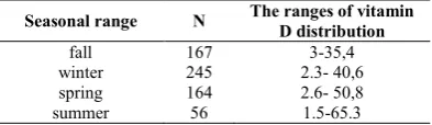
Table 2 shows range distribution of vitamin D values in pregnant women in different seasons. Seasonal vitamin D deficiency distributions are shown in tables 3,4,5,6. VDD reaches a summit in winter and decreases progressively until late summer. When VD values of pregnant patients in winter season were compared to fall VD values there is a statistically significant difference (p 0.5) Vitamin D values rose progressively through summer and reached a peak in fall self evidently
Health organizations recommendations for VD varies between 200 IU/day and 400IU/day for maintenance. But many researchers advocate optimum VD levels 30 ng/ML or higher. To attain this levels they recommend 1000-1600 IU/day VD intake.(76)(Dawson-Hughes 2005)
The requirement in pregnancy for both therapy and maintenance is not clear. Some researchers propose 1000 IU daily intake to reach levels above 20 ng/ml. (77) (HOLLİS 2004)
The Endocrine Society suggests that pregnant and lactating women require at least 600 IU/d of vitamin D and recognize that at least 1500–2000 IU/d may be needed tomaintain sufficient blood concentrations of 25(OH)D.(78)Holick
On the other hand daily clinical practice does not incorporate holistic approach and guidelines foroptimal sun exposure for pregnant women.(79) Beltran AJ
CONCLUSIONS AND RECOMMENDATIONS
VDD seems highly prevalent at the present time so health authorities should search and take the necessary steps to prevent the still high VDD in pregnancy when compared with former years.
References
1. Holden JM1, Lemar LE, Exler J. Vitamin D in foods: development of the US Department of Agriculture database Am J Clin Nutr. 2008 Apr; 87(4):1092S-6S. 2. Holick MF. Vitamin D deficiency. New England
Journal ofMedicine 2007;357(3):266-281;
3. Holick MF. Vitamin D deficiency: a worldwide problemwith health consequences. American Journal of ClinicalNutrition 2008; 87(4):1080S-1086S.
4. Hollis B. Vitamin D requirement during pregnancy andlactation. Journal of Bone and Mineral Research 2007; 22Suppl 2:V39-V44.
5. DeLuca HF. Overview of general physiologic featuresand functions of vitamin D. American Journal of ClinicalNutrition 2004; 80(6 Suppl):1689S-1696S. 6. Godar DE, Pope SJ, Grant WB, Holick MF. Solar UV
doses of youngAmericans and vitamin D3 production. Environ Health Perspect 2012; 120:139-143.
7. Juzeniene A, Brekke P, Dahlback A. Solar radiation and human health. Rep Prog Phys 2011; 74: 1–56.
8. Webb AR, Kift R, Berry JL, Rhodes LE. The vitamin D debate: translating controlledexperiments into reality for human sun exposure times. Photochem Photobiol 2011; 87: 741–745.
9. 9Armas LA, Hollis BW, Heaney RP. Vitamin D2 is much lesseffective than vitamin D3 in humans. Journal of Clinical Endocrinology and Metabolism 2004; 89(11):5387-91
10. 10Logan VF, Gray AR, Peddie MC, Harper MJ, Houghton LA. Long-term vitamin D3 supplementation is more effective than vitamin D2 in maintaining serum 25-hydroxyvitamin D status over the winter months. British Journal of Nutrition 2013;109(6):1082-8
11. McCullough M. Vitamin D deficiency in pregnancy: bringing the issues to light. Journal of Nutrition 2007; 137:305-6.
12. Theodoropoulos C, Demers C, Delvin E, Ménard D, Gascon-Barré M. Calcitriol regulates the expression of thegenes encoding the three key vitamin D3 hydroxylases andthe drug-metabolizing enzyme CYP3A4 in the human fetalintestine. Clinical Endocrinology 2003;58(4):489-99.
Table 1 Distribution of vitamin D values in all pregnant women between 15-43 years old (Total 632 pregnant
women)
Vitamin D value Number of pregnant
women %
0 - 5 ng/ml 163 25,8
5 -10 ng/ml 263 41,6
10 -15 ng/ml 98 15,5
15 - 20 ng/ml 52 8,2
>20 ng/ml 56 8,9
Table 2 Seasonal ranges of vitamin D values
Seasonal range N The ranges of vitamin
D distribution
fall 167 3-35,4
winter 245 2.3- 40,6
spring 164 2.6- 50,8
summer 56 1.5-65.3
Table 3
Vitamin D values in spring %
0 - 10 ng/ml 109 66,5
10 - 20 ng/ml 42 25,6
>20 ng/ml 13 7,9
Total n 164 pregnant
Table 4
Vitamin D values in summer %
10-10 ng/ml 25 44,6
10 - 20 ng/ml 18 32,1
>20 ng/ml 13 23,2
Total n 56 pregnant
Table 5
Vitamin D values in fall %
0 -10 ng/ml 108 64,7
10- 20 ng/ml 40 24,0
Table 6
Vitamin D values in winter %
0 - 10 ng/ml 182 74,3
10 - 20 ng/ml 51 20,8
>20 ng/ml 12 4,9
17554 |
P a g e
13. Clifton-Bligh RJ, McElduff P, McElduff A. Maternalvitamin D deficiency, ethnicity and gestational diabetes. Diabetic Medicine 2008; 25(6):678-84. 14. Maghbooli Z, Hossein-Nezhad A, Karimi F, Shafaei
AR, Larijani B. Correlation between vitamin D3 deficiency and insulin resistance in pregnancy. Diabetes/ Metabolism Research and Reviews 2008; 24(1):27-32. 15. Palomer X, González-Clemente JM, Blanco-Vaca F,
Mauricio D. Role of vitamin D in the pathogenesis of type2 diabetes mellitus. Diabetes, Obesity and Metabolism 2008; 10(3):185-97.
16. Xuan Y, Zhao HY, Liu JM. Vitamin D and type 2 diabetes mellitus (D2). Journal of Diabetes 2013;5(3):261-7
17. Institute of Medicine 2010Institute of Medicine 2010 Food, Nutrition Board. Institute of Medicine. Dietary Reference Intakes for Calcium and Vitamin D. WashingtonDC: National Academy Press, 2010.
18. Dawson-Hughes B, Heaney RP, HolickMF, Lips P, MeunierPJ, Vieth R. Estimates of optimal vitamin D status. Osteoporosis International 2005; 16:713-6. 19. Dawson-Hughes B. Serum 25-hydroxyvitamin D and
functional outcomes in the elderly. American Journal of Clinical Nutrition 2008;88(2):527S-540S.
20. Drincic AT, Armas LA, Van Diest EE, Heaney RP.Volumetric dilution, rather than sequestration best explainsthe low vitamin D status of obesity. Obesity (Silver Spring, Md.) 2012;20(7):1444-8.
21. Arunabh S, Pollack S, Yeh J, Aloia JF. Body fat content and25-hydroxyvitamin D levels in healthy women. Journal of Clinical Endocrinology and Metabolism 2003;88(1):157-61.
22. Vilarrasa N, Maravall J, Estepa A, Sánchez R, MasdevallC, Navarro MA, et al. Low 25-hydroxyvitamin D concentrations in obese women: their clinical significance and relationship with anthropometric and body composition variables. Journal of Endocrinological Investigation 2007;30(8):653-8. 23. Vimaleswaran KS, Berry DJ, Lu C, Tikkanen E, Pilz
S,Hiraki LT, et al. Causal relationship between obesity andvitamin D status: bi-directional Mendelian randomization analysis of multiple cohorts.. PLoS Medicine 2013; 10(2):e1001383.
24. Wortsman J, Matsuoka LY, Chen TC, Lu Z, Holick MF. Decreased bioavailability of vitamin D in obesity. AmericanJournal of Clinical Nutrition 2000; 72(3):690-3.
25. Nassar N, Halligan GH, Roberts CL, Morris JM, Ashton AW. Systematic review of first-trimester vitamin D normative levels and outcomes of pregnancy. Am J Obstet Gynecol 2011; 205: 208
26. Aghajafari F, Nagulesapillai T, Ronksley PE, Tough SC, O’ Beirne M, Rabi DM. Association between maternalserum 25-hydroxyvitamin D level and pregnancy and neonatal outcomes: systematic review and meta-analysis of observational studies. BMJ 2013;346:f1169
27. Bodnar LM, Catov JM, Simhan HN, Holick MF, PowersRW, Roberts JM. Maternal vitamin D deficiency increasesthe risk of preeclampsia. Journal of Clinical Endocrinology and Metabolism 2007;92(9):3517-22
28. Cardus A, Parisi E, Gallego C, Aldea M, Fernandez E, Valdivielso JM. 1, 25 Dihydroxyvitamin D3 stimulates vascular smooth muscle cell proliferation through a VEGF mediated pathway. Kidney International 2006; 69:1377-84.
29. Evans KN, Bulmer JN, Kilby MD, Hewison M. Vitamin Dand placental-decidual function. Journal of the Society for Gynecologic Investigation 2004; 11(5):263-71. 30. Hewison M. Vitamin D and the immune system. Journal
of Endocrinology 1992;132(2):173-6.
31. Li 2002Li Y, Kong J, Wei M, Chen ZF, Liu S, Cao LP. 1,25- dihydroxy vitamin D3 is a negative endocrine regulatorof the renin-angiotensin system. Journal of ClinicalInvestigation 2002;110(2):229-39.
32. Farrant HJ, Krishnaveni GV, Hill JC, Boucher BJ, FisherDJ, Noonan K, et al. Vitamin D insufficiency is commonin Indian mothers but is not associated with gestational diabetes or variation in newborn size. European Journal of Clinical Nutrition 2009;63(5):646-52.
33. Zhang C, Qiu C, Hu FB, David RM, Van Dam RM, Bralley A, et al. Maternal plasma 25-hydroxyvitamin Dconcentrations and the risk for gestational diabetes mellitus. PLoS ONE 2008;3(11):e3753.
34. Merewood A, Mehta SD, Chen TC, Bauchner H, HolickMF. Association between vitamin D deficiency and primary cesarean section. Journal of Clinical Endocrinology and Metabolism 2009;94(3):940-5. 35. Scholl 2012Scholl TO, Chen X, Stein P. Maternal
vitamin D status and delivery by cesarean. Nutrients 2012;4(4):319-30.
36. El Koumi MA, Ali YF, Abd El Rahman RN. Impact of maternal vitamin D status during pregnancy on neonatal vitamin D status. Turkish Journal of Pediatrics 2013;55(4): 371-7.
37. Glerup H, Mikkelsen K, Poulsen L, Hass E, Overbeck S,Andersen H, et al. Hypovitaminosis D myopathy without biochemical signs of osteomalacic bone involvement. Calcified Tissue International 2000;66(6):419-24.
38. Lips P. Vitamin D deficiency and secondary hyperparathyroidism in the elderly: consequences for bone loss and fractures and therapeutic implications. Endocrine Reviews 2001;22(4):477-501.
39. Dawodu A, Nath R. High prevalence of moderately severe vitamin D deficiency in pre-term infants. Pediatrics International 2011; Vol. 53, issue 2:207-10. 40. Morley R, Carlin JB, Pasco JA, Wark JD. Maternal
25-hydroxyvitamin D and parathyroid hormone concentrations and offspring birth size. Journal of Clinical Endocrinology and Metabolism 2006;91(3):906-12.
41. Wei SQ, Qi HP, Luo ZC, Fraser WD. Maternal vitamin Dstatus and adverse pregnancy outcomes: a systematic reviewand meta-analysis. Journal of Maternal-fetal & Neonatal Medicine 2013; 26(9):889-99.
43. Miller J, Gallo RL. Vitamin D and innate immunity. Dermatologic Therapy 2010;23(1):13-22.
44. Walker VP, Modlin RL. The vitamin D connection to pediatric infections and immune function. Pediatrics Research 2009;65(5 Pt 2):106R-113R
45. McGrath J. Does ’imprinting’ with low prenatal vitamin Dcontribute to the risk of various adult disorders?. MedicalHypotheses 2001; 56(3):367-71.
46. Moon R, Harvey N, Cooper C. Endocrinology in pregnancy: influence of maternal vitamin D status on obstetric outcomes and the foetal skeleton. Eur J Endocrinol 2015; 173: R69-R83
47. Bener A, Alsaied A, Al-Ali M, Al-Kubaisi A, Basha B,A braham A, et al. High prevalence of vitamin D deficiencyin type 1 diabetes mellitus and healthy children. Acta Diabetologica 2009;4(2):183-9.
48. Miller J, Gallo RL. Vitamin D and innate immunity. Dermatol Ther 2010 Jan feb ; 23(1):13-22
49. Pierrot-Deseilligny C, Souberbielle JC. Is hypovitaminosis D one of the environmental risk factors for multiplesclerosis?. Brain 2010; 133(Pt 7):1869-88. 50. Meral G, Uslu A, Yozgatli AU Tuna HTYilmazbas
NPAkcay F et al The Relationship between Vitamin D, Asthma and Total IgE in Children Ethno Med,11:91-97, 2017
51. Chocano-Bedoya P, Ronnenberg AG. Vitamin D andtuberculosis. Nutrition Reviews 2009; 67(5):289-93. 52. Hall WB, Sparks AA, Aris RM. Vitamin D deficiency in
cystic fibrosis. International Journal of Endocrinology 2010 [Epub ahead of print].
53. Nnoaham KE, Clarke A. Low serum vitamin D levelsand tuberculosis: a systematic review and meta-analysis. International Journal of Epidemiology 2008; 37(1):113-9.
54. Williams B, Williams AJ, Anderson ST. Vitamin Ddeficiency and insufficiency in children with tuberculosis. Pediatric Infectious Disease Journal 2008; 27(10):941-2.
55. Barker DJ. The fetal and infant origins of adult disease. BMJ: British Medical Journal.1990; 301:1111.
56. Devereux G, Litonjua AA, Turner SW, Craig LC, McNeillG, Martindale S, et al. Maternal vitamin D intake duringpregnancy and early childhood wheezing. American Journal of Clinical Nutrition 2007; 85(3):853-9.
57. Litonjua AA. Childhood asthma may be a consequence of vitamin D deficiency. Current Opinion in Allergy and linical immunology 2009; 9(3):202-7.
58. Eyles DW, Smith S, Kinobe R, Hewison M, McGrath JJ. Distribution of the Vitamin Dreceptor and 1α-hydroxylase in human brain. Journal of Chemical Neuroanatomy. 2005; 29:21-30.
59. Eyles DW, Burne THJ, McGrath JJ. Vitamin D, effects on brain development, adult brain function and the links between low levels of vitamin D and neuropsychiatric disease. Frontiers in Neuroendocrinology. 2013; 34:47-64.
60. Brown J, Bianco JI, McGrath JJ, Eyles DW. 1, 25-dihydroxyvitamin D3 induces nerve growth factor, promotes neurite outgrowth and inhibits mitosis in
embryonic rat hippocampalneurons. Neuroscience letters. 2003; 343:139-43.
61. Veenstra TD, Prufer K, Koenigsberger C, Brimijoin SW, Grande JP, Kumar R. 1,25-Dihydroxyvitamin D3 receptors in the central nervous system of the rat embryo. Brain research.1998; 804:193-205.
62. DeLuca G, Kimball S, Kolasinski J, Ramagopalan S, Ebers G. Review: the role of vitaminD in nervous system health and disease. Neuropathology and applied neurobiology.2013;39:458-84.
63. Moon R, Harvey N, Cooper C. Endocrinology in pregnancy: influence of maternal vitamin D status on obstetric outcomes and the foetal skeleton. Eur J Endocrinol 2015; 173
64. Ota K, Dambaeva S, Han AR, Beaman K, Gilman-Sachs A, Kwak-Kim J. Vitamin D deficiency may be a risk factor for recurrent pregnancy losses by increasing cellular immunity and autoimmunity. Hum Reprod 2014;29:208-219.
65. Ota K, Dambaeva S, KimMW, Han AR, Fukui A, Gilman-Sachs A, et al. 1,25 Dihydroxyvitamin D3 regulates NK-cell cytotoxicity, cytokine secretion, and degranulation in women with recurrent pregnancy losses. Eur J Immunol 2015;45:3188-3199
66. Bandeira F, Griz L, Dreyer P, Eufrazino C, Bandeira C,Freese E. Vitamin D deficiency: a global perspective [Deficiência de vitamina D: uma perspectiva global]. Arquivos Brasileiros de Endocrinologia e Metabologia 2006; 50(4):640-6.;
67. Palacios C, Gonzalez L. Is vitamin D deficiency amajor global public health problem?. Journal of Steroid Biochemistry and Molecular Biology 2014; 144(Pt A):138–45.
68. van Schoor NM, Lips P. Worldwide vitamin D status. BestPractice & Research. Clinical Endocrinology & Metabolism2011; 25(4):671-80.
69. Jiang L, Xu J, Pan S, Xie E, Hu Z, Shen H. High prevalence of hypovitaminosis D among pregnant women in southeast China. Acta Paediatr 2012; 101: e192–e194
70. Halicioglu O, Aksit S, Koc F, Akman SA, Albudak E, Yaprak I et al. Vitamin D deficiency in pregnant women and their neonates in spring time inwestern Turkey. Paediatr Perinat Epidemiol 2012; 26: 53–60.
71. Haliloglu B, Ilter E, Aksungar FB, Celik A, Coksuer H, Gunduz T et al. Bone turnoverand maternal 25(OH) vitamin D3 levels during pregnancy and the postpartumperiod: should routine vitamin D supplementation be increased inpregnant women? Eur J Obstet Gynecol Reprod Biol 2011; 158: 24-27.
72. Parildar H, Dogruk Unal A, Aksan Desteli G, Cigerli O, Guvener Demirag N. Frequency of vitamin D deficiency in pregnant diabetics at Baskent University Hospital, Istanbul. Pak J Med Sci 2013; 29: 15-20.
73. Ustuner I, Keskin HL, Tas EE, Neselioglu S, Sengul O, Avsar AF. Maternal serum 25(OH)D levels in the third trimester of pregnancy during the winter season. J Matern Fetal Neonatal Med 2011; 24: 1421-1426. 74. Pehlivan I, Hatun S, Aydoğan M, Babaoğlu K, Gökalp
17556 |
P a g e
supplementation in healthy infants. Turk J Pediatr 2003; 45: 315-320.
75. Meral G, Guven A, Uslu A Can G, Yaprak P, Akcay F et al. The prevalence of Vitamin D Deficiency in Children, Adolescents and Adults in a sample of Turkish Population. Ethno Med, 2016:10829;249-254
76. Dawson-Hughes B, Heaney RP, Hollick MF, Lips P,Meurier PJ, Vieth R. Estimates of vitamin D status. Osteoporosis international 2005 ;16: 713-6
77. Hollis BW, Wagner CL. Vitamin D requirements during lactation: high dose maternal supplementation as therapy to prevent hypovitaminosos for both the mother and the nursing infant. D. American Journal of Clinical Nutrition:2004;80 17525-17585
78. Holick MF, Binkley NC, Bischoff-Ferrari HA, Gordon CM, Hanley DA, Heaney RP et al. Evaluation, treatment, and prevention of vitamin D deficiency: an Endocrine Society clinical practice guideline. J Clin Endocrinol Metab 2011; 96: 1911-1930
79. Beltran AJ, Wu J, Laurent O. Associations of meteorology with adverse pregnancy outcomes: a systematic review of preeclampsia, preterm birth and birth weight. Int J Environ Res Public Health 2014; 11: 91-172
*******
How to cite this article: