7-Methoxyindan-1-one
Yuan Jay Changaand Kew-Yu Chenb*
aDepartment of Chemistry, Tung Hai University, 407 Taichung, Taiwan, and bDepartment of Chemical Engineering, Feng Chia University, 40724 Taichung,
Taiwan
Correspondence e-mail: [email protected]
Received 26 September 2012; accepted 27 September 2012
Key indicators: single-crystal X-ray study;T= 297 K; mean(C–C) = 0.002 A˚; Rfactor = 0.037;wRfactor = 0.116; data-to-parameter ratio = 15.1.
In the title compound, C10H10O2, the 1-indanone unit is essentially planar (r.m.s. deviation = 0.028 A˚ ). In the crystal, molecules are linkedviaC—H O hydrogen bonds, forming layers lying parallel to the ab plane. This two-dimensional structure is stabilized by a weak C—H interaction. A second weak C—H interaction links the layers, forming a three-dimensional structure.
Related literature
For the preparation of the title compound, see: Liet al.(2011). For applications of indanone derivatives, see: Borge et al.
(2010); Caiet al.(2005); Cuiet al.(2009); Fu & Wang (2008); Li
et al.(2009); Sousaet al.(2011); Tanget al.(2011). For related structures, see: Aliet al.(2010a,b,c,d); Chenet al.(2011a,b). For C—H O hydrogen bonds, see: Liet al.(2011a,b); Wang & Chen (2011); Xiet al.(2010).
Experimental
Crystal data
C10H10O2
Mr= 162.18
Orthorhombic,Pbca a= 8.5386 (7) A˚
b= 10.4949 (9) A˚
c= 18.8536 (16) A˚
V= 1689.5 (2) A˚3
Z= 8
MoKradiation = 0.09 mm1
T= 297 K
0.640.550.32 mm
Data collection
Bruker SMART CCD area-detector diffractometer
Absorption correction: multi-scan (SADABS; Bruker, 2001)
Tmin= 0.683,Tmax= 1.000
8807 measured reflections 1663 independent reflections 1278 reflections withI> 2(I)
Rint= 0.032
Refinement
R[F2> 2(F2)] = 0.037
wR(F2) = 0.116
S= 1.02 1663 reflections
110 parameters
H-atom parameters constrained max= 0.20 e A˚3
min=0.13 e A˚3
Table 1
Hydrogen-bond geometry (A˚ ,).
Cg1 is the centroid of the C1/C5–C9 ring.
D—H A D—H H A D A D—H A
C3—H3B O2i
0.97 2.60 3.5183 (18) 159
C7—H7A O1ii
0.93 2.57 3.4802 (18) 167
C10—H10B O1iii
0.96 2.59 3.486 (2) 156
C4—H4A Cg1iv 0.97 2.80 3.6430 (16) 146 C10—H10A Cg1v
0.96 2.82 3.6260 (16) 143
Symmetry codes: (i) xþ1 2;y
1
2;z; (ii) x1;y;z; (iii) x 1 2;y;zþ
1 2; (iv)
x;yþ1;z; (v)x;yþ1 2;zþ
1 2.
Data collection:SMART(Bruker, 2001); cell refinement:SAINT
(Bruker, 2001); data reduction:SAINT; program(s) used to solve structure:SHELXS97(Sheldrick, 2008); program(s) used to refine structure: SHELXL97 (Sheldrick, 2008); molecular graphics:
ORTEP-3 for Windows(Farrugia, 1997); software used to prepare material for publication: WinGX publication routines (Farrugia, 1999).
This work was supported by the National Science Council, Tung Hai University and Feng Chia University in Taiwan.
Supplementary data and figures for this paper are available from the IUCr electronic archives (Reference: ZL2507).
References
Ali, M. A., Ismail, R., Choon, T. S., Rosli, M. M. & Fun, H.-K. (2010a).Acta Cryst.E66, o2878.
Ali, M. A., Ismail, R., Tan, S. C., Quah, C. K. & Fun, H.-K. (2010b).Acta Cryst.
E66, o2875.
Ali, M. A., Ismail, R., Tan, S. C., Yeap, C. S. & Fun, H.-K. (2010c).Acta Cryst.
E66, o2753.
Ali, M. A., Ismail, R., Tan, S. C., Yeap, C. S. & Fun, H.-K. (2010d).Acta Cryst.
E66, o2864.
Borge, J., Cadierno, V., Dı´ez, J., Garcı´a-Garrido, S. E. & Gimeno, J. (2010).
Dyes Pigm.87, 209–217.
Bruker (2001).SMART,SAINTandSADABS. Bruker AXS Inc., Madison, Wisconsin, USA.
Cai, X., Wu, K. & Dolbier, W. R. Jr (2005).J. Fluor. Chem.126, 479–482. Chen, K.-Y., Fang, T.-C. & Chang, M.-J. (2011a).Acta Cryst.E67, o992. Chen, K.-Y., Wen, Y.-S., Fang, T.-C., Chang, Y.-J. & Chang, M.-J. (2011b).Acta
Cryst.E67, o927.
Cui, Y., Ren, H., Yu, J., Wang, Z. & Qian, G. (2009).Dyes Pigm.81, 53–57. Farrugia, L. J. (1997).J. Appl. Cryst.30, 565.
Farrugia, L. J. (1999).J. Appl. Cryst.32, 837–838. Fu, T. L. & Wang, I. J. (2008).Dyes Pigm.76, 590–595.
Li, C. J., Feng, Y. Q., Liu, X. J. & Zhang, T. Y. (2011a).Chin. Chem. Lett.22, 539–542.
Li, X., Kim, S.-H. & Son, Y.-A. (2009).Dyes Pigm.82, 293–298.
Li, Z., Lin, Y., Xia, J.-L., Zhang, H., Fan, F., Zeng, Q., Feng, D., Yin, J. & Liu, S. H. (2011).Dyes Pigm.90, 245–252.
Li, H. Q., Zhang, Z. B. & Li, L. (2011b).Chin. Chem. Lett.22, 280–283. Sheldrick, G. M. (2008).Acta Cryst.A64, 112–122.
Sousa, C. M., Berthet, J., Delbaere, S. & Coelho, P. J. (2011).Dyes Pigm.92, 537–541.
Tang, K.-C., Chang, M.-J., Lin, T.-Y., Pan, H.-A., Fang, T.-C., Chen, K.-Y., Hung, W.-Y., Hsu, Y.-H. & Chou, P.-T. (2011).J. Am. Chem. Soc.133, 17738– 17745.
Wang, E. J. & Chen, G. Y. (2011).Chin. Chem. Lett.22, 847–850. Xi, H. T., Yi, T. T. & Sun, X. Q. (2010).Chin. Chem. Lett.21, 633–636.
Structure Reports Online
supporting information
Acta Cryst. (2012). E68, o3063 [doi:10.1107/S1600536812040743]
7-Methoxyindan-1-one
Yuan Jay Chang and Kew-Yu Chen
S1. Comment
Indanone and its derivatives are some of the most widely used organic compounds (Tang et al., 2011). They are used as
dyes and pigments (Cui et al., 2009; Li et al., 2009), intermediates in organic synthesis (Fu & Wang, 2008; Borge et al.,
2010) and exhibit a wide variety of biological activities (Sousa et al., 2011). In addition, 1-indanones were important
precursors in the regiospecific synthesis of 2-fluoro-1-naphthols (Cai et al., 2005).
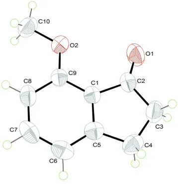
The molecular structure of the title compound is shown in Figure 1. The 1-indaneone moiety is essentially planar (r.m.s.
deviation = 0.028 Å), which is consistent with previous studies (Chen et al., 2011a,b; Ali et al., 2010a,b,c,d). There are
three different kinds of C—H···O (Li et al., 2011a,b; Wang et al., 2011; Xi et al., 2010) hydrogen bonds (Table 1) in the
crystal structure (Figure 2). In addition, C—H···π hydrogen bonds further stabilize the crystal structure (2.80 Å for the C4
—H4A···Cg1 distance and 146° for the C4—H4A—Cg1i angle; 2.82 Å for the C10—H10A···Cg1 distance and 143° for
the C10—H10A—Cg1ii angle; Cg1 is the centroid of the C1/C5—C9 ring; symmetry codes: (i): -x, 1 - y,- z (ii): -x, 1/2 +
y, 1/2 - z).
S2. Experimental
The title compound was synthesized by the methylation of 7-hydroxyindan-1-one with methyl iodide (Li et al., 2011).
Colorless parallelepiped-shaped crystals suitable for the crystallographic study reported here were isolated over a period
of six weeks by slow evaporation from a chloroform solution.
S3. Refinement
The C bound H atoms were positioned geometrically (C—H = 0.93–0.97 Å) and allowed to ride on their parent atoms,
Figure 1
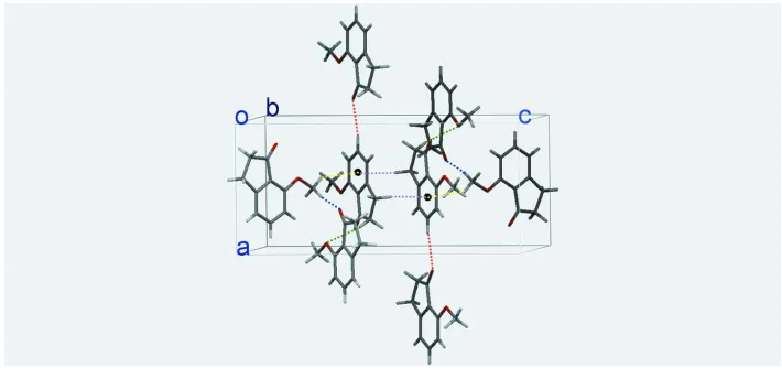
Figure 2
A section of the crystal packing of the title compound, viewed along the b axis. Blue, green and red dashed lines denote
the intermolecular C10—H10B···O1, C3—H3B···O2 and C7—H7A···O1 hydrogen bonds, respectively. Yellow and
purple dashed lines denote the intermolecular C10—H10A···π and C4—H4A···π hydrogen bonds, respectively. Cg1
(black circles) is the centroid of the C1/C5—C9 ring. For symmetry operators, see Table 1.
7-Methoxyindan-1-one
Crystal data
C10H10O2
Mr = 162.18
Orthorhombic, Pbca
Hall symbol: -P 2ac 2ab
a = 8.5386 (7) Å
b = 10.4949 (9) Å
c = 18.8536 (16) Å
V = 1689.5 (2) Å3
Z = 8
F(000) = 688
Dx = 1.275 Mg m−3
Mo Kα radiation, λ = 0.71073 Å Cell parameters from 3629 reflections
θ = 2.9–26.0°
µ = 0.09 mm−1
T = 297 K
Parallelepiped, colorless 0.64 × 0.55 × 0.32 mm
Data collection
Bruker SMART CCD area-detector diffractometer
Radiation source: fine-focus sealed tube Graphite monochromator
phi and ω scans
Absorption correction: multi-scan (SADABS; Bruker, 2001)
Tmin = 0.683, Tmax = 1.000
8807 measured reflections 1663 independent reflections 1278 reflections with I > 2σ(I)
Rint = 0.032
θmax = 26.0°, θmin = 2.2°
h = −10→10
k = −12→12
l = −16→23
Refinement
Refinement on F2
Least-squares matrix: full
R[F2 > 2σ(F2)] = 0.037
wR(F2) = 0.116
S = 1.02 1663 reflections 110 parameters 0 restraints
Primary atom site location: structure-invariant direct methods
Secondary atom site location: difference Fourier map
Hydrogen site location: inferred from neighbouring sites
H-atom parameters constrained
Δρmax = 0.20 e Å−3
Δρmin = −0.13 e Å−3
Fc*=kFc[1+0.001xFc2λ3/sin(2θ)]-1/4
Extinction coefficient: 0.0068 (15)
Special details
Geometry. All e.s.d.'s (except the e.s.d. in the dihedral angle between two l.s. planes) are estimated using the full covariance matrix. The cell e.s.d.'s are taken into account individually in the estimation of e.s.d.'s in distances, angles and torsion angles; correlations between e.s.d.'s in cell parameters are only used when they are defined by crystal symmetry. An approximate (isotropic) treatment of cell e.s.d.'s is used for estimating e.s.d.'s involving l.s. planes.
Refinement. Refinement of F2 against ALL reflections. The weighted R-factor wR and goodness of fit S are based on F2,
conventional R-factors R are based on F, with F set to zero for negative F2. The threshold expression of F2 > σ(F2) is used
only for calculating R-factors(gt) etc. and is not relevant to the choice of reflections for refinement. R-factors based on F2
are statistically about twice as large as those based on F, and R- factors based on ALL data will be even larger.
Fractional atomic coordinates and isotropic or equivalent isotropic displacement parameters (Å2)
x y z Uiso*/Ueq
O1 0.34168 (11) 0.54158 (11) 0.13950 (7) 0.0741 (4)
O2 0.07222 (10) 0.67528 (9) 0.20144 (5) 0.0545 (3)
C1 0.06609 (14) 0.50056 (11) 0.12296 (6) 0.0412 (3)
C2 0.23519 (16) 0.47945 (13) 0.11460 (7) 0.0480 (3)
C3 0.25583 (19) 0.36302 (14) 0.06788 (8) 0.0637 (4)
H3A 0.3207 0.3835 0.0272 0.076*
H3B 0.3057 0.2948 0.0943 0.076*
C4 0.09387 (18) 0.32271 (14) 0.04396 (8) 0.0606 (4)
H4A 0.0829 0.3318 −0.0070 0.073*
H4B 0.0734 0.2348 0.0568 0.073*
C5 −0.01569 (17) 0.41149 (12) 0.08242 (7) 0.0475 (3)
C6 −0.17764 (18) 0.41110 (14) 0.08112 (8) 0.0610 (4)
H6A −0.2322 0.3522 0.0538 0.073*
C7 −0.25596 (18) 0.49998 (16) 0.12127 (8) 0.0643 (5)
H7A −0.3649 0.5006 0.1207 0.077*
C8 −0.17749 (16) 0.58882 (14) 0.16256 (7) 0.0560 (4)
H8A −0.2340 0.6470 0.1896 0.067*
C9 −0.01507 (15) 0.59128 (12) 0.16371 (6) 0.0431 (3)
C10 −0.0085 (2) 0.76805 (14) 0.24349 (8) 0.0662 (4)
H10A 0.0664 0.8210 0.2674 0.099*
H10B −0.0729 0.7255 0.2779 0.099*
H10C −0.0730 0.8198 0.2134 0.099*
Atomic displacement parameters (Å2)
U11 U22 U33 U12 U13 U23
O1 0.0400 (6) 0.0783 (8) 0.1039 (9) 0.0010 (5) 0.0001 (6) −0.0157 (6)
O2 0.0529 (6) 0.0558 (6) 0.0548 (6) 0.0059 (4) −0.0009 (4) −0.0125 (4)
C1 0.0415 (7) 0.0426 (6) 0.0395 (6) 0.0022 (5) 0.0007 (5) 0.0071 (5)
C2 0.0418 (7) 0.0503 (7) 0.0520 (7) 0.0065 (6) 0.0016 (6) 0.0054 (6)
C3 0.0651 (9) 0.0637 (9) 0.0624 (8) 0.0227 (8) 0.0009 (7) −0.0052 (7)
C5 0.0565 (8) 0.0429 (7) 0.0432 (7) −0.0009 (6) −0.0041 (6) 0.0052 (5)
C6 0.0548 (9) 0.0610 (9) 0.0674 (9) −0.0121 (7) −0.0116 (7) 0.0017 (7)
C7 0.0380 (7) 0.0802 (11) 0.0747 (10) −0.0061 (7) −0.0015 (7) 0.0120 (8)
C8 0.0435 (8) 0.0664 (9) 0.0583 (8) 0.0090 (6) 0.0094 (6) 0.0041 (7)
C9 0.0440 (7) 0.0458 (7) 0.0393 (6) 0.0026 (5) 0.0020 (5) 0.0043 (5)
C10 0.0800 (10) 0.0591 (8) 0.0594 (9) 0.0161 (8) 0.0020 (8) −0.0128 (7)
Geometric parameters (Å, º)
O1—C2 1.2134 (17) C4—H4B 0.9700
O2—C9 1.3560 (15) C5—C6 1.383 (2)
O2—C10 1.4321 (16) C6—C7 1.375 (2)
C1—C5 1.3948 (17) C6—H6A 0.9300
C1—C9 1.4061 (17) C7—C8 1.387 (2)
C1—C2 1.4692 (18) C7—H7A 0.9300
C2—C3 1.517 (2) C8—C9 1.387 (2)
C3—C4 1.515 (2) C8—H8A 0.9300
C3—H3A 0.9700 C10—H10A 0.9600
C3—H3B 0.9700 C10—H10B 0.9600
C4—C5 1.5063 (19) C10—H10C 0.9600
C4—H4A 0.9700
C9—O2—C10 117.90 (11) C6—C5—C4 127.63 (13)
C5—C1—C9 120.43 (12) C1—C5—C4 111.55 (12)
C5—C1—C2 109.40 (11) C7—C6—C5 118.33 (13)
C9—C1—C2 130.17 (12) C7—C6—H6A 120.8
O1—C2—C1 127.87 (13) C5—C6—H6A 120.8
O1—C2—C3 124.79 (13) C6—C7—C8 122.01 (14)
C1—C2—C3 107.34 (12) C6—C7—H7A 119.0
C4—C3—C2 106.97 (12) C8—C7—H7A 119.0
C4—C3—H3A 110.3 C7—C8—C9 120.27 (13)
C2—C3—H3A 110.3 C7—C8—H8A 119.9
C4—C3—H3B 110.3 C9—C8—H8A 119.9
C2—C3—H3B 110.3 O2—C9—C8 124.74 (12)
H3A—C3—H3B 108.6 O2—C9—C1 117.13 (11)
C5—C4—C3 104.53 (11) C8—C9—C1 118.14 (12)
C5—C4—H4A 110.8 O2—C10—H10A 109.5
C3—C4—H4A 110.8 O2—C10—H10B 109.5
C5—C4—H4B 110.8 H10A—C10—H10B 109.5
C3—C4—H4B 110.8 O2—C10—H10C 109.5
H4A—C4—H4B 108.9 H10A—C10—H10C 109.5
C6—C5—C1 120.81 (13) H10B—C10—H10C 109.5
C5—C1—C2—O1 176.75 (14) C1—C5—C6—C7 −0.6 (2)
C9—C1—C2—O1 −3.7 (2) C4—C5—C6—C7 179.00 (13)
C5—C1—C2—C3 −3.15 (14) C5—C6—C7—C8 −0.1 (2)
C9—C1—C2—C3 176.41 (12) C6—C7—C8—C9 0.8 (2)
C2—C3—C4—C5 −4.24 (15) C7—C8—C9—O2 179.03 (12)
C9—C1—C5—C6 0.42 (18) C7—C8—C9—C1 −0.93 (19)
C2—C1—C5—C6 −179.97 (12) C5—C1—C9—O2 −179.63 (10)
C9—C1—C5—C4 −179.21 (11) C2—C1—C9—O2 0.84 (18)
C2—C1—C5—C4 0.41 (14) C5—C1—C9—C8 0.33 (17)
C3—C4—C5—C6 −177.12 (14) C2—C1—C9—C8 −179.19 (12)
C3—C4—C5—C1 2.47 (15)
Hydrogen-bond geometry (Å, º)
Cg1 is the centroid of the C1/C5–C9 ring.
D—H···A D—H H···A D···A D—H···A
C3—H3B···O2i 0.97 2.60 3.5183 (18) 159
C7—H7A···O1ii 0.93 2.57 3.4802 (18) 167
C10—H10B···O1iii 0.96 2.59 3.486 (2) 156
C4—H4A···Cg1iv 0.97 2.80 3.6430 (16) 146
C10—H10A···Cg1v 0.96 2.82 3.6260 (16) 143