Accepted Manuscript
Two-dimensional 1H and 1H-detected NMR study of a heterogeneous biocatalyst using fast MAS at high magnetic fields
Sabu Varghese, Peter J. Halling, Daniel Häussinger, Stephen Wimperis
PII: S0926-2040(18)30013-4 DOI: 10.1016/j.ssnmr.2018.03.003 Reference: YSNMR 832
To appear in: Solid State Nuclear Magnetic Resonance
Received Date: 5 February 2018 Revised Date: 13 March 2018 Accepted Date: 13 March 2018
Please cite this article as: S. Varghese, P.J. Halling, D. Häussinger, S. Wimperis, Two-dimensional 1H and 1H-detected NMR study of a heterogeneous biocatalyst using fast MAS at high magnetic fields,
Solid State Nuclear Magnetic Resonance (2018), doi: 10.1016/j.ssnmr.2018.03.003.
M
AN
US
CR
IP
T
AC
CE
PT
ED
ACCEPTED MANUSCRIPT
1
H MAS NMR at high spinning rates and high magnetic fields is
M
AN
US
CR
IP
T
AC
CE
PT
ED
Two-dimensional
1H and
1H-detected NMR study of a
heterogeneous biocatalyst using fast MAS at high magnetic fields
Sabu Varghese,*a Peter J. Halling,b Daniel Häussingerc and Stephen Wimperis*a
a
Department of Chemistry, Lancaster University, Lancaster LA1 4YB, UK
b
WestCHEM, Department of Pure & Applied Chemistry, University of Strathclyde, Glasgow G1 1XL, UK
c
Department of Chemistry, University of Basel, CH-4056 Basel, Switzerland
Keywords:
immobilized enzymes; heterogeneous biocatalysts; solid-state NMR, biocatalysis; 1H
MAS NMR; epoxy-functionalized silica; human carbonic anhydrase II; hCA II;
M
AN
US
CR
IP
T
AC
CE
PT
ED
Abstract
Nuclear magnetic resonance (NMR) is a powerful tool for investigating atomic-scale
structure in heterogeneous or composite materials where long-range order is absent.
In this work solid-state 1H and 1H-detected NMR experiments were performed with
fast magic angle spinning (νR = 75 kHz) and at high magnetic fields (B0 = 20 T) and
used to gain structural insight into a heterogeneous biocatalyst consisting of an
enzyme, human carbonic anhydrase II (hCA II), covalently immobilized on
epoxy-functionalized silica. Two-dimensional 1H-1H NOESY-type correlation experiments
were able to provide information on 1H environments in silica, epoxy-silica and the
immobilized enzyme. Two distinct signals originating from water protons were
observed: water associated with the surface of the silica and the water associated
with the immobilized enzyme. Additional two-dimensional 1H-1H double–single
quantum (DQ-SQ) correlation experiments suggested that the immobilized enzyme is
not in close contact with the silica surface. Most significantly, comparison of
two-dimensional 1H-15N spectra of the immobilized enzyme and the solution-state
enzyme confirmed that the structural integrity of the protein is well preserved upon
M
AN
US
CR
IP
T
AC
CE
PT
ED
Introduction
Protein immobilization on solid supports and surfaces plays a crucial role in a
range of technological applications including industrial biocatalysis, drug delivery,
medical diagnosis, and biosensing.1-2 Heterogeneous biocatalysis involves the
conversion of chemical or biological substances using immobilized enzymes or cells
3-4
and is employed in industrial applications for the large scale synthesis of a wide
variety of fine chemicals.5 Little is known about the atomic-level structure of
heterogeneous biocatalysts as conventional structural characterization methods,
such as X-ray crystallography and solution-state NMR, cannot be directly employed.
However, magic angle spinning NMR (MAS NMR) can be used to characterize
heterogeneous systems and has been demonstrated in the study of a variety of
immobilized enzymes and supports.6-15 In a similar context, it should be noted that
MAS NMR has also very been successfully employed in understanding the molecular
level interactions of peptides and proteins with non-biological surfaces involved in
biomineralization.16-26
Among the various means of immobilizing enzymes on solid supports,
covalent immobilization has proven to be the most stable as leaching of the enzyme
from the support is minimized. However, the state of the enzyme and changes in its
dynamics upon immobilization are not well understood. Recently, we have been able
to show that the structural integrity of an enzyme was not drastically changed upon
immobilization and was comparable to that in the lyophilized state by using a model
enzyme human carbonic anhydrase II (hCA II) covalently immobilized on
epoxy-silica.27 Since the lyophilized state of a protein may not necessarily be identical to the
native structure in solution, better insight into the native fold of the protein before and
M
AN
US
CR
IP
T
AC
CE
PT
ED
solution-state 1H-15N HSQC NMR spectrum with that of the immobilized enzyme. In
this research work, solid-state 1H and 1H-detected NMR experiments utilizing fast
MAS (νR = 75 kHz) at high magnetic fields (B0 = 20 T)28 have been employed to
characterize a model enzyme (hCA II) covalently immobilized on epoxy-silica. Protein
samples were prepared in both isotopic natural abundance and in isotopically
enriched (15N) states (uniformly labelled samples termed hereafter as [U-15N]/hCA II).
Furthermore, to reduce spectral overcrowding in multidimensional MAS NMR
experiments, hCA II samples were selectively 15N labelled for the most abundant
amino acid residue leucine (samples termed hereafter as [15N Leu]/hCA II). Our
results show that two-dimensional 1H MAS NMR methods can be successfully
employed with fast MAS to characterize the different proton environments in the silica
support, covalent linker, and the immobilized enzyme. Comparison of
two-dimensional 1H-15N spectrum from the immobilized enzyme and the solution-state
NMR spectrum confirms that the structural integrity of the protein is well preserved
upon covalent immobilization.
Experimental
Materials and sample preparation
The hCA II plasmid (pACA) used for the production of hCA II mutants was a
generous gift from Carol A. Fierke (University of Michigan, USA).29 Expression,
purification, and characterization of [U-15N]/hCA II and [15N Leu]/hCA II were
performed as described previously.27, 30 Synthesis of epoxy-silica using SP-100-15-P
Daiso silica gel and (3-glycidyloxypropyl)trimethoxysilane (GLYMO) as the covalent
linker, immobilization of [U-15N]/hCA II and [15N Leu]/hCA II on epoxy-silica, and the
M
AN
US
CR
IP
T
AC
CE
PT
ED
Solution and solid-state NMR experiments
Solution-state NMR experiments were carried out on a Bruker Avance III HD
spectrometer operating at 600 MHz 1H frequency, equipped with a cryogenic QCI
probe 1H/13C/15N/19F with z-axis pulsed field gradients. Solid-state NMR experiments
were performed on a Bruker Avance III 850 MHz spectrometer with a widebore 20 T
magnet (850 MHz 1H frequency). The dry powdered samples were packed into 1.0
mm ZrO2 rotors and were spun at a frequency of 75 kHz. Chemical shifts were
referenced externally relative for 1H (adamantane: 1.87 ppm) and 15N (glycine: 32.4
ppm). Two-dimensional 1H-1H correlation experiments were performed either with a
NOESY-type (Nuclear Overhauser Effect SpectroscopY) sequence consisting of
three simple 90° pulses31 or with a double–single quantum (DQ-SQ) sequence
utilizing BABA (BAck-to-BAck)32 recoupling. For two-dimensional 1H-15N correlation,
pulse sequences utilized linearly ramped cross-polarization (CP)33-35 and small phase
incremental alternation (SPINAL)36 1H decoupling. All experiments were performed at
room temperature. All NMR data were processed using TopSpin software. Further
experimental and processing details can be found in the text and figure captions.
Results and discussion
To gain greater insight into the 1H environments present during the different
stages of enzyme immobilization, we performed two-dimensional 1H-1H correlation
experiments on silica, epoxy-silica and immobilized hCA II using NOESY-type pulse
sequences. Fig. 1 shows two-dimensional 1H-1H NOESY spectra of (a) silica, (b)
epoxy-silica, (c) [U-15N]/hCA IIimmobilized on epoxy-silica and (d) [15N Leu]/hCA II
immobilized on epoxy-silica recorded using a mixing time of 100 ms on a B0 = 20 T
M
AN
US
CR
IP
T
AC
CE
PT
ED
diagonal peaks and off-diagonal or cross peaks; the latter may arise from either
chemical exchange or from spin diffusion, both incoherent processes, during the
mixing time. Cross peaks ascribed to the spin diffusion mechanism and with high
intensity may cautiously be interpreted as arising from two nuclei that possess a
strong mutual dipolar coupling and hence are close in space. Indeed, the 100 ms
mixing time duration was selected as a compromise between cross-peak intensity
and the desire to facilitate this interpretation. It has been reported that the use of
radiofrequency-driven recoupling (RFDR) during the NOESY mixing time can
increase the rate of 1H-1H polarization transfer.37
The NOESY spectrum of bare silica (Fig. 1a) features diagonal peaks
appearing from isolated silanol groups (iOH, 1.1 ppm), physisorbed water (H2O, 4.0
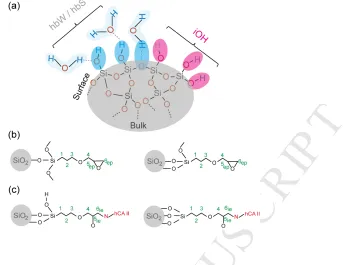
ppm) and hydrogen-bonded silanol groups (hBS, 4.5 to 9.0 ppm). The chemical shift
assignments use the labelling scheme shown in Fig. 2, where the simplified
representation of the hydrogen bonding of water with surface silanol groups is based
on previous reports.38 The peak from physisorbed water (4.0 ppm) is relatively
narrow when compared with the broad peak from the hBS (4.5 to 9.0 ppm) indicating
the presence of dynamics to average out anisotropic spin interactions. The hBS peak
broadening (4.5 to 9.0 ppm) can be ascribed to the absence of dynamics and the
inhomogeneous broadening associated with a wide range of chemical shifts
contributed by the different modes of hydrogen bonding between surface silanol
groups.39 Intense cross peaks can be observed (4.0 ppm – 4.5 to 9.0 ppm) between
physisorbed water and hBS (4.0 ppm – 4.5 to 9.0 ppm) and between physisorbed
water and iOH (1.1 – 4.0 ppm), indicating that all silanol groups are associated with
M
AN
US
CR
IP
T
AC
CE
PT
ED
The two-dimensional NOESY spectrum of epoxy-silica (Fig. 1b) is well
resolved and shows diagonal peaks from the epoxy-linker (0 – 3.5 ppm), physisorbed
water (4.0 ppm, a shoulder on the intense peak at 3.25 ppm from the epoxy-linker),
and hBS groups (4.5 – 10.0 ppm). The spectrum also reveals cross peaks between
protons in the epoxy-linker (with the green labels); these cross peaks may be
ascribed to spin diffusion as the linker protons are non-exchangeable. Cross peaks
are also observed between hBS and the 1, 2, 3, 4, 5 and SiOCH3 protons in the
epoxy-linker. This is probably a consequence of the relatively long mixing time (100
ms) used, with spin diffusion producing long-range magnetization transfer. It is worth
noting the weak cross peak (highlighted in yellow, but below the plotted contour
levels on the other side of the diagonal in Fig. 1b) between proton 6 from the
epoxy-ring (2.4 ppm) and the hBS groups (5.5 ppm), highlighted in yellow. This indicates
that some of the epoxy groups have opened to form diols and that the hydroxyl
protons are either in close proximity or chemical exchange with hBS groups on the
surface of silica. Finally, partly resolved cross peaks (0.8 – 4.0 ppm), (1.4 – 4.0 ppm),
(3.0 – 4.0 ppm) can be observed in Fig. 1b between the 1, 2, 3, 4, 5 and SiOCH3
protons in the epoxy-linker with the physisorbed water (highlighted in red). It is worth
noting the 0.8 ppm shift of the 1 protons in the cross peak with the water, rather than
the 0.4 ppm seen elsewhere, indicating a possible specific interaction with the
physisorbed water. However, no noticeable changes in chemical shifts could be
observed for the 2, 3, 4, 5 and SiOCH3 protons in the epoxy-linker. Note that,
although synthesis of epoxy-silica was performed under non-aqueous conditions, the
peak at 4.0 ppm can be ascribed to water physisorbed from the atmosphere due to
M
AN
US
CR
IP
T
AC
CE
PT
ED
The 1H NOESY spectra of [U-15N]/hCA II (Fig. 1c) and [15N Leu]/hCA II (Fig.
1d) immobilized on epoxy-silica are very similar, as expected, and reveal diagonal
peaks from the epoxy-linker (0 – 3.5 ppm), water (4.0, 5.0 ppm) and from the
aromatic and amide protons in the immobilized enzyme (6 – 10 ppm), highlighted
with green, blue and red labels respectively. It is worth noting the two distinct water
peaks at 4 and 5 ppm. The partly resolved water peak at 4 ppm can be ascribed to
water physisorbed onto the surface of the epoxy-silica, while the water peak at 5 ppm
can be ascribed to water associated with the immobilized enzyme
(protein-associated water is usually observed at ~4.7 ppm).40 These spectra also reveal cross
peaks between protons from the epoxy-linker (0 – 4 ppm), epoxy-linker and water (0
to 4 ppm – 5 ppm), epoxy-linker and hCA II (0 to 4 ppm – 6 to 10 ppm), and also
between water and hCA II (5 – 6 to 10 ppm).
To ascertain spatial proximities utilizing the 1H-1H dipolar coupling in a
coherent process, two-dimensional 1H double–single quantum (DQ-SQ) experiments
using BABA recoupling were performed. The DQ-SQ NMR experiments selectively
excite pairs of 1H nuclei that are strongly dipolar coupled and, as a result, mobile or
isolated protons are normally not observed. Figure 3 shows the 1H DQ-SQ NMR
spectra of (a) epoxy-silica, (b) [U-15N]/hCA IIimmobilized on epoxy-silica and (c) [15N
Leu]/hCA II immobilized on epoxy-silica. These spectra were recorded at room
temperature with one rotor period of recoupling on a B0 = 20 T magnet at a MAS
frequency of νR = 75 kHz. Our DQ-SQ experiments on silica were unsuccessful as
the recoupling was inefficient owing to the mobility of the water molecules (data not
shown).
The DQ-SQ spectrum of epoxy-silica (Fig. 3a) is well resolved and reveals
M
AN
US
CR
IP
T
AC
CE
PT
ED
diagonal, arising from protons that are close in space. The DQ-SQ correlation peaks
from protons in the epoxy-linker are shown connected by dotted green lines, while
the correlation peaks from protons in the epoxy-linker and the hBS groups are shown
connected by dotted cyan lines. Well-resolved pairs of correlation peaks are
observed for the 1 and 2 protons (F2: 0.4 –1.4 ppm), the 1 proton and SiOCH3 (F2:
0.4 – 3.1 ppm) and the 2 and 3 protons (F2: 1.4 – 3.3 ppm) corresponding to their
close proximities in space. The unsymmetrical peaks appearing around 3 ppm in F2
probably arise as a result of t1 noise from the adsorbed water.
The DQ-SQ spectra of [U-15N]/hCA IIimmobilized on epoxy-silica (Fig. 3b) and
[15N Leu]/hCA II immobilized on epoxy-silica (Fig. 3c) are very similar and consist of
pairs of correlation peaks from the epoxy-linker, protons 1 and 2 (F2: 0.4 – 1.4 ppm),
2 and 3 (F2: 1.4 – 3.3 ppm), plus a range of correlation peaks connecting protons 3,
4 and 5 and the immobilized hCA II (a few pairs are highlighted with dotted red lines).
Interestingly, we do not observe any correlation peaks connecting 1 and H2 protons
in the epoxy-linker with protons in the immobilized hCA II, indicating that these
protons are not close in space. These 1 and 2 protons in the epoxy linker are
expected to be found close to the silica surface and the absence of any correlation
peaks with the hCA II may indicate that immobilized enzyme is not in close contact
with the silica surface. Thus the epoxy-linker may be acting as a cushion between the
silica surface and the protein, thereby preventing any strong charge interactions that
might lead to protein denaturation. Similar studies involving reconstitution of
transmembrane proteins anchored into polymer-supported cushioned lipid bilayers
have shown increased incorporation and enhanced enzymatic activity when
M
AN
US
CR
IP
T
AC
CE
PT
ED
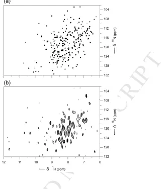
Finally, in order to help understand the state of the enzyme after
immobilization, a 1H-detected MAS NMR (1H-15N) correlation experiment was
performed at B0 = 20 T and a MAS frequency of νR = 75 kHz. Figure 4 compares the
resulting spectrum of [U-15N]/hCA II in the immobilized state (Fig. 4b) with the
solution-state TROSY-HSQC (Transverse Relaxation-Optimized Spectroscopy –
Heteronuclear Single Quantum Coherence)42 NMR spectrum of hCA II (Fig. 4a).
Comparison of the two spectra reveals that majority of the peaks in the central region
are well preserved, while the overall distribution of the peaks from the immobilized
hCA II indicates that the structural integrity of the enzyme is well preserved on
immobilization. Additional high frequency peaks (> 11 ppm in the 1H dimension) in
the immobilized state can be ascribed to strong hydrogen bonds in the solid that are
broken upon dissolution.
Overall, a smaller number of resonances are observed from the hCA II in the
immobilized state (Fig. 4b) compared with the solution-state HSQC NMR spectrum
(Fig. 4a). The solution-state HSQC NMR experiment is based on INEPT-type
(Insensitive Nuclei Enhanced by Polarization Transfer)43 coherence transfer through
J couplings, which works efficiently for many liquid samples. In solid-state NMR,
INEPT signals from proteins are normally observed from residues that have sufficient
mobility to average out anisotropic interactions and yield narrow resonances. We did
not observe any signals from immobilized hCA II using INEPT-based experiments
(data not shown). In contrast, 1H-detected MAS NMR experiments rely on the
method of cross polarization (CP) via heteronuclear through-space dipolar
couplings,33-35 which works efficiently for rigid solid samples. It seems likely that the
absence of many of the cross peaks in Fig. 4b can be attributed to static disorder
M
AN
US
CR
IP
T
AC
CE
PT
ED
previously in the case of proteins entrapped in bioinspired silica.23 It should be noted
that, although we are suggesting that the structural integrity of the enzyme is well
preserved on immobilization, this does not mean that the sample is not highly
disordered. In addition to some likely disorder of the tertiary structure, individual
protein molecules can be expected to be tethered to the silica surface by a variable
number of linker molecules and to have a very wide range of orientations with
respect to the surface, which itself will be highly heterogeneous. 1H-detected
experiments on [15N Leu]/hCA II immobilized on epoxy-silica were not successful
(data not shown) owing to the very much smaller number of expected resonances
(26 Leu residues) consequent upon selective 15N isotopic labelling and to the limited
experimental sensitivity arising from the smaller amount of protein grafted onto the
surface of the epoxy-silica support (9.9 mg of protein per 100 mg of support).27
Conclusions
In conclusion, we have demonstrated that 1H and 1H-detected solid-state NMR
experiments performed at high magnetic fields and utilizing fast MAS can be
successful in gaining insight into 1H environments in silica, epoxy-silica and enzymes
covalently immobilized on epoxy-silica. Most importantly, our results confirm our
earlier result that the structural integrity of the protein is not drastically changed, but
is well preserved upon covalent immobilization.27 This had added significance when
related to our previous observation that this immobilized enzymatic system retains
71% of its effective specific activity when compared with free hCA II in solution.27 We
believe that the outcomes of this present work are not limited to the better
M
AN
US
CR
IP
T
AC
CE
PT
ED
biotechnological processes and applications involving interactions of proteins with
solid surfaces and supports.
Conflicts of interest
There are no conflicts to declare.
Acknowledgements
We thank the Leverhulme Trust (award RPG-2013-361) for financial support. We are
indebted to Dr Kaspar Zimmermann for preparation of all protein constructs, Dr Elisa
Nogueira for assistance, and the Swiss National Science Foundation for a grant to
DH (SNF 200021_130263). The UK 850 MHz Solid-State NMR Facility used in this
research was funded by EPSRC and BBSRC (contract reference PR140003), as well
as the University of Warwick including via partial funding through Birmingham
Science City Advanced Materials Projects 1 and 2 supported by Advantage West
Midlands (AWM) and the European Regional Development Fund (ERDF).
Collaborative assistance from the 850 MHz Facility Manager (Dr. Dinu Iuga,
University of Warwick) is gratefully acknowledged. We also thank Daiso Chemical
Co. Ltd, Japan, for donating the silica support used in this research.
References
1. F. Rusmini, Z. Zhong and J. Feijen, Biomacromolecules, 2007, 8, 1775-1789.
2. A. Kuchler, M. Yoshimoto, S. Luginbuhl, F. Mavelli and P. Walde, Nat.
Nanotechnol., 2016, 11, 409-420.
3. D. N. Tran and K. J. Balkus, ACS Catal., 2011, 1, 956-968.
M
AN
US
CR
IP
T
AC
CE
PT
ED
5. R. DiCosimo, J. McAuliffe, A. J. Poulose and G. Bohlmann, Chem. Soc. Rev.,
2013, 42, 6437-6474.
6. P. V. Bower, E. A. Louie, J. R. Long, P. S. Stayton and G. P. Drobny,
Langmuir, 2005, 21, 3002-3007.
7. P. Xue, F. Xu and L. Xu, Appl. Surf. Sci., 2008, 255, 1625-1630.
8. M. Park, S. S. Park, M. Selvaraj, D. Zhao and C.-S. Ha, Microporous
Mesoporous Mater., 2009, 124, 76-83.
9. S. B. Hartono, S. Z. Qiao, J. Liu, K. Jack, B. P. Ladewig, Z. Hao and G. Q. M.
Lu, J. Phys. Chem. C, 2010, 114, 8353-8362.
10. T. Weidner, N. F. Breen, K. Li, G. P. Drobny and D. G. Castner, Proc. Natl.
Acad. Sci. U.S.A., 2010, 107, 13288-13293.
11. N. E. Fauré, P. J. Halling and S. Wimperis, J. Phys. Chem. C, 2014, 118,
1042-1048.
12. Z. Zhou, F. Piepenbreier, V. R. R. Marthala, K. Karbacher and M. Hartmann,
Catal. Today, 2015, 243, 173-183.
13. C. Guo and G. P. Holland, J. Phys. Chem. C, 2015, 119, 25663-25672.
14. J. M. Bolivar, I. Eisl and B. Nidetzky, Catal. Today, 2016, 259, Part 1, 66-80.
15. L. Cerofolini, S. Giuntini, A. Louka, E. Ravera, M. Fragai and C. Luchinat, J.
Phys. Chem. B, 2017, 121, 8094-8101.
16. G. Goobes, P. S. Stayton and G. P. Drobny, Prog. Nucl. Magn. Reson.
Spectrosc., 2007, 50, 71-85.
17. W. Y. Chow, R. Rajan, K. H. Muller, D. G. Reid, J. N. Skepper, W. C. Wong,
R. A. Brooks, M. Green, D. Bihan, R. W. Farndale, D. A. Slatter, C. M.
Shanahan and M. J. Duer, Science, 2014, 344, 742-746.
M
AN
US
CR
IP
T
AC
CE
PT
ED
19. M. J. Duer, J. Magn. Reson., 2015, 253, 98-110.
20. K. H. Mroue, Y. Nishiyama, M. K. Pandey, B. Gong, E. McNerny, D. H. Kohn,
M. D. Morris and A. Ramamoorthy, Sci. Rep. 2015, 5, 11991.
21. E. Ravera, V. K. Michaelis, T.-C. Ong, E. G. Keeler, T. Martelli, M. Fragai, R.
G. Griffin and C. Luchinat, ChemPhysChem, 2015, 16, 2751-2754.
22. T. Martelli, E. Ravera, A. Louka, L. Cerofolini, M. Hafner, M. Fragai, C. F. W.
Becker and C. Luchinat, Chem. Eur. J., 2016, 22, 425-432.
23. E. Ravera, L. Cerofolini, T. Martelli, A. Louka, M. Fragai and C. Luchinat, Sci.
Rep., 2016, 6, 27851.
24. E. Ravera, T. Martelli, Y. Geiger, M. Fragai, G. Goobes and C. Luchinat,
Coord. Chem. Rev., 2016, 327, 110-122.
25. S. I. Brückner, S. Donets, A. Dianat, M. Bobeth, R. Gutiérrez, G. Cuniberti and
E. Brunner, Langmuir, 2016, 32, 11698-11705.
26. X. Yang, F. Huang, X. Xu, Y. Liu, C. Ding, K. Wang, A. Guo, W. Li and J. Li,
Chem. Mater., 2017, 29, 5663-5670.
27. S. Varghese, P. J. Halling, D. Häussinger and S. Wimperis, J. Phys. Chem. C,
2016, 120, 28717-28726.
28. R. Zhang, K. H. Mroue and A. Ramamoorthy, Acc. Chem. Res., 2017, 50,
1105-1113.
29. S. K. Nair, T. L. Calderone, D. W. Christianson and C. A. Fierke, J. Biol.
Chem., 1991, 266, 17320-17325.
30. L. Zheng, U. Baumann and J.-L. Reymond, Nucleic Acids Res., 2004, 32,
e115.
31. J. Jeener, B. H. Meier, P. Bachmann, and R. R. Ernst, J. Chem. Phys., 1979,
M
AN
US
CR
IP
T
AC
CE
PT
ED
32. W. Sommer, J. Gottwald, D. E. Demco and H. W. Spiess, J. Magn. Reson.,
Ser. A, 1995, 113, 131-134.
33. A. Pines, M. G. Gibby and J. S. Waugh, J. Chem. Phys., 1973, 59, 569-590.
34. J. Schaefer and E. O. Stejskal, J. Am. Chem. Soc., 1976, 98, 1031-1032.
35. G. Metz, X. L. Wu and S. O. Smith, J. Magn. Reson., 1994, 110, 219-227.
36. B. M. Fung, A. K. Khitrin and K. Ermolaev, J. Magn. Reson., 2000, 142,
97-101.
37. M. K. Pandey, S. Vivekanandan, K. Yamamoto, S. Im, L. Waskell and A.
Ramamoorthy, J. Magn. Reson., 2014, 242, 169-179.
38. B. Grünberg, T. Emmler, E. Gedat, I. Shenderovich, G. H. Findenegg, H.-H.
Limbach and G. Buntkowsky, Chem. Eur. J, 2004, 10, 5689-5696.
39. G. E. Maciel, J. Am. Chem. Soc., 1996, 118, 401-406.
40. A. Lesage and A. Böckmann, J. Am. Chem. Soc., 2003, 125, 13336-13337.
41. L. Renner, T. Pompe, R. Lemaitre, D. Drechsel and C. Werner, Soft Matter,
2010, 6, 5382-5389.
42. K. Pervushin, R. Riek, G. Wider and K. Wüthrich, Proc. Natl. Acad. Sci.
U.S.A., 1997, 94, 12366-12371.
M
AN
US
CR
IP
T
AC
CE
PT
ED
Figure Legends
[image:18.595.103.499.129.532.2]Fig. 1. Two-dimensional 1H-1H NOESY-type spectra of (a) silica, (b) epoxy-silica, (c)
[U-15N]/hCA II immobilized on epoxy-silica and (d) [15N Leu]/hCA II immobilized on
epoxy-silica. Spectra were recorded using 100 ms mixing time at B0 = 20 T and a
MAS frequency of νR = 75 kHz. Spectra were acquired using 8 transients for each of
300 t1 increments of 53.3 µs using a recycle interval of 2 s. Spectrum (a) was
processed with 50 Hz line broadening in both F1 and F2 dimensions; the remainder
were processed using a resolution enhancement function (Bruker parameters LB = –
M
AN
US
CR
IP
T
AC
CE
PT
ED
use the scheme shown in Fig. 2. The cross peaks highlighted in red and yellow are
M
AN
US
CR
IP
T
AC
CE
PT
ED
Fig. 2. Simplified schematic representation of (a) bare silica, (b) epoxy-silica before
immobilization and (c) epoxy-silica after immobilization with hCA II. Two possible
[image:20.595.153.496.74.339.2]M
AN
US
CR
IP
T
AC
CE
PT
ED
Fig. 3. Two-dimensional 1H DQ-SQ spectra of (a) epoxy-silica, (b) [U-15N]/hCA II
immobilized on epoxy-silica and (c) [15N Leu]/hCA II immobilized on epoxy-silica
recorded at B0 = 20 T and a MAS rate of νR = 75 kHz. Spectra were acquired using
16 transients for each of the 128 t1 increments of 13.3 µs using a recycle interval of 2
s. Spectra were processed using a resolution enhancement function (Bruker
parameters LB = –150 Hz and GB = 0.2) in both F1 and F2 dimensions. The
[image:21.595.164.499.68.586.2]M
AN
US
CR
IP
T
AC
CE
PT
ED
Fig. 4. (a) Solution-state TROSY36-HSQC NMR spectrum of hCA II and (b) 1
H-detected (1H-15N) MAS NMR spectrum of [U-15N]/hCA II in the immobilized state. The
solution-state NMR spectrum of [U-15N]/hCA II was acquired at B0 = 14.1 T using 64
transients for each of the 256 t1 increments of 500 µs using a recycle interval of 1 s
magnet. The spectrum of the [U-15N]/hCA II in the immobilized state was acquired at
B0 = 20 T and a MAS frequency of νR = 75 kHz using 1024 transients for each of the
40 t1 increments of 290 µs using a recycle interval of 2 s. The spectra were
[image:22.595.175.499.75.451.2]M
AN
US
CR
IP
T
AC
CE
PT
ED
Two-dimensional 1H and 1H-detected NMR study of a heterogeneous biocatalyst using fast MAS at high magnetic fields
Highlights:
1. Solid-state 1H and 1H-detected NMR experiments were performed with fast magic angle
spinning (νR = 75 kHz) and at high magnetic fields (B0 = 20 T) and used to gain structural
insight into a heterogeneous biocatalyst
2. Two-dimensional 1H-1H NOESY-type correlation experiments were able to provide
information on 1H environments in silica, epoxy-silica and the immobilized enzyme.
3. Additional two-dimensional 1H-1H double–single quantum (DQ-SQ) correlation
experiments suggested that the immobilized enzyme is not in close contact with the silica surface.
4. Comparison of two-dimensional 1H-15N spectra of the immobilized enzyme and the

![Fig. 3. Two-dimensional ACCEPTED1H DQ-SQ spectra of (a) epoxy-silica, (b) [U-15N]/hCA II](https://thumb-us.123doks.com/thumbv2/123dok_us/9352971.437411/21.595.164.499.68.586/fig-two-dimensional-accepted-spectra-epoxy-silica-hca.webp)