organic papers
o586
Quet al. C7H7FN2O4 doi:10.1107/S1600536806000584 Acta Cryst.(2006). E62, o586–o588
Acta Crystallographica Section E Structure Reports Online
ISSN 1600-5368
5-Fluorouracil-1-propionic acid
Jian-Qiang Qu,a* Xiao-Fei Jia,a Jian-Zhong Cui,aHong Zhanga and Liu-Fang Wangb
aDepartment of Chemistry, Tianjin University,
Tianjin 300072, People’s Republic of China, andbState Key Laboratory of Applied Organic
Chemistry, Lanzhou University, Lanzhou 730000, People’s Republic of China
Correspondence e-mail: [email protected]
Key indicators
Single-crystal X-ray study
T= 294 K
Mean(C–C) = 0.004 A˚ Disorder in main residue
Rfactor = 0.047
wRfactor = 0.145
Data-to-parameter ratio = 12.8
For details of how these key indicators were automatically derived from the article, see http://journals.iucr.org/e.
Received 12 December 2005 Accepted 5 January 2006
#2006 International Union of Crystallography All rights reserved
In the title compound, C7H7FN2O4, the propionic acid group is
twisted out of the pyrimidine plane. In the crystal structure, molecules are connected by intermolecular N—H O and O—H O hydrogen bonds, forming columns.
Comment
5-Fluorouracil is a normal antitumor medicine which has been used in clinics for 40 years; it can be used to treat breast cancer, gastric carcinoma and bladder cancer (Duschinsky et al., 1957; Heidelberger et al., 1957; Correale et al., 2005). However, the toxic side effects, such as marrow inhibition and a little harmful to liver, kidney and digestive system, limit its wider applicability (Wasterack & Bettina, 1987). Searching for compounds with high antitumor activity and low toxicity is an urgent task for scientists. In order to reduce the side effects, many derivatives of 5-fluorouracil have been synthesized and some of these compounds have better biological activity (Zhuo et al., 1986). 5-Fluorouracil-1-propionic acid, (I), is a member of the family. Its rare earth metal complexes have been reported to have prooxidative and antitumor activity (Liuet al., 2000).
The propionic acid group is twisted out of the pyrimidine plane [torsion angles C7—N1—C3—C2 and C4—N1—C3— C2 are 88.0 (3) and 94.4 (2), respectively] (Fig. 1). C—F, C—O and C—N bond distances are given in Table 1.
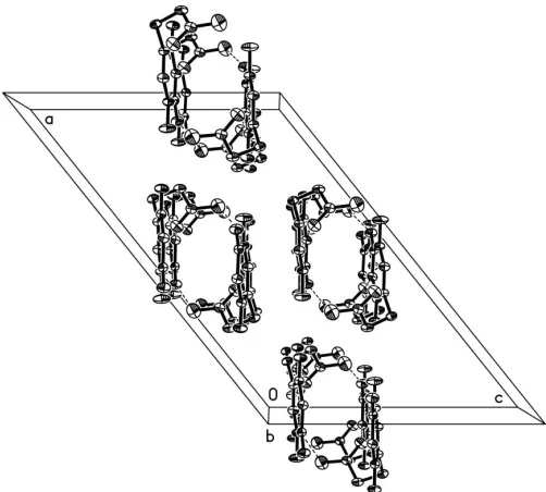
Inter-molecular N—H O and O—H O hydrogen bonds
(Table 2) form columns along thebaxis (Fig. 2).
Experimental
obtained by slow evaporation of an ethanol solution. IR (KBr,
cm1): 3284, 1694, 1416;1H NMR (d
6-DMSO,, p.p.m.): 11.70 (s, 1H),
12.65 (b, 1H), 7.85 (d, 1H), 3.72 (t, 2H), 2.54 (t, 2H); analysis calcu-lated for C7H7FN2O4: C 41.58, H 3.49, N 13.86%; found: C 41.50,
H 3.62, N 13.77%.
Crystal data
C7H7FN2O4 Mr= 202.15
Monoclinic,C2=c a= 20.279 (6) A˚
b= 8.137 (2) A˚
c= 13.222 (4) A˚
= 128.673 (4)
V= 1703.3 (8) A˚3 Z= 8
Dx= 1.577 Mg m
3
MoKradiation Cell parameters from 1663
reflections
= 2.8–26.3
= 0.14 mm1 T= 294 (2) K Block, colourless 0.260.240.20 mm
Data collection
Bruker SMART CCD area-detector diffractometer
’and!scans
Absorption correction: multi-scan (SADABS; Sheldrick, 1996)
Tmin= 0.958,Tmax= 0.972
4556 measured reflections
1747 independent reflections 1245 reflections withI> 2(I)
Rint= 0.035
max= 26.5 h=20!25
k=7!10
l=16!16
Refinement
Refinement onF2 R[F2> 2(F2)] = 0.047 wR(F2) = 0.145
S= 1.05 1747 reflections 137 parameters
H atoms treated by a mixture of independent and constrained refinement
w= 1/[2(F
o2) + (0.0679P)2
+ 1.9948P]
whereP= (Fo2+ 2Fc2)/3
(/)max= 0.002 max= 0.30 e A˚
3 min=0.41 e A˚
3
Table 1
Selected bond lengths (A˚ ).
F1—C6 1.352 (3) O1—C1 1.229 (3) O2—C1 1.324 (3) O3—C4 1.217 (3) O4—C5 1.222 (3)
N1—C3 1.473 (3) N1—C4 1.380 (3) N1—C7 1.370 (3) N2—C4 1.377 (3) N2—C5 1.376 (3)
Table 2
Hydrogen-bond geometry (A˚ ,).
D—H A D—H H A D A D—H A
N2—H2E O1i
0.81 (3) 2.00 (3) 2.797 (3) 171 (3) O2—H2C O4ii
0.839 (10) 2.06 (2) 2.887 (3) 167 (8) O2—H2D O3iii 0.839 (10) 2.154 (14) 2.990 (3) 174 (8)
Symmetry codes: (i)x;yþ1;z; (ii)x;y1;zþ1
2; (iii)xþ12;yþ32;zþ1.
H atoms attached to O and N atoms were located in a difference map. The OH group is disordered over two positions with an occu-pancy ratio of 0.5:0.5 and the H atom was refined with a restraint of O—H = 0.82 (2) A˚ . The H atom of the NH group was refined freely. All other H atoms were placed in geometrically calculated positions (C—H = 0.93 or 0.97 A˚ ) and refined as riding atoms [Uiso(H) =
1.2Ueq(C)].
Data collection:SMART(Bruker, 1997); cell refinement:SAINT
(Bruker, 1997); data reduction: SAINT; program(s) used to solve structure: SHELXS97(Sheldrick, 1997); program(s) used to refine
structure: SHELXL97 (Sheldrick, 1997); molecular graphics:
SHELXTL (Bruker, 1997); software used to prepare material for publication:SHELXTL.
This work was supported by the Scientific Research Foun-dation of Tianjin University.
References
Bruker (1997). SMART (Version 5.06a), SAINT (Version 5.051) and
SHELXTL(Version 5.10). Bruker AXS Inc., Madison, Wiscosin, USA.
organic papers
Acta Cryst.(2006). E62, o586–o588 Quet al. C
[image:2.610.338.544.73.301.2] [image:2.610.314.565.358.584.2]7H7FN2O4
o587
Figure 1
The molecular structure of (I), showing the atom-labelling scheme. Displacement ellipsolids are drawn at the 35% probability level. Both disorder components of the OH group are shown.
Figure 2
Correale, P., Fulfaro, F., Marsili, S., Cicero, G., Bajardi, E., Intrivici, C., Vuolo, G., Carli, A. F., Caraglia, M., Prete, S. D., Greco, E., Gebbia, N. & Francini, G. (2005).Cancer Chemother. Pharmacol.56, 563–568.
Duschinsky, R., Pleven, E. & Heidelberger, C. (1957).J. Am. Chem. Soc.79, 4559–4560.
Heidelberger, C., Chaudhuri, M. S., Danneberg, P., Mooren, D., Duschinsky, R., Schnitzer, R. J., Pleven, E. & Scheiner, J. (1957).Nature (London),179, 663–666.
Liu, Y.-M., Kang, J.-H., Wang, Z.-P., Wang, L.-F., Gao, L., Xia, C.-G. & Cui, J.-R. (2000).J. Coord. Chem.52, 1–13.
Sheldrick, G. M. (1996).SADABS. University of Go¨ttingen, Germany. Sheldrick, G. M. (1997). SHELXS97 and SHELXL97. University of
Go¨ttingen, Germany.
Wasterack, C. & Bettina, H. (1987).Pharmazie,12, 73–75.
Zhuo, R.-X., Fan, C.-L. & Zhou, R.-L. (1986).Chem. J. Chin. Univ.7, 508– 510.
organic papers
o588
Quet al. Csupporting information
sup-1 Acta Cryst. (2006). E62, o586–o588
supporting information
Acta Cryst. (2006). E62, o586–o588 [https://doi.org/10.1107/S1600536806000584]
5-Fluorouracil-1-propionic acid
Jian-Qiang Qu, Xiao-Fei Jia, Jian-Zhong Cui, Hong Zhang and Liu-Fang Wang
5-Fluorouracil-1-propionic acid
Crystal data
C7H7FN2O4
Mr = 202.15 Monoclinic, C2/c a = 20.279 (6) Å
b = 8.137 (2) Å
c = 13.222 (4) Å
β = 128.673 (4)°
V = 1703.3 (8) Å3
Z = 8
F(000) = 832
Dx = 1.577 Mg m−3
Melting point: 457 K
Mo Kα radiation, λ = 0.71073 Å Cell parameters from 1663 reflections
θ = 2.8–26.3°
µ = 0.14 mm−1
T = 294 K Block, colourless 0.26 × 0.24 × 0.20 mm
Data collection
Bruker SMART CCD area-detector diffractometer
Radiation source: fine-focus sealed tube Graphite monochromator
φ and ω scans
Absorption correction: multi-scan (SADABS; Sheldrick, 1996)
Tmin = 0.958, Tmax = 0.972
4556 measured reflections 1747 independent reflections 1245 reflections with I > 2σ(I)
Rint = 0.035
θmax = 26.5°, θmin = 2.6°
h = −20→25
k = −7→10
l = −16→16
Refinement
Refinement on F2
Least-squares matrix: full
R[F2 > 2σ(F2)] = 0.047
wR(F2) = 0.145
S = 1.05 1747 reflections 137 parameters 14 restraints
Primary atom site location: structure-invariant direct methods
Secondary atom site location: difference Fourier map
Hydrogen site location: inferred from neighbouring sites
H atoms treated by a mixture of independent and constrained refinement
w = 1/[σ2(F
o2) + (0.0679P)2 + 1.9948P]
where P = (Fo2 + 2Fc2)/3
(Δ/σ)max = 0.002
Δρmax = 0.30 e Å−3
Δρmin = −0.41 e Å−3
Special details
supporting information
sup-2 Acta Cryst. (2006). E62, o586–o588
Refinement. Refinement of F2 against ALL reflections. The weighted R-factor wR and goodness of fit S are based on F2,
conventional R-factors R are based on F, with F set to zero for negative F2. The threshold expression of F2 > σ(F2) is used
only for calculating R-factors(gt) etc. and is not relevant to the choice of reflections for refinement. R-factors based on F2
are statistically about twice as large as those based on F, and R- factors based on ALL data will be even larger.
Fractional atomic coordinates and isotropic or equivalent isotropic displacement parameters (Å2)
x y z Uiso*/Ueq Occ. (<1)
F1 −0.11918 (9) 0.74550 (19) −0.00859 (18) 0.0595 (5) O1 0.09860 (12) 0.3482 (2) 0.23697 (17) 0.0488 (5) O2 0.16000 (17) 0.3696 (3) 0.4480 (3) 0.0767 (7)
H2C 0.130 (4) 0.291 (6) 0.439 (8) 0.092* 0.50 H2D 0.199 (3) 0.411 (10) 0.520 (4) 0.092* 0.50 O3 0.19940 (10) 0.9631 (2) 0.30681 (17) 0.0455 (5)
O4 −0.08222 (10) 1.0748 (2) 0.04855 (17) 0.0453 (5) N1 0.10767 (11) 0.7502 (2) 0.19657 (18) 0.0301 (5) N2 0.05845 (12) 1.0148 (2) 0.18074 (19) 0.0315 (5) H2E 0.0676 (17) 1.110 (4) 0.201 (3) 0.042 (8)* C1 0.15073 (15) 0.4128 (3) 0.3432 (2) 0.0348 (6) C2 0.20949 (14) 0.5458 (3) 0.3625 (2) 0.0391 (6)
H2A 0.2185 0.6243 0.4252 0.047*
H2B 0.2637 0.4969 0.3982 0.047*
C3 0.17716 (14) 0.6360 (3) 0.2399 (2) 0.0360 (6)
H3A 0.2233 0.6972 0.2539 0.043*
H3B 0.1577 0.5564 0.1718 0.043*
C4 0.12708 (14) 0.9128 (3) 0.2337 (2) 0.0300 (5) C5 −0.02572 (14) 0.9731 (3) 0.0961 (2) 0.0302 (5) C6 −0.03845 (14) 0.7997 (3) 0.0698 (2) 0.0345 (6) C7 0.02560 (14) 0.6967 (3) 0.1174 (2) 0.0351 (6)
H7 0.0145 0.5857 0.0965 0.042*
Atomic displacement parameters (Å2)
U11 U22 U33 U12 U13 U23
supporting information
sup-3 Acta Cryst. (2006). E62, o586–o588
Geometric parameters (Å, º)
F1—C6 1.352 (3) N2—C5 1.376 (3)
O1—C1 1.229 (3) N2—H2E 0.81 (3)
O2—C1 1.324 (3) C1—C2 1.509 (3)
O2—H2C 0.839 (10) C2—C3 1.506 (3)
O2—H2D 0.839 (10) C2—H2A 0.9700
O3—C4 1.217 (3) C2—H2B 0.9700
O4—C5 1.222 (3) C3—H3A 0.9700
N1—C3 1.473 (3) C3—H3B 0.9700
N1—C4 1.380 (3) C5—C6 1.437 (3)
N1—C7 1.370 (3) C6—C7 1.328 (3)
N2—C4 1.377 (3) C7—H7 0.9300
C1—O2—H2C 116 (6) N1—C3—C2 113.14 (19)
C1—O2—H2D 119 (6) N1—C3—H3A 109.0
H2C—O2—H2D 124 (8) C2—C3—H3A 109.0
C7—N1—C4 121.10 (19) N1—C3—H3B 109.0
C7—N1—C3 120.44 (18) C2—C3—H3B 109.0
C4—N1—C3 118.42 (18) H3A—C3—H3B 107.8
C5—N2—C4 127.6 (2) O3—C4—N2 122.3 (2)
C5—N2—H2E 114.8 (19) O3—C4—N1 122.6 (2)
C4—N2—H2E 118 (2) N2—C4—N1 115.05 (19)
O1—C1—O2 122.7 (2) O4—C5—N2 122.6 (2)
O1—C1—C2 121.6 (2) O4—C5—C6 124.9 (2)
O2—C1—C2 115.7 (2) N2—C5—C6 112.51 (19)
C3—C2—C1 113.6 (2) C7—C6—F1 121.0 (2)
C3—C2—H2A 108.8 C7—C6—C5 122.0 (2)
C1—C2—H2A 108.8 F1—C6—C5 117.01 (19)
C3—C2—H2B 108.8 C6—C7—N1 121.5 (2)
C1—C2—H2B 108.8 C6—C7—H7 119.2
H2A—C2—H2B 107.7 N1—C7—H7 119.2
O1—C1—C2—C3 23.2 (3) C4—N2—C5—O4 −175.6 (2) O2—C1—C2—C3 −157.7 (2) C4—N2—C5—C6 3.5 (3) C7—N1—C3—C2 −88.0 (3) O4—C5—C6—C7 175.1 (2) C4—N1—C3—C2 94.4 (2) N2—C5—C6—C7 −3.9 (3) C1—C2—C3—N1 73.9 (3) O4—C5—C6—F1 −3.4 (4) C5—N2—C4—O3 179.1 (2) N2—C5—C6—F1 177.6 (2) C5—N2—C4—N1 −0.3 (3) F1—C6—C7—N1 179.8 (2) C7—N1—C4—O3 177.8 (2) C5—C6—C7—N1 1.3 (4)
C3—N1—C4—O3 −4.6 (3) C4—N1—C7—C6 2.3 (3)
supporting information
sup-4 Acta Cryst. (2006). E62, o586–o588
Hydrogen-bond geometry (Å, º)
D—H···A D—H H···A D···A D—H···A
N2—H2E···O1i 0.81 (3) 2.00 (3) 2.797 (3) 171 (3)
O2—H2C···O4ii 0.84 (1) 2.06 (2) 2.887 (3) 167 (8)
O2—H2D···O3iii 0.84 (1) 2.15 (1) 2.990 (3) 174 (8)