ISSN Online: 2333-9721 ISSN Print: 2333-9705
Correlation between Three Pregnancy
Characteristics (Age, Parity, βhCG Level)
and pAkt Immunoexpression on Complete
Hydatidiform Mole
Zulvayanti
1, Sofie R. Krisnadi
1, Achadiyani
21Department of Obstetrics and Gynecology, Dr. Hasan Sadikin General Hospital, Faculty of Medicine, Universitas Padjadjaran, Bandung, Indonesia
2Department of Biology Cell, Faculty of Medicine, Universitas Padjadjaran, Bandung, Indonesia
Abstract
The incidence of hydatidiform mole as pregnancy failure is still high in Asia, leading to high mortality and morbidity. Several risk factors play roles in the occurrence of hydatidiform mole, including age and parity. Until now, β-human chronic gonadotropin (βhCG) is used to predict the risk and devel-opment of hydatidiform mole. Akt, also known as protein kinase B (PKB), is a downstream effector of the intracellular phosphatidylinositol 3-kinase (PI3K) signaling pathway, which is a central regulatory pathway for cell proliferation, growth, differentiation, and survival. This study aimed to analyze the correla-tion between three pregnancy characteristics i.e. age, parity, levels of βhCG and pAkt immunoexpression. Cross sectional study was conducted on 30 samples of complete hydatidiform mole (CHM). The βhCG content was measured by ELISA method. The immunoexpression of pAkt was measured by immunohistochemical staining using the phospho-Akt antibody (Ser473) (736E11) Rabbit mAb # 3787 (CST). Cells with positive pAkt immunoex- pression showed brown color. The stronger the intensity of the visible brown color, the higher the level of expression. pAkt immunoexpression level was then expressed as histoscore value, which was calculated using staining inten-sity and positively-stained-cell distribution number. When the immuno- expression level was high, its histoscore value would be high too. Results of this study showed that the histoscore of pAkt samples ranged from 0 - 16 with a median value 4, and the most samples (56.7%) had histoscore ≤ 4. Correla-tion coefficient (p) value that was used in this study was 0.01. CorrelaCorrela-tion be-tween two variables was significant if p < 0.01. Correlation coefficient (p) val-ue between age and pAkt immunoexpression was 0.260 and thus was not
sta-How to cite this paper: Zulvayanti, Krisnadi, S.R. and Achadiyani (2017) Correlation between Three Pregnancy Characteristics (Age, Parity, βhCG Level) and pAkt Immu- noexpression on Complete Hydatidiform Mole. Open Access Library Journal, 4: e3634. https://doi.org/10.4236/oalib.1103634
Received: April 25, 2017 Accepted: May 8, 2017 Published: May 11, 2017
Copyright © 2017 by authors and Open Access Library Inc.
This work is licensed under the Creative Commons Attribution International License (CC BY 4.0).
tistically significant, neither was the number of parity and pAkt immuno- expression (p = 0.524). However, βhCG level and pAkt immunoexpression showed statistically significant correlation (p = 0.00). It was concluded that there was no significant correlation between maternal age and parity and pAkt immunoxpression (p > 0.01), but there was a significant correlation between βhCG levels and pAkt immunoxpression (p < 0.01).
Subject Areas
Gynecology & Obstetrics
Keywords
Complete Hydatidiform Mole, Akt, βhCG, Age, Parity
1. Introduction
There are two types of hydatidiform mole as pregnancy failure, i.e. complete hydatidiform mole (CHM) and partial hydatidiform mole (PHM), which are cy-togenetically different. CHM exhibits androgenetic properties, where all chro-mosomes in CHM tissue are derived from paternal cells. CHM pregnancy occurs because an empty ovum is fertilized by a 23x haploid sperm, resulting in concep-tion with 23x chromosome. This chromosome then undergoes self doubling (endoreduplication) to 46xx. PHM is generally triploid or trisomic on a single chromosome. In CHM, all chorionic villi are hydropic degenerated so that no fetal element is found, whereas in PHM there is fetus, umbilical cord, normal amniotic membrane, and placenta with hydropic degenerated chorionic villi [1]
[2][3][4].
The incidence of hydatidiform mole in the world varies depending on the region, differences can even be found in the regions of the same continent. Based on the report of The National Coordination Research Group of Chorioma (NCRG) hydatidiform mole incidence in China was 0.78 per 1000 live births. The incidence of hydatidiform mole in Japan was much higher, reaching 2.0 per 1000 live births, 3 times higher than the incidence in European or North American countries (0.6 - 1.1 per 1000 pregnancies). Other studies showed that hydatidiform mole incidence in Japan ranged from 2.83 to 3.05 per 1000 live births. The highest incidence of hydatidiform mole was reported in Indonesia, i.e. 1 in 77 pregnancies or 1 in 57 deliveries [5]. Martaadisoebrata et al. in Hasan Sadikin Hospital Bandung, Indonesia, showed that the incidences of gestational trophoblast disease over three periods (1971-1976, 1996-2000, 2001-2005) were still high (19.8, 30.8, 23.6 per 1000 births), and the percentages of hydatidiform mole were 83%, 67%, and 45% respectively [6].
with higher risk for hydatidiform mole are those who are pregnant at under 20 and over 35 years old. Within the age groups above 40 years old, hydatidiform mole incidence is known 4 - 10 times higher than those at age 20 - 40 years old.
In addition to age, parity can be a risk factor for hydatidiform mole, although some studies showed different results. A study in Italy showed an increased risk of hydatidiform mole in nullipara with miscarriage history. However, other studies that were also conducted in Italy and the Rhode islands, United States, showed that parity was not associated with hydatidiform mole risk [5]. Mar-taadisoebrata [7] in Bandung demonstrated a positive correlation between parity and hydatidiform mole incidence. Several other studies found an association between parity and hydatidiform mole development to gestational trophoblast tumor. Aziz et al. [8], Khrismawan et al., [9] and Prajatmo [10] found a signifi-cant correlation between parity and the incidence of post-CHM gestational tro-phoblast tumor malignancy. In contrast to these studies, Yudi et al. showed no significant relationship between parity and hydatidiform mole in persistent CHM cases and CHM cases with spontaneous regression [11].
Hydatidiform mole causes high morbidity and mortality. Complications that often accompany hydatidiform mole may be early complications such as bleed-ing, preeclampsia, hyperthyroidism, and thyrotoxicosis, or advanced complica-tions such as gestational trophoblast tumor [3]. Therefore, early detection of CHM is essential to prevent the onset of these complications.
Until now, the level of βhCG has been widely used to predict the risk and de-velopment of hydatidiform mole [12] [13]. βhCG level is also used as a funda-mental consideration for treatment, both prophylaxis and definitive therapy for gestational trophoblast tumor [3][4][13]. βhCG level > 100,000 mIU/ mL indi-cates a high risk of hydatidiform mole, hence some treatment centers establish prophylactic chemotherapy [3] [4] [13]. Monitoring of post-evacuation serum βhCG level in CHM patient management guidelines at Hasan Sadikin Hospital Bandung was performed using Mochizuki regression curve [14]. Evaluation of serum βhCG level is performed at week 2, 4, 6, 8, and 12 post-evacuation of CHM. Deviation of the normal regression curve or the distortion toward ele-vated serum βhCG level indicates persistent trophoblastic diseases or transfor-mation of post-CHM malignancy into gestational trophoblast tumor [14].
βhCG is a subunit of human chorionic gonadotropin (hCG) hormone, a gly-coprotein consisting of two subunits, i.e. α subunit (identical to α subunits of LH, FSH and TSH glycoprotein hormones) and β subunits (specific to hCG) with 8 sugar chains. The α subunit has 92 amino acids, while the β subunit has 145 amino acids. Based on composition of these structures, there are several variants of hCG hormone. The four major variants of hCG structure commonly detected in serum are hCG free β subunit, nicked hCG, hCG missing the β sub-unit C-terminal peptide, and hyperglycosylated hCG. Measurement of βhCG level is a common examination to assess the presence of hCG hormones in se-rum, urine, or other body fluids [12].
the excessive proliferation of syncytiotrophoblasts will increase hCG level [12] [15]. In CHM pregnancy, the proliferation rate of trophoblast cells, both syncyti-otrophoblast and cytsyncyti-otrophoblast, is higher than in normal pregnancy, hence hCG level in CHM pregnancy is generally higher than in normal pregnancy [1] [2]. During pregnancy, hCG stimulates corpus luteum cells to produce proges-terone in order to maintain pregnancy. hCG is luteotropic hormone and corpus luteum has a high-affinity receptor for hCG. Thus, under conditions of very high hCG level, cysts are frequently found in corpus luteum [3][11].
Akt or protein kinase B (PKB) is a downstream effector of the intracellular phosphatidylinositol 3-kinase (PI3K) signaling pathway, which is the central regulatory pathway for cell proliferation, growth, differentiation, and survival
[16] [17]. Akt will convey the intracellular transduction signals stimulated by growth factors such as platelet-derived growth factor (PDGF), epidermal growth factor (EGF), basic fibroblast growth factor (BFGF), and insulin-like growth factor (IGF-I), which will then activate thousands of downstream substances. Akt activity will change the subcellular location or modify the stability of pro-tein, which further affects the activity of downstream substances in regulating cell proliferation, differentiation, invasion, and apoptosis [16].
In trophoblast cells, Akt regulates the phenotype differentiation of the cells. Akt also plays role in coding proteins that potentially cause direct trophoblast invasion to maternal/ uterine environment (MMP9, IGF2, Serpine1), affecting immunity and vascular cells (Cgm4, Faslg), regulating androgen biosynthesis, and activating trophoblast invasion process [17][18].
Choi et al. showed an association between Akt and age on rats. Akt plays role in FoxO3a phosphorylation, a transcription factor that plays an important role in aging processes. FoxO3a’s activity decreases with age, and negatively regu-lated through phosphorylation by PI3K/Akt signaling pathway [19]. Prast et al. showed that hCG increases the phosphorylation of Akt so that Akt becomes ac-tive and phosphorylates the downstream substances [15].
This study aimed to analyze the correlation between demographic characteris-tics: age, parity, βhCG levels and pAkt in CHM pregnancy.
2. Methods
This was a cross sectional study on 30 patients that were diagnosed with CHM based on the examination results conducted at Anatomy & Pathology Laboratory of Hasan Sadikin Hospital, Faculty of Medicine, Padjadjaran University. The CHM patients were selected based on sample criteria that were recorded in the last five years at Obstetrics and Gynecology Department and Anatomy & Pa- thology Laboratory of Hasan Sadikin Hospital, Faculty of Medicine, Padj- adjaran University. Ethical clearance for this study was obtained from Health Research Ethics Committee, Faculty of Medicine, Padjadjaran University, Ban- dung, Indonesia (Registration number: 0515020111).
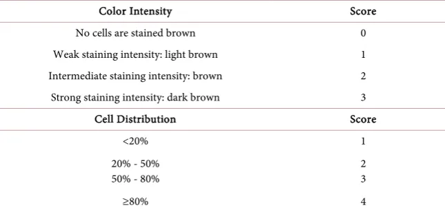
phospho-Akt antibody (Ser473) (736E11) Rabbit mAb # 3787 (CST). The research was performed at Anatomy & Pathology Laboratory of Hasan Sadikin Hospital, Faculty of Medicine, Padjadjaran University, Bandung, Indonesia. Immunoexpression assessment of pAkt was determined based on staining in- tensity and cell distribution as described in Table 1.
The histoscore from the staining results were calculated using following formula: [20]
(
)
Histoscore= ∑ +i 1 Pi i: intensity
Pi: cell distribution with positive staining
Data were then analyzed statistically using chi square analysis, with correla-tion coefficient (p) value 0.01. Correlacorrela-tion between two variables was significant if p < 0.01.
3. Results
A total of 30 CHM cases were collected in this study. Data collection on demo-graphic characteristics (maternal age and parity) and βhCG levels were con-ducted. Immunohistochemical staining was then performed to assess the pAkt immunoexpression. Histoscore calculation of pAkt immunoexpression was ap-plied on all subjects. Immunoexpression assessment of pAkt was determined based on staining intensity and cell distribution.
Table 2 shows that most of patients involved in the study were in the age of
20 - 34 years old which are reproductive age. This can happen because most pregnancies occur at that range of age.
From 30 examined CHM samples, histoscore values ranged from 0 - 16 with median 4. Most samples (56.7%) had histoskor values ≤ 4.
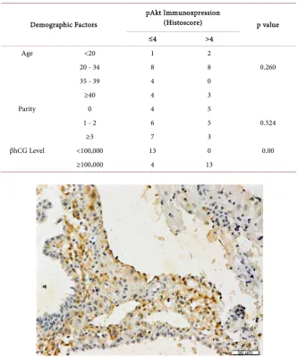
Cells with positive pAkt immunoexpression showed brown color. The strong-er the intensity of the visible brown color, the highstrong-er the level of expression
(Figure 1).
Table 3 shows no significant correlation between age and parity and pAkt
[image:5.595.208.531.579.733.2]immunoxpression (p > 0.01). However, βhCG level and pAkt immunoexpression
Table 1. Immunohistochemical scores determination.
Color Intensity Score
No cells are stained brown 0
Weak staining intensity: light brown 1 Intermediate staining intensity: brown 2 Strong staining intensity: dark brown 3
Cell Distribution Score
<20% 1
20% - 50%
50% - 80% 2 3
Table 2. Distribution table of the results.
Characteristics Number Percentage (%)
Age <20 3 1
20 - 34 16 53.3
35 - 39 4 13.3
≥40 7 23.3
Parity 0 9 30
1 - 2 11 36.7
≥3 10 33.3
βhCG Level <100,000 13 43.3
≥100,000 17 56.7
[image:6.595.206.540.316.716.2]pAkt Histoscore Median Range 0 - 16 4
Table 3. Correlation between demographic factors (Age, parity, βhCG Level) and pAkt immunoxpression.
Demographic Factors
pAkt Immunoxpression
(Histoscore) p value
≤4 >4
Age <20 1 2
20 - 34 8 8 0.260
35 - 39 4 0
≥40 4 3
Parity 0 4 5
1 - 2 6 5 0.524
≥3 7 3
βhCG Level <100,000 13 0 0.00
≥100,000 4 13
was significantly correlated (p < 0.01).
4. Discussion
1) Correlation between Age and Parity and pAkt Immunoexpression
It is known that age is one of the important risk factors in hydatidiform mole incidence as well as the transformation of post-hydatidiform mole malignancies. The incidence of CHM increases at extreme age (under 20 years, and over 35 years). There is a progressive increase in risk (more than 10 times) in women over the age of 40. Prajatmo showed that the age group ≥35 years old had 2 times greater risk when compared with age <35 years. [10] Theoretically, at that age, the risk of sex chromosome division (X chromosome) failure in ovum mei-ogenesis is higher. Failure in the process can produce ovum without X chromo-some (empty ovum). Fertilization on the empty ovum will produce an abnormal zygote with sex chromosomes that only originate from the father (paternal/ spermatozoa). Without maternal sex chromosome, trophoblast cells of the zy-gote will undergo self reduplication i.e. endoreduplication, which is the patho-genesis of CHM (Androgenetic theory) [4].
Similarly, previous epidemiological studies conducted by Martaadisoebrata et al. [7] in Bandung showed a positive correlation between parity and hydatidi-form mole incidence. In pregnancy, trophoblast cells always circulate in the blood circulation throughout mother's body, hence an immunological reaction between trophoblast cells and immune system occurs in mother’s body. Under these conditions, the shorter the time of the pregnancy interval, mother’s im-munologic reaction disorder will more likely increase, thus increasing the inci-dence of hydatidiform mole pregnancy in mother with high parity [19].
Akt plays role in FoxO3a phosphorylation, a transcription factor that plays an important role in the aging process. FoxO3a expression decreases with age, and are negatively regulated through phosphorylation of PI3K/Akt signaling path- way. The expression of Akt itself increases with age. Choi et al. [19] showed that ferulate administration in rats can suppress Akt activity, and thus inhibit the aging process.
However, the results of this study indicated that age was not statistically cor-related with pAkt immunoexpression (p > 0.01). This might happen because in CHM the relationship between Akt expression and age does not follow the usual pattern of normal cells, yet this suposition needs to be investigated further. The same presumption can be applied for parity factor. Age and parity are generally linearly related, where the older women usually have higher parity. Therefore, this study also showed no significant correlation between parity with pAkt immunoexpression (p > 0.01).
2) Correlation between βhCG Level and pAkt Immunoexpression
disease case [3][13].
In CHM, the proliferated trophoblast cells fill uterine cavity. Along with the increased proliferation rate of trophoblast cells, especially syncytiotrophoblasts, serum βhCG level will tend to increase, and potentially reach above 5,000,000 mIU/mL. High level of βhCG is an indicator of high trophoblast activity. Experts agree that elevated level of βhCG can be used as specific and sensitive marker to predict CHM post-evacuation malignancy [13]. FIGO, ISSTD, and WHO estab-lished pre-evacuation serum βhCG level ≥100,000 mIU/ mL as a parameter that indicates a high risk of CHM transformation into malignancy [13]. Marta- adisoebrata, [7] Khrismawan et al. [9], and Pradjatmo [10] obtained similar results that pre-evacuation βhCG serum level ≥100,000 mIU/mL was a risk factor for CHM post-evacuation malignancy.
This study showed significant correlation between βhCG level and pAkt immunoexpression (p < 0.01). The high value of pAkt histoscore was associated with high level of βhCG. This finding was in line with previous research showing that Akt will be activated/phosphorylated by hCG administration. Thus, the higher βhCG level, the higher pAkt expression [15]. It has been known that hCG has a synergistic effect on PI3K-Akt, ERK, and MAPK signalling pathways against cytotrophoblast invasion [16].
5. Conclusion
From the results obtained in this study, it was concluded that maternal age and parity were not significantly correlated with pAkt immunoxpression (p > 0.01). However, there was a significant correlation between βhCG levels and pAkt immunoxpression (p < 0.01).
References
[1] Lurain, J.R. (2010) Gestational Trophoblastic Disease I: Epidemiology, Pathology, Clinical Presentation and Diagnosis of Gestational Trophoblastic Disease, and Management of Hydatidiform Mole. American Journal of Obstetrics and Gynecology, 203, 531-539.
[2] Sebire, N.J., Linsdsay, I. and Paradinas, F. (2009) Pathology. In: Hancock, B.W., Seckl, M.J., Berkowitz, R.S. and Cole, L.A., Eds., Gestational Trophoblastic Disease, 3rd Edition, ISSTD, McLean, 97-147.
[3] Bratakoesoema, D.S. (2006) Penyakit Trofoblas Gestasional. In: Aziz, M.F. and Andrijono, S.A.B., Eds., Buku Acuan Nasional Onkologi Ginekologi, Yayasan Bina Pustaka Sarwono Prawirohardjo, Jakarta, 532-568.
[4] Fisher, R.A. (2009) Genetics. In: Hancock, B.W., Seckl, M.J., Berkowitz, R.S. and Cole, L.A., Eds., Gestational Trophoblastic Disease, 3rd Edition, ISSTD, McLean, 6- 48.
[5] Lee, C., Smith, H.O. and Kim, S.J. (2009) Epidemiology. In: Hancock, B.W., Seckl, M.J., Berkowitz, R.S. and Cole, L.A., Eds., Gestational Trophoblastic Disease, 3rd Edition, ISSTD, McLean, 49-97.
2001-2005. 16th World Congress on Gestational Trophoblastic Disease, Budapest, 16-19 October 2011, 54.
[7] Martaadisoebrata, D. (1998) Perkembangan penyakit trofoblas gestasional di Jawa Barat dan peranan RSHS dalam upaya penanggulangannya. Risalah seminar sehari penyakit trofoblas gestasional, Bandung.
[8] Aziz, F.M., Kampono, N. and Sjamsuddin, S. (1984) Neoplasma trofoblas: Faktor risiko tinggi dan prognosis. In: Aziz, F.M. and Pen, Y., Eds., Neoplasma trofoblas gestational epidemiologi diagnosis, pengobatan, pencegahan dan prognosis, Bagian Obstetri Ginekologi FKU, Jakarta, 35-54.
[9] Khrismawan, S.A.Z. and Sanif, R. (2004) Theodorus. Efficacy of NETDC (New England Trophoblastic Disease Center) Prognostic Index Score to Predict Gestational Trophoblastic Tumor from Hydatidiform Mole. Efficacy of NETDC Prognostic Index Score. Medical Journal of Indonesia, 13, 40-46.
[10] Pradjatmo, H. (2013) Malignancy Risk Scoring of Hydatidiform Moles ISSTD World Congress XVII. Departement of Obstetric and Gynecology, Sardjito Hospital/Faculty of Medicine, Gadjah Mada University, Chicago.
[11] Hidayat, Y.M. (2014) Peran ekspresi imunohistokimia Cyclin D1, D3, Retino- blastoma (Rb) dan faktor risiko klinis mola hidatidosa komplet sebagai faktor risiko mola persisten. Disertasi. Universitas Padjadjaran, Bandung.
[12] Cole, L.A. (2009) Structurally Related Molecules of Human Chorionic Gonadotropin (hCG) in Gestasional Trophoblastic Diseases. In: Hancock, B.W., Seckl, M.J., Berkowitz, R.S. and Cole, L.A., Eds., Gestational Trophoblastic Disease, 3rd Edition, ISSTD, McLean, 148.
[13] Ngan, H., Chui, L. and Ma, H.K. (2009) Staging and Classification Systems. In: Hancock, B.W., Seckl, M.J., Berkowitz, R.S. and Cole, L.A., Eds., Gestational Trophoblastic Disease, 3rd Edition, ISSTD, McLean, 184.
[14] Sub-bagian Onkologi, Bagian Obstetri GInekologi RSHS/FKUP. Protokol Pengelolaan Penyakit Trofoblas di Lab/UPF Obstetri dan Ginekologi. 2002.
[15] Prast, J., Saleh, L., Husslein, H., Sonderegger, S., Helmer, H. and Knöfler, M. (2008) Human Chorionic Gonadotropin Stimulates Trophoblast Invasion through Extracellularly Regulated Kinase and AKT Signaling. Endocrinology, 149, 979-987. https://doi.org/10.1210/en.2007-1282
[16] Liao, Y. and Hung, M.-C. (2010) Physiological Regulation of Akt Activity and Stability. American Journal of Translational Research, 2, 19-42.
[17] Kent, L.N., Konno, T. and Soares, M.J. (2010) Phosphatidylinositol 3-Kinase Modulation of Trophoblast Cell Differentiation. BMC Developmental Biology, 10, 97-125. https://doi.org/10.1186/1471-213X-10-97
[18] Kamei, T., Jones, S.R., Chaoman, B.M., et al. (2002) The Phosphatidylinositol 3- Kinase/Akt Signaling Pathway Modulates the Endocrine Differentiation of Trophoblast Cells. Molecular Endocrinology, 16, 1469-1481.
https://doi.org/10.1210/mend.16.7.0878
[19] Choi, Y.J., Kim, D.H., Lee, E.K., et al. (2012) Attenuation of Age-Related Changes in FOXO3a Activity and the PI3K/Akt Pathway by Short-Term Feeding of Ferulate.
AGE, 34, 317-327. https://doi.org/10.1007/s11357-011-9235-3
Submit or recommend next manuscript to OALib Journal and we will pro-vide best service for you:
Publication frequency: Monthly
9 subject areas of science, technology and medicine Fair and rigorous peer-review system
Fast publication process
Article promotion in various social networking sites (LinkedIn, Facebook, Twitter, etc.)
Maximum dissemination of your research work
Submit Your Paper Online: Click Here to Submit