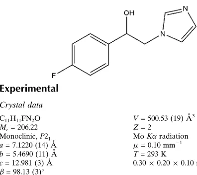
1-(4-Fluorophenyl)-2-(1
H
-imidazol-1-yl)-ethanol
Dong-liang Liu, Chen Li, Xin Tian, Song Li and Tao Xiao*
Department of Applied Chemistry, College of Science, Nanjing University of Technology, Nanjing 210009, People’s Republic of China
Correspondence e-mail: [email protected]
Received 13 November 2011; accepted 12 December 2011
Key indicators: single-crystal X-ray study;T= 293 K; mean(C–C) = 0.005 A˚;
Rfactor = 0.041;wRfactor = 0.124; data-to-parameter ratio = 7.4.
In the title compound, C11H11FN2O, the dihedral angle between the mean planes of the two rings is 1.30 (4). In the
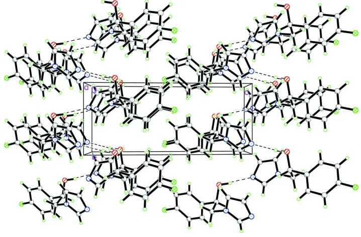
crystal, O—H N hydrogen bonds link the molecules into chains along thebaxis.
Related literature
For related compounds containing a 2-(1H-imidazol-1-yl)-1-phenylethanol fragment, see: Porrettaet al.(1993). For related structures, see: Taoet al.(2007); Liuet al.(2011). For standard bond lengths, see: Allenet al.(1987).
Experimental
Crystal data
C11H11FN2O
Mr= 206.22 Monoclinic,P21
a= 7.1220 (14) A˚
b= 5.4690 (11) A˚
c= 12.981 (3) A˚ = 98.13 (3)
V= 500.53 (19) A˚3
Z= 2
MoKradiation = 0.10 mm 1
T= 293 K
0.300.200.10 mm
Data collection
Enraf–Nonius CAD-4 diffractometer
Absorption correction: multi-scan (Northet al., 1968)
Tmin= 0.970,Tmax= 0.990
1992 measured reflections
1024 independent reflections 876 reflections withI> 2(I)
Rint= 0.029
3 standard reflections every 200 reflections
intensity decay: 1%
Refinement
R[F2> 2(F2)] = 0.041
wR(F2) = 0.124
S= 1.00 1024 reflections 139 parameters 1 restraint
H atoms treated by a mixture of independent and constrained refinement
max= 0.13 e A˚ 3
[image:1.610.46.244.442.618.2]min= 0.18 e A˚ 3
Table 1
Hydrogen-bond geometry (A˚ ,).
D—H A D—H H A D A D—H A
O—H0A N2i
0.87 (4) 1.90 (4) 2.762 (4) 171 (5)
Symmetry code: (i) x;y 1 2; z.
Data collection: CAD-4 EXPRESS (Enraf–Nonius, 1994); cell refinement:CAD-4 EXPRESS; data reduction:XCAD4(Harms & Wocadlo, 1995); program(s) used to solve structure: SHELXS97 (Sheldrick, 2008); program(s) used to refine structure:SHELXL97 (Sheldrick, 2008); molecular graphics:PLATON(Spek, 2009); soft-ware used to prepare material for publication:SHELXTL(Sheldrick, 2008).
The authors thank the Center of Testing and Analysis, Nanjing University, for support.
Supplementary data and figures for this paper are available from the IUCr electronic archives (Reference: ZQ2137).
References
Allen, F. H., Kennard, O., Watson, D. G., Brammer, L., Orpen, A. G. & Taylor, R. (1987).J. Chem. Soc. Perkin Trans. 2, pp. S1–19.
Enraf–Nonius (1994).CAD-4 EXPRESS. Enraf–Nonius, Delft, The Nether-lands.
Harms, K. & Wocadlo, S. (1995).XCAD4. University of Marburg, Germany. Liu, D., Li, C., Yu, G. & Xiao, T. (2011).Acta Cryst.E67, o2036.
North, A. C. T., Phillips, D. C. & Mathews, F. S. (1968).Acta Cryst.A24, 351– 359.
Porretta, G. C., Fioravanti, R., Biava, M., Cirilli, R., Simonetti, N., Villa, A., Bello, U., Faccendini, P. & Tita, B. (1993).Eur. J. Med. Chem.28, 749–760. Sheldrick, G. M. (2008).Acta Cryst.A64, 112–122.
Spek, A. L. (2009).Acta Cryst.D65, 148–155.
Tao, X., Yuan, L., Zhang, X.-Q., Jing, C. & Wang, J.-T. (2007).Acta Cryst.E63, o1330–o1331.
Acta Crystallographica Section E
Structure Reports
Online
supporting information
Acta Cryst. (2012). E68, o152 [doi:10.1107/S1600536811053505]
1-(4-Fluorophenyl)-2-(1
H
-imidazol-1-yl)ethanol
Dong-liang Liu, Chen Li, Xin Tian, Song Li and Tao Xiao
S1. Comment
The title compound, C11H11ON2F, is the key intermediate in the synthesis of a new kind of antifungal drug (Porretta et al.,
1993). As a part of our ongoing studies (Tao et al., 2007; Liu et al., 2011), the crystal structure determination of the title compound has been carried out in order to elucidate its molecular conformation. The molecular structure of the title compound is shown in Fig. 1. Bond lengths (Allen et al., 1987) and angles are within normal ranges. The dihedral angle between the mean planes of the two rings is 1.30 (4)°. In the crystal structure, intermolecular O—H···N hydrogen bonds (Table 1) link the molecules in chains along the b axis.
S2. Experimental
A mixture of 1-(4-fluorophenyl)-2-(1H-imidazol-1-yl)ethanone (2.04 g, 10 mol), sodium borohydride (0.756 g, 20 mmol) and 30 ml dry ethanol was refluxed for 3 h. After solvent evaporation, the mixture was neutralized with dilute hydro-chloric acid and then refluxed for 30 min. After the mixture was cooled, the solution was alkalinized with sodium hydroxide, the precipitate was collected, recrystallized with ethanol, and a yellow deposit was obtain (m.p. 410–412 K). Crystals suitable for a X-ray analysis were obtained by dissolving the crude product (1.0 g) in ethanol (30 ml) and then allowing the solution to evaporate slowly at room temperature for about 7 d.
S3. Refinement
The H atom of the hydroxy group was located in a difference Fourier map and was freely refined with Uiso(H) =
1.5Ueq(O). The other H atoms were positioned geometrically with C—H = 0.93 Å (aromatic), 0.97 Å (methylene), and
0.98 Å (methine) and constrained to ride on their parent atoms with Uiso(H) = 1.2Ueq(C). The absolute structure parameter
Figure 1
The molecular structure of the title compound with displacement ellipsoids drawn at the 50% probability level.
Figure 2
A packing diagram of the title compound viewed down the b axis. Hydrogen bonds are shown as dashed lines.
1-(4-Fluorophenyl)-2-(1H-imidazol-1-yl)ethanol
Crystal data
C11H11FN2O Mr = 206.22
Monoclinic, P21
Hall symbol: P 2yb
a = 7.1220 (14) Å
b = 5.4690 (11) Å
c = 12.981 (3) Å
β = 98.13 (3)°
V = 500.53 (19) Å3 Z = 2
F(000) = 216
Dx = 1.368 Mg m−3
Melting point: 410 K
Mo Kα radiation, λ = 0.71073 Å Cell parameters from 25 reflections
θ = 9–14°
[image:3.610.125.484.280.516.2]Data collection
Enraf–Nonius CAD-4 diffractometer
Radiation source: fine-focus sealed tube Graphite monochromator
ω/2θ scans
Absorption correction: multi-scan (North et al., 1968)
Tmin = 0.970, Tmax = 0.990
1992 measured reflections
1024 independent reflections 876 reflections with I > 2σ(I)
Rint = 0.029
θmax = 25.4°, θmin = 1.6° h = 0→8
k = −6→6
l = −15→15
3 standard reflections every 200 reflections intensity decay: 1%
Refinement
Refinement on F2
Least-squares matrix: full
R[F2 > 2σ(F2)] = 0.041 wR(F2) = 0.124 S = 1.00 1024 reflections 139 parameters 1 restraint
Primary atom site location: structure-invariant direct methods
Secondary atom site location: difference Fourier map
Hydrogen site location: inferred from neighbouring sites
H atoms treated by a mixture of independent and constrained refinement
w = 1/[σ2(F
o2) + (0.090P)2]
where P = (Fo2 + 2Fc2)/3
(Δ/σ)max < 0.001
Δρmax = 0.13 e Å−3
Δρmin = −0.18 e Å−3
Special details
Geometry. All e.s.d.'s (except the e.s.d. in the dihedral angle between two l.s. planes) are estimated using the full covariance matrix. The cell e.s.d.'s are taken into account individually in the estimation of e.s.d.'s in distances, angles and torsion angles; correlations between e.s.d.'s in cell parameters are only used when they are defined by crystal symmetry. An approximate (isotropic) treatment of cell e.s.d.'s is used for estimating e.s.d.'s involving l.s. planes.
Refinement. Refinement of F2 against ALL reflections. The weighted R-factor wR and goodness of fit S are based on F2,
conventional R-factors R are based on F, with F set to zero for negative F2. The threshold expression of F2 > σ(F2) is used
only for calculating R-factors(gt) etc. and is not relevant to the choice of reflections for refinement. R-factors based on F2
are statistically about twice as large as those based on F, and R- factors based on ALL data will be even larger.
Fractional atomic coordinates and isotropic or equivalent isotropic displacement parameters (Å2)
x y z Uiso*/Ueq
C7 0.3052 (4) 0.2069 (6) 0.2080 (2) 0.0426 (7) H7A 0.3311 0.2836 0.1432 0.051* C8 0.1259 (4) 0.3188 (7) 0.2395 (2) 0.0499 (8) H8A 0.1010 0.2448 0.3041 0.060* H8B 0.1460 0.4924 0.2518 0.060* C9 −0.1609 (5) 0.0911 (7) 0.1518 (3) 0.0538 (9) H9A −0.1592 −0.0411 0.1970 0.065* C10 −0.2855 (5) 0.1288 (8) 0.0645 (3) 0.0538 (9) H10A −0.3853 0.0254 0.0395 0.065* C11 −0.0933 (5) 0.4306 (7) 0.0793 (3) 0.0518 (8) H11A −0.0336 0.5770 0.0672 0.062*
Atomic displacement parameters (Å2)
U11 U22 U33 U12 U13 U23
F 0.0595 (12) 0.0851 (18) 0.0844 (15) 0.0039 (12) −0.0237 (11) −0.0152 (13) O 0.0673 (15) 0.0462 (14) 0.0498 (13) 0.0048 (12) −0.0049 (12) −0.0048 (11) N1 0.0383 (13) 0.0482 (15) 0.0465 (14) 0.0036 (13) 0.0049 (11) 0.0024 (12) C1 0.0580 (19) 0.0450 (19) 0.0544 (18) 0.0012 (16) 0.0083 (16) 0.0045 (15) C2 0.050 (2) 0.0474 (18) 0.074 (2) −0.0045 (16) 0.0051 (17) −0.0051 (19) N2 0.0467 (15) 0.065 (2) 0.0539 (15) 0.0065 (14) 0.0007 (12) 0.0065 (15) C3 0.0438 (17) 0.054 (2) 0.0572 (19) 0.0073 (16) −0.0035 (14) −0.0126 (17) C4 0.063 (2) 0.056 (2) 0.0449 (18) 0.0105 (18) −0.0012 (15) 0.0034 (16) C5 0.0486 (17) 0.0496 (19) 0.0452 (16) 0.0010 (16) 0.0057 (14) 0.0024 (15) C6 0.0384 (15) 0.0433 (18) 0.0426 (16) 0.0063 (13) 0.0084 (12) −0.0042 (13) C7 0.0459 (16) 0.0440 (17) 0.0374 (15) 0.0043 (13) 0.0041 (13) 0.0003 (13) C8 0.0492 (18) 0.053 (2) 0.0474 (17) 0.0050 (17) 0.0047 (14) −0.0054 (16) C9 0.0556 (19) 0.053 (2) 0.0545 (19) −0.0056 (16) 0.0128 (16) 0.0074 (16) C10 0.0409 (16) 0.066 (2) 0.0550 (18) −0.0048 (16) 0.0077 (15) −0.0031 (18) C11 0.0437 (16) 0.0496 (19) 0.062 (2) 0.0009 (16) 0.0054 (15) 0.0104 (16)
Geometric parameters (Å, º)
C7—O—H0A 106 (3) C1—C6—C7 121.2 (3) C11—N1—C9 106.3 (3) O—C7—C6 111.0 (3) C11—N1—C8 126.6 (3) O—C7—C8 109.6 (3) C9—N1—C8 127.0 (3) C6—C7—C8 109.2 (2) C2—C1—C6 120.6 (3) O—C7—H7A 109.0 C2—C1—H1A 119.7 C6—C7—H7A 109.0 C6—C1—H1A 119.7 C8—C7—H7A 109.0 C3—C2—C1 118.6 (4) N1—C8—C7 112.4 (3) C3—C2—H2A 120.7 N1—C8—H8A 109.1 C1—C2—H2A 120.7 C7—C8—H8A 109.1 C11—N2—C10 105.0 (3) N1—C8—H8B 109.1 C2—C3—F 118.8 (3) C7—C8—H8B 109.1 C2—C3—C4 122.5 (3) H8A—C8—H8B 107.9 F—C3—C4 118.6 (3) C10—C9—N1 106.9 (3) C3—C4—C5 118.2 (3) C10—C9—H9A 126.6 C3—C4—H4A 120.9 N1—C9—H9A 126.6 C5—C4—H4A 120.9 C9—C10—N2 109.5 (3) C6—C5—C4 120.9 (3) C9—C10—H10A 125.3 C6—C5—H5A 119.5 N2—C10—H10A 125.3 C4—C5—H5A 119.5 N2—C11—N1 112.4 (3) C5—C6—C1 119.1 (3) N2—C11—H11A 123.8 C5—C6—C7 119.7 (3) N1—C11—H11A 123.8
C6—C1—C2—C3 −1.7 (5) C1—C6—C7—C8 −89.0 (3) C1—C2—C3—F −177.9 (3) C11—N1—C8—C7 −87.2 (4) C1—C2—C3—C4 1.5 (5) C9—N1—C8—C7 89.0 (4) C2—C3—C4—C5 −0.3 (5) O—C7—C8—N1 −59.6 (4) F—C3—C4—C5 179.0 (3) C6—C7—C8—N1 178.6 (3) C3—C4—C5—C6 −0.6 (5) C11—N1—C9—C10 0.1 (3) C4—C5—C6—C1 0.3 (5) C8—N1—C9—C10 −176.7 (3) C4—C5—C6—C7 −178.1 (3) N1—C9—C10—N2 0.2 (4) C2—C1—C6—C5 0.9 (5) C11—N2—C10—C9 −0.5 (4) C2—C1—C6—C7 179.3 (3) C10—N2—C11—N1 0.6 (4) C5—C6—C7—O −31.6 (4) C9—N1—C11—N2 −0.5 (4) C1—C6—C7—O 150.0 (3) C8—N1—C11—N2 176.3 (3) C5—C6—C7—C8 89.4 (3)
Hydrogen-bond geometry (Å, º)
D—H···A D—H H···A D···A D—H···A
O—H0A···N2i 0.87 (4) 1.90 (4) 2.762 (4) 171 (5)