Original Article
Regulation of exogenous P53 combined with
Dickkopf-1 on human osteosarcoma MG-63 cells
Fuyu Gao, Benjun Bi, Guangjun Liao
Department of Orthopedic Surgery, Yantaishan Hospital, Yantai, Shandong, China
Received March 8, 2017; Accepted April 22, 2017; Epub June 15, 2017; Published June 30, 2017
Abstract: Objective: To regulate the growth of human osteosarcoma cell strain MG-63 in vitro through the combined drugs of exogenous P53 gene and Dickkopf-1 and to explore its possible mechanisms in order to provide the refer-ence for clinical treatment of osteosarcoma. Methods: Human osteosarcoma MG-63 cell strain was subcultured in vitro, and exogenous P53 and Dickkopf-1 were used separately or jointly to act on MG-63 cells. The proliferation and apoptosis of MG-63 cells were respectively detected by trypan blue exclusion and flow cytometry; the expres-sion levels of tumor suppressor gene P53 and oncogene c-fos and c-myc were detected by real-time fluorescent quantitative and western blot in mRNA and protein levels. Results: The proliferation test results showed that the combination of exogenous P53 and Dickkopf-1 could significantly inhibit the growth of MG-63 cells, compared with the single drug using (P<0.05), and cell cycle assay found that the combined drugs caused the cell cycle arrest at G0/G1 phase (P<0.05). Flow cytometry assay indicated the apoptosis of MG-63 cells caused by the combined drugs was significantly higher than that of using exogenous P53 or Dickkopf-1 alone, and the results of western blot manifested the combined drugs could down-regulate the expression levels of c-fos and c-myc whichwere related to the proliferation and differentiation of MG-63 cells, and increase the expression of tumor suppressor gene P53. Conclusion: The exogenous P53 gene transfer combined with Dickkopf-1 may induce cell apoptosis and inhibit cell proliferation by inhibiting the expression levels of c-fos and c-myc and promoting the expression of tumor suppressor gene P53. Thus, it can play an inhibitory role in osteosarcoma in vitro. The combined drugs are expected to become a new treatment method for osteosarcoma.
Keywords: Human osteosarcoma cell strain MG-63, P53, Dickkopf-1, apoptosis, P53
Introduction
Osteosarcoma is a malignant bone tumor which has a high incidence in children and adoles-cents. According to statistics, the annual inci-dence rate is about 0.00003% and osteosar-coma accounts for 4% of primary malignant tumors among children and adolescents in China [1, 2]. Before 1970, osteosarcoma was generally treated by amputation, but the inci-dence of lung metastasis was relatively high and the survival rate was extremely low [3]. Until 1993, G Rosen used a large dose of meth-otrexate to treat osteosarcoma, which started with the medication treatment for osteosarco-ma [4]. However, the chemical drugs in conven-tional treatment all have the characteristics of strong cytotoxicity and severe side effects in liver and kidney, which seriously affects the survival and quality of life of patients with
osteosarcoma. Therefore, the research on the regulation of drugs on proliferation of osteosar-coma cells can help to find an effective combi -nation therapy with low side effects, which is of great significance for the treatment of human bone tumor diseases.
transferred into tumor cells to express P53 pro-tein to produce anti-tumor effects [8].
Currently, Dickkopf-1 (DKK1) is the most fre-quently studied DKK family member. Studies have shown that DKK1 plays an important role in inducing tumor cells apoptosis and inhibiting tumor cell growth and proliferation [9, 10]. Chamorro et al found that DKK1 could regulate the growth of endometrioid adenocarcinoma cells through Wnt signaling pathway [11]. Patil et al also found DKK1 had high expression in hepatocellular carcinoma through DNA micro-array [12]. However, there are no related reports about the combined drugs of P53 and DKK1 now.
In this study, we investigate the regulatory effects of combined use of exogenous P53 and Dickkopf-1 on human osteosarcoma cell strain MG-63, so as to provide other feasible thera-peutic methods for the treatment of osteo- sarcoma.
Materials and methods
Materials
Experimental cells: Human osteosarcoma cell strain MG-63 was purchased from Shanghai Institutes for Biological Sciences Cell Resource Center (ATCC number: CRL-1427).
Main reagents: We purchased 1*1012 VP/mL of Exogenous P53 (Gendicine) from Shenzhen Saibainuo Company and 4 µg/mL of DKK1 from AmyJet Scientific Inc (PRO-1566). Dulbeco’s modified eagle medium (DMEM) and fetal bovine serum (FBS) were purchased from American Gibco Company. Trypan blue was from Beyotime. Penicillin and streptomycin anti-biotics and dimethyl sulfoxide were purchased from American Solarbio Company. Exogenous P53 and DKK1 were supplied by Shanghai Bio-light Technology Company. Primary antibodies including β-actin, c-fos, c-myc, and P53 and secondary antibody including goat anti-rabbit antibody which contains horseradish peroxi-dase (HRP) were bought from American Abcam Company.
Methods
Cell thawing and culture: The human osteosar-coma cell strain MG-63 was put in a 37°C con-stant temperature water bath and was thawed.
After that, it was added into DMEM medium (complete medium) containing 10% FBS, dou-ble antibiotics (containing 100 U/mL penicillin and 100 U/mL streptomycin) and they were fully mixed. After the strain centrifuged for 5 minutes at the speed of 1000 rpm and super-natant removed, it was added in complete medium, fully mixed, put into a 25 cm2 sterile culture bottle with the concentration of 1*106/ mL, and incubated in a 37°C incubator contain-ing 5% CO2. The cells at logarithmic phase were used for experiments.
Growth curve determination: Cells were seeded in 25 mL culture flasks with the density of 5*104/mL and placed in a 37°C incubator con-taining 5% CO2 for 24 h. The control group was replaced with fresh complete medium, and the experimental group was replaced with the cul-ture medium containing P53 alone, Dickkopf-1 alone and the combined drugs. While the cells were cultured for 1-7 days, three bottles were taken from each group every day, trypan blue exclusion was used to count viable cells and cell growth curve was drawn.
Cell cycle determination: When cells grew adhering to the wall until 80% fusion, trypsin was used to digest them, and the cells were col-lected after centrifugation for 5 minutes at the speed of 1000 rpm. Pre-cold PBS was used to wash the cells twice, which were then incubat-ed in 100 µg/mL RNase for 20-30 min at 37°C, and then immediately put in an ice bath. Meanwhile, 0.5% v/v propidium iodide (PI) was added in the tube to stain in the dark surround-ings for 30 minutes. Then flow cytometry was used to take a cell cycle dynamic detection and analyze the phase distribution of cell cycle (Flowjo Software).
Western blot: MG-63 cells were spread in a 6-well plate with the density of 1*106/mL and cultured for 16 hours. Each was given exoge-nous P53 protein alone, DKK1 alone or the combined drugs, with the complete medium without drugs as a control, and the cells were collected after cultured for 24 hours. A total of 80 μL of radio immunoprecipitation assay (RIPA) protein lysis buffer was added in each well to extract total protein, and 10% of sodium dodecyl sulfate polyacrylamide gel electropho-resis (SDS-PAGE) was used to isolate proteins, which were transferred to a polyvinylidene fluo -ride (PDVF) membrane, then they were incubat-ed with primary antibodies like c-fos, c-myc, P53 (the diluted concentration was 1:1000) and internal reference β-actin (the diluted con -centration was 1:600) at 4°C overnight, and washed with Tirs-buffered Satine with Tween 20 (TBST) for three times and 15 minutes each time. The proteins and secondary antibody (the diluted concentration was 1:3000) were incu-bated for 90 minutes at room temperature. We used enhanced chemiluminescence to expose
and develop the image after membrane rinsing with TBST, took photos and recorded (Image Lab).
Statistical processing: Graphpad Prism 6 soft-ware was applied to analyze the data. The mea-surement data were expressed as _x±S. Com- parison between two groups was compared by t test (ANOVA). P<0.05 was considered as sta-tistically significant.
Results
Inhibition effects of exogenous P53 combined with DKK1 on MG-63 cells growth
The growth curve obtained by trypan blue exclu-sion showed the rapid proliferation of human osteosarcoma cell strain MG-63. After seven days, the number of inoculated cells in the con-trol group increased from 5.0*104/mL to 82.0*104/mL, which increased by nearly 16 times, and the doubling time was 48 h. However, the growth rate of MG-63 cells was inhibited when they were treated with exogenous P53 or Dickkopf-1, the number of cells increased from 5.0*104/mL to 69*104/mL and 60*104/mL respectively, and the number of cells was still more than 10 times than the original number. But the growth rate of MG-63 cells was signifi -cantly inhibited by the combined use of exoge-nous P53 and DKK1. The cell number was 36*104/mL, and the inhibition ratio was 43.9% at the seventh day (Figure 1).
Effects of exogenous P53 and DKK1 on the cell cycle of MG-63 cells
[image:3.612.91.285.71.183.2]Cell cycle determination results showed that after the combined treatment of exogenous P53 and DKK1, cell cycle distribution of the human osteosarcoma cell strain MG-63 app- eared obvious changes, G0/G1 phase of the control group accounted for 49.6%, S phase accounted for 19.5%, and G2/M phase acc- ounted for 30.9%. After the combined use, G0/ G1 phase accounted for 64.2% which indicated a significant block compared with the control group (P<0.05), S phase accounted for 10.1%, and G2/M phase accounted for 25.7% which showed a decrease compared with the control group. However, compared with the control group, there was no obvious difference after the use of exogenous P53 or DKK1 alone (Figure 2).
Figure 1. MG-63 cells growth curve after treated with exogenous P53 and Dickkopf-1.
[image:3.612.92.285.240.386.2]Regulatory effects of exogenous P53, DKK1 or the combined drugs on apoptosis of MG-63 cells
The results of flow cytometer detection showed that the percentage of apoptotic cells was 16.73% in the human osteosarcoma cell strain MG-63 treated with exogenous P53 and DKK1, which was significantly higher than that of the control group, 0.56% (P<0.05). Compared with the control group, apoptotic cells increased slightly after the treatment of exogenous P53 or DKK1 alone (Figure 3).
Effects of exogenous P53 and DKK1 on the expression of oncogenes and tumor suppres-sor gene associated with proliferation and differentiation
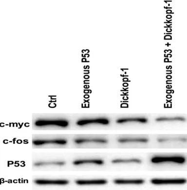
Western blot detection results showed that the combined use of exogenous P53 and DKK1 could significantly inhibit the protein expression of the cancer suppressor genes c-myc and c-fos, promote the protein expression of cancer suppressor gene P53, and these effects on protein also existed after the treatment of P53 or DKK1 alone, but, which was less strong than those after the treatment of the combined drugs (Figure 4).
Discussion
Osteosarcoma is a kind of malignant tumor originated from mesenchymal cells, which is
common in adolescents and children with the characteristic of early metastasis, bad progno-sis and high morality. Research found that cell proliferation and apoptosis were two important factors that could not be ignored in the occur-rence and development of tumor, and continu-ous proliferation and division were important biological characteristics which differed tumor cells from normal cells [13-15]. Therefore, we explored the inhibitory effects of the exogenous P53 and DKK1 on the proliferation of human osteosarcoma cell strain MG-63 cells which can be considered as an effective means to find anti-tumor drugs.
In this study, the cell growth curve showed that the growth of MG-63 cells increased by 16 times after 7 days. After the treatment of exog-enous P53 combined with DKK1, the growth rate of MG-63 cells was significantly inhibited, and the inhibition rate was 43.9% in the sev-enth days (Figure 1), indicating the drug combi-nation therapy can significantly inhibit the pro -liferation of osteosarcoma cell strain MG-63, and the difference was statistically significant. Through PI staining and flow cytometer detec -tion, we found that combined use of exogenous P53 and DKK1 significantly blocked MG-63 cells in G0/G1 phase (Figure 2, P<0.05), and it was consistent with what HanKi Park et al reported that green tea polyphenols could cause contact inhibition of osteosarcoma cells and the increase of cell distribution in G0/G1 phase [16]. We also found that the percentage of apoptosis of MG-63 cells treated by exoge-nous P53 combined with DKK1 (16.73%) was significantly increased (Figure 3, P<0.05), com-pared with the control group (0.56%), which fur-ther proved that the combined use of exoge-nous P53 and DKK1 could inhibit the growth and proliferation of osteosarcoma cells.
With the process of molecular biology and molecular technology, the research on the dis-ease develops gradually from cells to molecular mechanisms. Different cancer related genes exist in normal cells of human body, and they are divided into proto-oncogenes and tumor suppressor genes, which maintain dynamic bal-ance to regulate cell growth and differentiation, and henogenesis. The proto-oncogene c-myc was found firstly in the v-myc gene separated from chicken virus, and it is a kind of nuclear transcription factor, proto-oncogene, which can promote cell proliferation and inhibit cell
[image:5.612.94.286.75.270.2]entiation, and play an important role in the regulation of cell proliferation, differentiation and apoptosis, meanwhile, there are c-myc overexpression phenomena in a variety of human cancer tissues [17]. C-fos, a kind of endonuclear proto-oncogene, is called tran-sient gene for its quick expression to external stimulus in the cell. Related research showed c-fos played a key role in the occurrence and development of multiple tumors [18, 19]. But expression of c-myc and c-fos in osteosarco- ma is rarely reported [20, 21], so this study explored the effects of the combined use of exogenous P53 and DKK1 on the protein expression of proto-oncogene. The results shows that the combined use of P53 and DKK1 could obviously inhibit the expression of cancer gene c-myc and c-fos protein, and increase the expression of tumor suppressor gene P53 pro-tein, which suggests that exogenous P53 com-bined with DKK1 could regulate cell prolifera-tion by affecting the expression of cancer related genes to strengthen its anti-tumor effects.
In summary, exogenous P53 combined with DKK1 has obvious inhibitory effects on the pro-liferation and differentiation of human osteo-sarcoma MG-63 cells, which might be closely related with the inhibition of c-myc and c-fos gene and the increasing expression of tumor suppressor gene P53. The specific regulation mechanisms required in-depth study.
Disclosure of conflict of interest
None.
Address correspondence to: Guangjun Liao, De- partment of Orthopedic Surgery, Yantaishan Ho- spital, No. 91 Jiefang Road, Yantai 264000, Shandong, China. Tel: +86-0535-6602001; E-mail: [email protected]
References
[1] Unni KK. Osteosarcoma of bone. J Orthop Sci 1998; 3: 287-294.
[2] Lam KY. Characteristics of malignant tumors in young people, with particular emphasis on carcinomas and sarcomas. Cancer Detection & Prevention 2001; 25: 223-230.
[3] Tang S, Guo W, Yang RL, Tang XD, Li DS and Dong S. [Surgical treatment and prognostic analysis of osteosarcoma in adults older than 40 years]. Journal of Peking University 2015; 47: 165-169.
[4] Rosen G. An opinion supporting the role of high-dose methotrexate in the treatment of os-teosarcoma. Cancer Treat Res 1993; 62: 49-54.
[5] Bieging KT, Mello SS and Attardi LD. Unravel-ling mechanisms of p53-mediated tumour suppression. Nat Rev Cancer 2014; 14: 359. [6] Oda S. p53-dependent suppression of genome
instability in germ cells. Mutat Res 2014; 760: 24-32.
[7] Zhang T, Wang YY, Fan X, Ma J, Li S, Jiang T and Wang L. Anatomical localization of p53 mutat-ed tumors: a radiographic study of human glio-blastomas. J Neurol Sci 2014; 346: 94-98. [8] Muller PA and Vousden KH. Mutant p53 in
can-cer: new functions and therapeutic opportuni-ties. Cancer Cell 2014; 25: 304.
[9] Hirata H, Hinoda Y, Nakajima K, Kawamoto K, Kikuno N, Ueno K, Yamamura S, Zaman MS, Khatri G and Yi C. Wnt antagonist DKK1 acts as a tumor suppressor gene that induces apoptosis and inhibits proliferation in human renal cell carcinoma. Int J Cancer 2011; 128: 1793-1803.
[10] Shan M and Zhou FL. Advances in the role of Dickkopf (DKK1) in myeloma bone disease and its targeted therapy. Tumor 2011; 31: 273-276.
[11] Chamorro MN, Schwartz DR, Vonica A, Brivan-lou AH, Cho KR and Varmus HE. FGF-20 and DKK1 are transcriptional targets of beta-catenin and FGF-20 is implicated in cancer and development. EMBO J 2005; 24: 73-84. [12] Patil MA, Chua MS, Pan KH, Lin R, Lih CJ,
Cheung ST, Ho C, Li R, Fan ST, Cohen SN and Chen X. An integrated data analysis approach to characterize genes highly expressed in he-patocellular carcinoma. Oncogene 2005; 24: 3737.
[13] Kenny LM, Vigushin DM, Alnahhas A, Osman S, Luthra SK, Shousha S, Coombes RC and Abo-agye EO. Quantification of cellular proliferation in tumor and normal tissues of patients with breast cancer by [18F] fluorothymidine-posi-tron emission tomography imaging: evaluation of analytical methods. Cancer Res 2005; 65: 10104-10112.
[14] Nicco C, Laurent A, Chereau C, Weill B and Bat-teux F. Differential modulation of normal and tumor cell proliferation by reactive oxygen spe-cies. Biomed Pharmacother 2005; 59: 169-174.
[15] Braeuning A. Liver cell proliferation and tumor promotion by phenobarbital: relevance for hu-mans? Arch Toxicol 2014; 88: 1771-1772. [16] Park HK, Han DW, Park YH and Park JC.
[17] Franco M, Shastri AJ, Boothroyd JC. Infection by toxoplasma gondii specifically induces host c-Myc and the genes this pivotal transcription factor regulates. Eukaryot Cell 2014; 13: 483. [18] Geng CJ, Zhang GQ, Wang ZX and Dermatology
DO. Expressions and significance of c-fos and c-myc protein in skin squamous cell carcino-ma. Hebei Medical Journal 2014; 6: 822-824. [19] Wang CM, Li XY, Shi QF and Chen Y. Correlation
of proto-oncogene c-myc and c-fos expression with the occurrence of hepatocellular carcino-ma. Journal of Hainan Medical University 2014; 4: 433-436.
[20] Gamberi G, Benassi MS, Bohling T, Ragazzini P, Molendini L, Sollazzo MR, Pompetti F, Merli M, Magagnoli G, Balladelli A and Picci P. C-myc and c-fos in human osteosarcoma: prognostic value of mRNA and protein expression. Oncol-ogy 1998; 55: 556-563.