Acta Crystallographica Section F Structural Biology and Crystallization Communications
ISSN 1744-3091
Crystallization and preliminary diffraction analysis
of Wzi, a member of the capsule export and
assembly pathway in
Escherichia coli
Simon R. Bushell,aHubing Lou,a Gregor D. Wallat,aKonstantinos Beis,a‡ Chris Whitfieldband James H. Naismitha*
aBiomedical Sciences Research Complex,
University of St Andrews, North Haugh, St Andrews KY16 9ST, Scotland, and
bDepartment of Molecular and Cell Biology,
University of Guelph, Ontario N1G 2W1, Canada
‡ Current address: Membrane Protein Laboratory, Diamond Light Source, Imperial College, Chilton OX11 0DE, England.
Correspondence e-mail: [email protected]
Received 20 September 2010 Accepted 9 October 2010
External polysaccharide capsules provide a physical barrier that is employed by many species of bacteria for the purposes of host evasion and persistence. Wzi is a 53 kDa outer membrane-barrel protein that is thought to play a role in the attachment of group 1 capsular polysaccharides to the cell surface. The purification and crystallization of an Escherichia coli homologue of Wzi is reported and diffraction data from native and selenomethionine-incorporated protein crystals are presented. Crystals of C-terminally His6-tagged Wzi diffracted to 2.8 A˚ resolution. Data processing showed that the crystals belonged to the orthorhombic space group C222, with unit-cell parameters a= 128.8,b= 152.8,c= 94.4 A˚ ,=== 90. A His-tagged
selenomethionine-containing variant of Wzi has also been crystallized in the same space group and diffraction data have been recorded to 3.8 A˚ resolution. Data processing shows that the variant crystal has similar unit-cell parameters to the native crystal.
1. Introduction
Extracellular polysaccharide capsules are produced by many species of bacteria to provide protection from the environment. In patho-gens, capsules are often considered to be antiphagocytic structures, but they play other diverse roles depending on the species and the structure of the polysaccharide (Horwitz, 1982). The capsule primarily consists of long chains of repeat-unit polysaccharides that are firmly attached to the bacterial outer membrane by processes that are frequently not understood. Structural variation inEscherichia coli
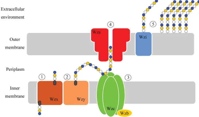
capsular polysaccharides (CPSs) gives rise to more than 80 different K (capsular) antigen types (Whitfield, 2006). Capsules are classified into four groups depending on various biochemical and genetic criteria (Whitfield, 2006; Whitfield & Roberts, 1999). The model system for group 1 capsules is theE. coliK30 serotype, which utilizes the Wzy-dependent pathway for capsular synthesis and export (Fig. 1).
InE. coliK30, the 16 kb capsule-biosynthesis (cps) gene cluster encodes activities required for the biosynthesis, export and assembly of the capsular structure. The locus contains 12 ORFs, four of which (wzi,wza,wzcandwzb) encode proteins involved in the assembly and export of CPS, while the remainder encode proteins responsible for CPS biogenesis. Undecaprenol diphosphate-linked CPS repeat units are synthesized at the cytoplasmic face of the inner membrane and are transported across the bilayer by the flippase Wzx. In the periplasm, these building blocks are polymerized by Wzy (Whitfield, 2006). Higher order polymerization is facilitated by Wzc, a trans-membrane tyrosine kinase whose function is moderated by Wzb, its cytoplasmic cognate phosphatase (Hagelueken et al., 2009; Wuge-ditsch et al., 2001). The completed polymer is then transported to the bacterial surface via Wza, which acts as the pathway’s outer membrane channel (Beiset al., 2004; Donget al., 2006; Nesperet al., 2003).
(which use virtually identical Wzi, Wza, Wzb and Wzc proteins; Rahn
et al., 1999) show a profoundly altered morphology of the capsule (Alvarez et al., 2000; Rahn et al., 2003). This is the result of a markedly increased secretion of unattached exopolysaccharide into the environment and a commensurate decrease in attached CPS (Alvarez et al., 2000; Rahnet al., 2003). Wzi proteins are found in representatives from pathogens including E. coli and the genera
Acinetobacter,Klebsiella,Providenciaand Serratia, as well as free-living bacteria such as Shewanella. Interestingly, several other bacteria utilize similar Wzy-dependent pathways for the biosynthesis of predominantly secreted, rather than attached, exopolysaccharides. Although these bacteria contain homologues of Wza, Wzb and Wzc (which are presumably needed to make and export polysaccharide), they lack any obvious homologue of Wzi. Examples of these exo-polysaccharides include amylovoran and stewartan from the plant pathogens Erwinia amylovora (Bugert & Geider, 1995) and
E. stewartii(Carlieret al., 2009), respectively, and colanic acid from
Escherichia coli(Stevensonet al., 1996). Taken together, the collec-tive evidence strongly suggests that Wzi plays a key role in the attachment of the capsule to the bacterial surface. By determining its structure, it may be possible to determine how it performs this vital role. In this report, we describe the purification and crystallization of recombinant Wzi and report on the progress of crystallographic diffraction studies.
2. Experimental
2.1. Expression and purification
DNA encoding the Wzi homologue fromE. coliB44 (O9:K30:H ) was amplified using PCR primers that introduced a C-terminal hexahistidine tag into the gene product and was cloned into a pBAD24 vector, forming the plasmid pWQ192, as described pre-viously (Rahnet al., 2003). N-terminal tagging was inappropriate as Wzi has a signal sequence that is cleaved during passage across the inner membrane. For the expression of native Wzi-His6, overnight
cultures ofE. coliDH5(transformed to ampicillin resistance with pWQ192) were grown at 310 K in Luria–Bertani (LB) medium supplemented with ampicillin (100mg ml 1) and used to inoculate 10 l LB broth. Expression of Wzi was inducedviathe addition of 0.02%(w/v) arabinose upon the cells reaching an OD600 of 0.6.
Expression continued for 4 h at 310 K before the cells were harvested by centrifugation and stored at 193 K until required.
For the expression of selenomethionine-variant Wzi-His6, the
methionine auxotroph E. coli B834 (DE3) strain was transformed to ampicillin resistance with pWQ192. Cultures of the transformed bacteria were grown overnight in LB in the presence of 100mg ml 1
ampicillin. Overnight cultures were harvested by centrifugation (4250gfor 20 min at 293 K) and washed twice with PBS before being used to inoculate 61 l Glucose-Free SelenoMet Medium (Mole-cular Dimensions, Newmarket, England) supplemented with 0.4%(v/v) glycerol as a carbon source and 100mg ml 1ampicillin. The
media contained 40mg ml 1 l(+)-selenomethionine and were
prepared according to the manufacturer’s instructions. Cells were grown at 310 K to an OD600of0.6 and expression was induced by
the addition of 0.02%(w/v) arabinose for 4 h at 310 K. Expression was continued overnight at 298 K before the cells were harvested by centrifugation at 7000gand frozen at 193 K until required.
Both native and selenomethionine-labelled recombinant Wzi-His6
were purified from harvested cells using a modified protocol adapted from Rahnet al.(2003). Frozen cells were thawed in bufferA(20 mM
[image:2.610.130.477.491.696.2]phosphate buffer pH 7.4, 50 mMNaCl) and mechanically lysed using a pre-chilled Constant Systems Z Plus series cell disrupter (Constant Systems, Northants, England). Unbroken cells and cell debris were removed by centrifugation at 6000g for 20 min. The membrane fraction was isolated from the resultant supernatant by ultra-centrifugation at 120 000g for 1 h. Inner membranes in the total membrane fraction were selectively solubilized by resuspending the pellet in buffer Acontaining 2%(w/v)N-lauroylsarcosine (Sigma– Aldrich UK, catalogue No. L5125) and incubating at 277 K with stirring for 3 h. The volume of this solubilization was 20 ml per litre of original culture. The outer membrane fraction was isolated as a pellet
Figure 1
after further ultracentrifugation at 120 000gfor 1 h and solubilized overnight in 120 ml bufferAcontaining 0.5%(w/v) SB3.14 [3-(N,N -dimethylmyristylammonio)propanesulfonate; Sigma–Aldrich UK,
catalogue No. T7763]. Insoluble material was removed by ultra-centrifugation at 120 000gfor 1 h. The supernatant was applied onto a 5 ml HisTrap nickel column (GE Healthcare, Uppsala, Sweden) pre-equilibrated in 20 mM sodium phosphate pH 7.4, 50 mM NaCl, 0.05%(w/v) SB3.14. Once Wzi-His6 was bound, the column was
washed with five column volumes of 20 mMsodium phosphate pH 7.4, 50 mMNaCl, 0.1%(w/v) LDAO (n-dodecyl-N,N
-dimethylamine-N-oxide; Anatrace, Maumee, USA, catalogue No. D360). Wzi-His6
was eluted by a step gradient of wash buffer with increasing amounts of imidazole (12.5, 25, 50, 100 and 500 mM). Wzi-His6readily eluted
in 50 mMimidazole, which is an unusually low concentration for a hexahistidine-tagged protein; however, SDS–PAGE analysis of peak fractions showed it to be nearly homogenous. Final purification was performedvia gel-filtration chromatography using a Superdex 200 10/300 size-exclusion column (GE Healthcare, Uppsala, Sweden) equilibrated in 20 mMTris pH 7.4, 0.1% LDAO. Gel filtration of the selenomethionine form of Wzi-His6 used the same buffer but
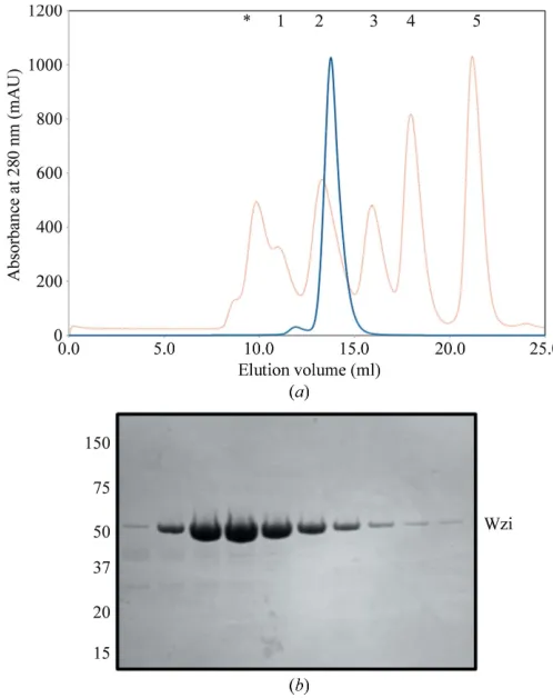
supplemented with 0.5 mMtris(2-carboxyethyl)phosphine (TCEP). The elution profile indicated a single oligomeric state of Wzi-His6
in solution. Comparison with known protein standards suggested that Wzi elutes as a monomer–detergent complex (Fig. 2a). When plotted on a calibration curve, the Wzi–detergent complex was calculated to have an approximate molecular weight of 80 kDa, which is in reasonable agreement with the molecular weight of Wzi of 53 kDa and an LDAO micelle size of25 kDa (Strop & Brunger, 2005). The purification purity was monitored at all stages via SDS–PAGE (Fig. 2b), with no other contaminants being visible on a Coomassie Blue-stained gel. The identity of recombinant Wzi-His6 was
con-firmed by Western blot using an antihexahistidine monoclonal anti-body andviaMALDI–TOF mass-spectrometric analysis of peptides derived from a tryptic digest. The final yield of the native form of the protein was approximately 0.4 mg per litre of culture. The yield of the selenomethionine variant was higher at 0.7 mg per litre of culture, most likely owing to the longer overnight expression. Wzi was con-centrated to12 mg ml 1for crystallization trials.
2.2. Crystallization
Several commercially available screens were employed to deter-mine the crystallization space of Wzi-His6. Crystallization trays were
[image:3.610.43.292.121.435.2]generally constructed using a Cartesian Honeybee 963 liquid-handing
Figure 2
Purification of native Wzi-His6. (a) Elution profile of native Wzi-His6(blue line) on
size-exclusion chromatography using a Superdex 200 10/300 column. The elution was standardized using a mixture containing (1) thyroglobulin (670 kDa), (2) -globulin (158 kDa), (3) ovalbumin (44 kDa), (4) myoglobin (17 kDa) and (5) vitamin B12(1350 Da) (red line). The protein aggregate peak (marked with an
asterisk) represents the void volume of the column. (b) SDS–PAGE analysis of peak fractions from SEC purification, showing the homogeneity of Wzi-His6
[image:3.610.108.505.535.724.2]immediately prior to crystallization. The protein runs to its true size of 52 kDa upon boiling.
Figure 3
Crystals of native and selenomethionine-containing forms of Wzi-His6. (a) Native crystals grown in 0.03MCaCl2, 25%(v/v) PEG 350 MME, 0.1MMES pH 6.5. (b)
robot in Innovadyne SD-2 96-well trays (IDEX Corp, Lake Forest, USA) with 60ml precipitant dispensed into the reservoir. Crystal-lization was performedviasitting-drop vapour diffusion at 293 K by mixing 150 nl Wzi-His6 solution (at 12 mg ml 1) with 150 nl well
solution. Native Wzi-His6crystallized in several different conditions,
with crystals taking between one and eight weeks to appear. Oval-shaped crystals grew in 12.5%(w/v) PEG 200 MME, 0.1Msodium cacodylate pH 6.5; however, these crystals proved to be difficult to reproduce and were marked by severe anisotropy, most likely owing
to their shape. The best crystals appeared in screens optimized for the crystallization of membrane proteins, specifically MembFac (Hampton Research, Aliso Vijeo, USA), MemGold and MemStart/ MemSys (Molecular Dimensions, Newmarket, England). Broad screen hits were improved by varying the pH, salt and PEG concentrations. The most tractable crystals grew in 0.03M CaCl2,
25%(v/v) PEG 350 MME, 0.1MMES pH 6.5. The crystals resembled flattened hexagonal rods and usually grew attached to the bottom of the crystallization well. Small crystals appeared after3 d and grew to approximately 30mm in size within 7–10 d (Fig. 3a).
Selenomethionine-labelled Wzi-His6 crystals grew under slightly
different conditions, with the best crystals forming in 0.15MCaCl2,
23% PEG 350 MME, 0.1MMES pH 6.0. These crystals were of a similar morphology to those of the native form and grew to a comparable size (Fig. 3b). However, these crystals grew more quickly, appearing overnight and growing to full size (30mm) within 2–3 d.
All crystals used the drop itself as a cryoprotectant. Crystals were flash-cooled in liquid N2, either through immersion in pre-cooled
robot pucks or by freezing the crystals directly in the cryostream prior to collection. Initial diffraction data showed that the majority of crystals frequently displayed moderate to severe anisotropy. Varia-tion of cryoprotecVaria-tion condiVaria-tions did not demonstrate any improve-ment of this problem. Rather, crystals were screened in advance in order to select the least anisotropic for further data collection.
2.3. Data collection
Data sets from native Wzi-His6 crystals were collected on the
ID14-4 beamline at the ESRF, Grenoble, France. A 2.8 A˚ resolution data set (Fig. 4) was collected from a single native Wzi-His6crystal.
The data were processed withXDS using the xia2 data-reduction pipeline (Kabsch, 2010; Winter, 2010).xia2 usedPOINTLESSand
LABELITto assist it in indexing the data (Collaborative Computa-tional Project, Number 4, 1994; Sauteret al., 2004). During proces-sing, several wedges of data were reduced separately in xia2 and subsequently merged withSCALA(Evans, 2006).
Data processing showed that the native crystals belonged to the orthorhombic space groupC222, with unit-cell parametersa= 128.8,
b= 152.8,c= 94.4 A˚ ,=== 90. The full details of this data set
are described in Table 1. Matthews coefficient analysis suggested that either one or two monomers of the protein are present in the asymmetric unit (VM = 4.46 or 2.23 A˚
3Da 1, respectively), with a
[image:4.610.314.566.103.247.2]solvent content of 72 or 45%, respectively (Kantardjieff & Rupp, 2003; Matthews, 1968). Although 72% solvent content is at the upper range of commonly observed values, the anisotropic nature of the
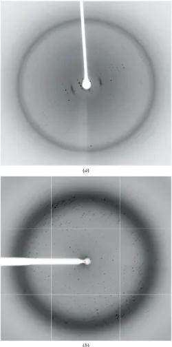
Figure 4
Native crystal diffraction. (a) Diffraction pattern from a typical native Wzi-His6
crystal showing high anisotropy. Data were collected in-house using a Saturn 944+ CCD detector attached to a Rigaku MicroMax-007 HFM X-ray source (= 1.54178 A˚ ). Such crystals were discarded without further experimentation. (b) Diffraction pattern from the native Wzi-His6crystal used to derive the data in
Table 1.
Table 1
Data-collection and processing statistics.
Values in parentheses are for the last shell.
Native Wzi SeMet Wzi
Source ESRF, ID14-4 DLS, I24
Wavelength (A˚ ) 0.9255 0.9778
Oscillation () 0.65 1.0
Space group C222 C222
Unit-cell parameters (A˚ ,)
a= 133.2,b= 152.3,c= 95.3, === 90
a= 128.8,b= 152.8,c= 94.4, === 90
Resolution (A˚ ) 50–2.8 53–3.8
Total reflections 132346 41664
Unique reflections 23861 9484
Mosaicity () 0.72 0.14
Anomalous correlation — 0.114 (0.047)
Completeness (%) 98.5 (98.7) 99.9 (100)
Multiplicity 5.5 (4.7) 4.4 (4.6)
MeanI/(I) 13.7 (2.7) 7.6 (3.0)
[image:4.610.46.296.184.688.2]diffraction, the absence of any noncrystallographic symmetry and the fragility of the crystals point towards the presence of a monomer in the asymmetric unit.
Although the sequence of Wzi suggests that it is a-barrel, we have not identified a matching structure and all attempts at molecular replacement failed. The selenomethionine variant of Wzi-His6was
crystallized in order to determine the phases using single-wavelength anomalous dispersion (SAD). Data sets from crystals that showed promising in-house diffraction were collected on beamline I24 at the Diamond Light Source (DLS), Didcot, England using a Pilatus 6M detector (= 0.9778 A˚ ). Several data sets were collected, with the best example containing diffaction to 3.8 A˚ resolution. These data were indexed and scaled usingXDSinxia2 using the same approach as described for the native form (Kabsch, 2010; Winter, 2010). Processing showed that these crystals also belonged to space group
C222 and they showed similar unit-cell parameters to the native protein (Table 1). Fluorescence scans of crystals of the seleno-methionine variant showed that selenoseleno-methionine had been incor-porated into the protein. Data reduction indicated the presence of anomalous scattering by measuring the difference between |I | and |I+|. We have not yet been able to solve the substructure of Se atoms.
3. Discussion
We have obtained crystals of the integral membraneE. coliprotein Wzi. Although crystals appeared in initial screens, extensive optimi-zation was required in order to obtain crystals which diffracted to beyond 6 A˚ resolution. The native crystals diffracted to 2.8 A˚ reso-lution, suggesting that it will be possible to report a well defined experimental structure for this protein. Sequence analysis suggests the protein will be a-barrel, but with a structure different to those observed previously. Gel filtration and crystal packing are consistent with a monomeric protein. We anticipate that solution of the struc-ture of the selenomethionine variant of Wzi-His6will require higher
quality diffraction data. Efforts to improve the crystal quality for the selenomethionine protein are continuing.
This work was supported by funding from the Wellcome Trust (program grant to JHN and CW). Access to the Diamond and ESRF synchrotrons is gratefully acknowledged. The authors would also like to gratefully acknowledge Ms Victoria Elder (St Andrews) for her valuable input.
References
Alvarez, D., Merino, S., Toma´s, J. M., Benedı´, V. J. & Albertı´, S. (2000).Infect. Immun.68, 953–955.
Beis, K., Collins, R. F., Ford, R. C., Kamis, A. B., Whitfield, C. & Naismith, J. H. (2004).J. Biol. Chem.279, 28227–28232.
Bugert, P. & Geider, K. (1995).Mol. Microbiol.15, 917–933.
Carlier, A., Burbank, L. & von Bodman, S. B. (2009).Mol. Microbiol.74, 903–913.
Collaborative Computational Project, Number 4 (1994).Acta Cryst. D50, 760–763.
Dong, C., Beis, K., Nesper, J., Brunkan-Lamontagne, A. L., Clarke, B. R., Whitfield, C. & Naismith, J. H. (2006). Nature (London), 444, 226– 229.
Drummelsmith, J. & Whitfield, C. (1999).Mol. Microbiol.31, 1321–1332. Evans, P. (2006).Acta Cryst.D62, 72–82.
Hagelueken, G., Huang, H., Mainprize, I. L., Whitfield, C. & Naismith, J. H. (2009).J. Mol. Biol.392, 678–688.
Horwitz, M. A. (1982).Rev. Infect. Dis.4, 104–123. Kabsch, W. (2010).Acta Cryst.D66, 125–132.
Kantardjieff, K. A. & Rupp, B. (2003).Protein Sci.12, 1865–1871. Matthews, B. W. (1968).J. Mol. Biol.33, 491–497.
Nesper, J., Hill, C. M. D., Paiment, A., Harauz, G., Beis, K., Naismith, J. H. & Whitfield, C. (2003).J. Biol. Chem.278, 49763–49772.
Rahn, A., Beis, K., Naismith, J. H. & Whitfield, C. (2003).J. Bacteriol.185, 5882–5890.
Rahn, A., Drummelsmith, J. & Whitfield, C. (1999).J. Bacteriol.181, 2307– 2313.
Sauter, N. K., Grosse-Kunstleve, R. W. & Adams, P. D. (2004).J. Appl. Cryst.
37, 399–409.
Stevenson, G., Andrianopoulos, K., Hobbs, M. & Reeves, P. R. (1996).J. Bacteriol.178, 4885–4893.
Strop, P. & Brunger, A. T. (2005).Protein Sci.14, 2207–2211. Whitfield, C. (2006).Annu. Rev. Biochem.75, 39–68.
Whitfield, C. & Roberts, I. S. (1999).Mol. Microbiol.31, 1307–1319. Winter, G. (2010).J. Appl. Cryst.43, 186–190.