INTRODUCTION
The first paper in this two part series reported relationships of form and function of the mechanosensory lateral line canal system in three stingray species; in the present study we explore similar relationships in the electrosensory system to further understand how these animals use sensory modalities other than vision to capture prey in their ventral mouths. Like the lateral line canal system, the electrosensory system is highly modified in batoid fishes. In stingrays the canals extend over the ventral body surface and out toward the wing tips with increased density surrounding the mouth (Chu and Wen, 1979; Raschi, 1986; Jordan, 2008). Electric signals have a short range relative to visual and olfactory signals and provide directional information for locating buried prey and directing the mouth strike to ingest prey. Previous studies of elasmobranchs have demonstrated feeding responses to weak electric signals (Kalmijn, 1971; Kalmijn, 1978; Kalmijn, 1982; Tricas, 1982; Johnson et al., 1984; Tricas and McCosker, 1984; Blonder and Alevizon, 1988; Kajiura and Holland, 2002; Whitehead, 2002); however, detection capabilities have rarely been related to interspecific anatomical differences.
The electrosensory system in marine elasmobranchs consists of pores at the skin’s surface, which lead through canals to sensory cells located in ampulla clusters in the head (Chu and Wen, 1979; Raschi, 1986; Tricas, 2001). This system enables detection of weak electric fields such as those generated by living organisms, which can be mimicked using dipole electrodes (Kalmijn, 1971; Kalmijn, 1982; Tricas, 1982; Johnson et al., 1984; Tricas and McCosker, 1984; Haine et al., 2001; Kajiura and Holland, 2002). Despite considerable interspecific variation in the number, density and distribution of electrosensory pores, few studies have compared the
detection capabilities of species with quantified morphological differences. Carcharhinid and sphyrnid sharks with similar pore densities yet different pore numbers showed similar behavioral-response thresholds to dipole electric fields (Kajiura and Holland, 2002). Benthic-feeding elasmobranchs typically have high electrosensory pore numbers and densities whereas pelagic species have a lower pore number and density with more similar dorso-ventral distributions (Raschi, 1986; Kajiura, 2001; Raschi et al., 2001; Cornett, 2006; Jordan, 2008).
The present paper reports differing behavioral responses to electric signals in three Eastern Pacific stingray species with significant differences in sensory morphology. Ventral electrosensory pore numbers range from 1200±27 and 1425±41 in Urobatis halleriand Myliobatis californica, respectively, to just 553±26 in Pteroplatytrygon violacea(Jordan, 2008). The density of pores within the ventral pore field in the two benthically feeding species is more than three times that of the pelagic stingray P. violacea(Jordan, 2008). While factors such as canal length and convergence ratios also influence sensitivity, high ventral electrosensory pore numbers and densities may provide enhanced electro-sensitivity to benthic-feeding elasmobranchs; therefore, we hypothesize that both U. halleriand M. californicawill demonstrate greater resolution in locating weak dipole electric signals relative to the pelagic stingray P. violacea.
MATERIALS AND METHODS Experimental animals
The experimental animals and study design are described in detail in part I (Jordan et al., 2009) and are briefly outlined here. Twenty-five round stingrays Urobatis halleriCooper [12 females, 13 males; The Journal of Experimental Biology 212, 3044-3050
Published by The Company of Biologists 2009 doi:10.1242/jeb.028738
Functional consequences of structural differences in stingray sensory systems.
Part II: electrosensory system
Laura K. Jordan
1,*, Stephen M. Kajiura
2and Malcolm S. Gordon
11Ecology and Evolutionary Biology, University of California at Los Angeles, 621 Charles E. Young Drive South, Los Angeles,
CA 90095, USA and 2Biological Sciences, Florida Atlantic University, 777 Glades Road, Boca Raton, FL 33431, USA
*Author for correspondence ([email protected])
Accepted 6 July 2009
SUMMARY
Elasmobranch fishes (sharks, skates and rays) possess highly sensitive electrosensory systems, which enable them to detect weak electric fields such as those produced by potential prey organisms. Different species have unique electrosensory pore numbers, densities and distributions. Functional differences in detection capabilities resulting from these structural differences are largely unknown. Stingrays and other batoid fishes have eyes positioned on the opposite side of the body from the mouth. Furthermore, they often feed on buried prey, which can be located non-visually using the electrosensory system. In the present study we test functional predictions based on structural differences in three stingray species (Urobatis halleri, Pteroplatytrygon violaceaandMyliobatis californica) with differing electrosensory system morphology. We compare detection capabilities based upon behavioral responses to dipole electric signals (5.3–9.6μA). Species with greater ventral pore numbers and densities were predicted to demonstrate enhanced electrosensory capabilities. Electric field intensities at orientation were similar among these species, although they differed in response type and orientation pathway. Minimum voltage gradients eliciting feeding responses were well below 1 nV cm–1for all species regardless of pore number and density.
Supplementary material available online at http://jeb.biologists.org/cgi/content/full/212/19/3044/DC1
disc width (DW)=9.5–24.0 cm], six pelagic stingrays Pteroplatytrygon violacea Bonaparte (five females, one male; DW=49.5–60.0 cm) and 14 bat rays Myliobatis californicaGill (five females, nine males; DW=26.5–38.5 cm) were held at Wrigley Marine Science Center (WMSC) on Santa Catalina Island, CA, USA (33°30⬘18.52⬙N, 118°30⬘36.32⬙W) in 2.4 m diameter, 1 m deep outdoor fiberglass tanks with flow through ambient seawater ranging from 18 to 25°C at 35 p.p.t. Rays were held for a total of 3–5 weeks and tested in behavioral trials only after normal feeding was observed in the holding tank, usually within one week after capture. All work with these animals was done during June through to September of 2006 and 2007 according to approved IACUC protocols at both USC and UCLA. Species were studied at the same time of year to avoid effects of breeding season (Sisneros and Tricas, 2000).
Study design
The experimental tank was fitted with an apparatus consisting of a 1⫻1 m acrylic plate with 6 mm holes fitted with polyethylene tubing (Tygon, Akron, OH, USA) underneath [see fig. 1 in part I (Jordan et al., 2009)]. Four dipole electrodes, with a 1 cm dipole separation distance, were connected to underwater electric cables (Impulse Enterprise, San Diego, CA, USA) and a 9 V battery source with controls to adjust the current, which was monitored by an ammeter in series (Meterman 35XP, Everett, WA, USA), and a switch to activate one of four electrodes at a time following Kajiura and Holland (Kajiura and Holland, 2002). The tank contained no metal and sat on a wooden platform for isolation from confounding electric signals.
Behavioral experiments
Prior to each trial, food was withheld for 1–2 days until rays showed sufficient motivation to search for food in the experimental tank. Strengths of electric signals were chosen to simulate signals from prey; invertebrate DC electric fields range from 1 to 100μV cm–1 (Kalmijn, 1974; Haine et al., 2001). Electric currents ranged from 5.3 to 9.6μA, producing weak dipole electric fields similar to those produced by small prey organisms. Current was varied during some early trials to ensure signals were representative of prey (i.e. larger rays feed on larger prey and might better associate a stronger signal with food).
Motivation for feeding was confirmed by observing active searching behavior on or near the experimental plate after introduction of a food odor at the start of each trial. One electric signal was activated at a time and responses were recorded on miniDV tapes at 30 frames s–1using a Sony DCR-PC109 mounted on a track above the center of the tank. Individual rays participated in up to seven 1-hour experimental trials and were exposed to both water jet and electric signals. Rays were fed only in the experimental tank after trials began and each ray was tested with 1 or 2 days in between trials. A training period was typically not necessary for electrical signals; once rays began searching on the experimental plate they readily responded to active electrodes.
Video and image analysis
Video sequences of each response to an electrode were captured and individual frames extracted using iMovie software. Measurements on these images were performed using ImageJ (NIH: http://rsb.info.nih.gov/ij). In total, 1640 electrode responses were analyzed and compared (493 U. halleri; 656 P. violacea; 491 M. californica). All statistical analyses were performed using SPSS version 15 and 16 (SPSS Inc., Chicago, IL, USA) with models
nesting multiple observations for each individual within each species. Variation in body size was standardized in statistical comparisons by centering the mean body size for each species. Differences were considered significant at P<0.05.
The frame immediately preceding the initiation of the orientation response to the electric signal was saved and analyzed using ImageJ. The distance and angle from the center of the electrode to the nearest ampulla cluster, located just posterior to the spiracle, were measured (Fig. 1). The angle relative to the dipole axis was calculated as the difference between the dipole angle and the orientation angle (Fig. 1). The electric field strength (E) at the point of orientation was calculated using the following equation: E=ρId/πr3cosθ, where ρis the resistivity of seawater (Ωcm), Iis the current (A), dis the dipole separation distance (cm), ris the distance from the dipole center (cm) and θis the orientation angle relative to the dipole axis in degrees (Kalmijn, 1982). The resistivity varied between 18.9 and 21.9Ωcm with fluctuations in temperature and salinity. In most cases the approach consisted of a right or left turn; however, when the animal was on a straight trajectory toward the dipole center the initial response was to brake with the pectoral and pelvic fins to allow a bite at the electrode. The frame immediately preceding the brake response was analyzed. Only initial responses where the ray initiated a right or left turn are included in interspecific comparisons, although rays occasionally made more than one turn to approach the center of the electrode and in some cases they passed over the dipole center and reversed backwards prior to biting. Responses were ranked from 1 to 5 for increasing intensity, where 1=slight pause and 5=bite, as described for water jets in part I (Jordan et al., 2009). No zero response rank was included as rays rarely approached an active electrode without exhibiting a response.
Initial responses to electric signals for bite responses were compared for each species using a two-level random effect regression model to compare the electric response thresholds, log transformed, between species. This model includes random and fixed effects to compare multiple observations, level 1, on each individual, level 2, between the three species. Responses at field intensities greater than 500 nV cm–1 were not included in interspecific comparisons. These responses account for less than 9% of the total number of responses for each species and are calculated at very short distances from the center of the dipole where angle measurements from ImageJ have low resolution thus increasing error in calculations of E. The minimum value of E and the maximum
Orientation distance Orientation angle Dipole axis
orientation distance for each individual with more than five total observations were compared by species using similar models to control for body size.
Spiral tracking
Dipole electrodes create electric fields with voltage equipotentials of decreasing strength with distance from the center of the electrode that extend farthest in line with the axis of the electrode. Perpendicular to this axis are current flow lines, which also decrease in intensity with distance from the electrode (Fig. 2). In order to determine which path the rays were following, two representative tracks of U. halleriindividuals were analyzed. The strength of the electric field on both right, E1, and left, E2, wing tips, near the extent of the farthest lateral electrosensory pores, were calculated for the initial orientation point and at every six video frames until the ray was close enough to the electrode that the distance, r, was less than 10d, the dipole separation distance. According to the model described by Kalmijn (Kalmijn, 1974), if a fish follows the current flow lines created by the dipole electrode, E1–E2 should be 0. Alternatively, if the fish follows the voltage equipotentials, E1–E2 should be constant along the spiral trajectory.
RESULTS
All three stingray species successfully acclimated to experimental conditions and readily searched for food during trials. Individuals of bothM. californicaand P. violaceatypically began active search and response patterns during their first experimental trial whereas individualU. hallerioften required two to three exposures to the experimental situation before they ventured onto the experimental plate. Only trials where rays attempted to feed on the plate are included in these results. Positive responses to electric signals were nearly always observed when rays passed within the range of the electric field created by an active dipole electrode. However, intra-individual variability in the electric field strength at orientation was common creating high overlap between individuals.
Electric field strength at orientation point
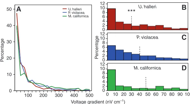
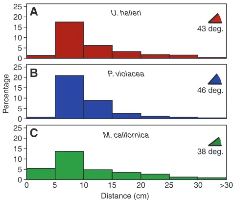
All three species showed a similar pattern with most bite responses at electric field strengths below 100 nV cm–1 (Fig. 3A). Without controlling for individual variations, a significantly lower median response was observed for U. halleri at 29 nV cm–1 (Fig. 3B) (Kruskal–Wallis, P<0.001) whereasP. violaceaand M. californica did not differ (Fig. 3B,C) (Mann–Whitney, P=0.114). When species were compared while controlling for individual variation and body size, U. halleriresponded at electric field intensities significantly lower than M. californicaand P. violacea(Table 1). The minimum electric field strength at orientation for any individual was calculated for M. californica(Table 2); however, when the minimum electric field strength recorded for each individual is averaged by species, no significant differences emerged (F=0.455, P=0.637). No intraspecific effect of body size on electric field strength at orientation was apparent for U. halleri, the species with the widest size range represented. Similarly, no consistent patterns with electrode number, turn direction and trial were observed (Table 1). Trends in orientation distance and angle varied between species; P. violaceahad a significantly greater number of responses at higher angles and tended to orient from shorter distances than the other two species even when tested at a higher current (Fig. 4). Over 50% of orientation angles were greater than 45 deg. while 92% of orientation distances were below 20 cm in P. violacea. The maximum orientation distance averaged near 30 cm for all species, although distances over 40 cm were measured for one individual of both U. halleriand M. californica.
Response levels
Overall, M. californica was the most likely to exhibit the level 5 bite response when encountering active electrodes (Table 3). The distribution of bite responses over the electric field gradient suggest that U. halleri and M. californica had a relatively high percentage of all bites at intensities below 10 nV cm–1 (30% and 26%, respectively). As field strength increased, U. halleri also displayed the greatest percentage of bites below 100 nV cm–1at 82% (Fig. 3).
Orientation pathways
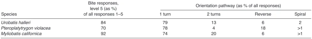
Interspecific differences were observed in the route taken to reach the center of the electrode. Individual stingrays most commonly displayed single turn orientations with a resulting trajectory leading directly to the center of the dipole, demonstrating their ability to determine the direction of the dipole source from afar (see Movie 1 in supplementary material). Urobatis halleriand M. californica typically made a single right or left turn or added a second turn to arrive at the dipole center (Table 3). Alternatively, individual P. violacea had a higher frequency of straight approaches and the largest percentage of reversals (Table 3). In reverse behaviors, the ray swam past the electrode, with some portion of the body directly overlapping with the dipole center, and then used their fins to brake and swim backwards to position the mouth over the electrode (see Movie 2 in supplementary material). While this behavior was occasionally observed in all species, U. halleriand M. californica overshot the electrode in only 6% of all bite responses (Table 3).
A small percentage of orientations included spiral tracking turns where rays appeared to follow curved voltage equipotentials to locate the center of the dipole (see Movie 3 in supplementary material) (Kajiura and Holland, 2002). This approach was observed most frequently for U. halleri in approximately 2% of orientations (Table 3). By quantifying the electric field strength on both the right and left sides of the body we determined that rays maintained a constant voltage gradient on either side of the body midline. The
difference in electric field strength calculated between right and left wing tips (E1–E2) for two tracks were fairly consistent across frames with a mean ± s.e.m. of 4.93±0.95 and 2.42±0.78 nV cm–1, respectively.
DISCUSSION
These results are the first to relate quantified morphological differences in electrosensory anatomy with behavioral differences in the detection capabilities of batoid fishes. Sensitivity to electric fields was similar for all three species despite differing electrosensory morphology; however, benthic feeders with high electrosensory pore numbers and densities, U. halleri and M. californica, responded with greater intensity and demonstrated greater resolution in locating the dipole center without first passing over it and reversing backwards.
The minimum sensitivities did not differ, implying that low thresholds may be consistent across species regardless of electrosensory pore number and distribution. This trend may exist across additional species tested for electrosensory capabilities (Table 2). Higher threshold values shown in earlier studies were
conservative estimates that did not benefit from the increased precision allowed by video analysis techniques (Table 2). It is likely that further study of those species and others with digital image analysis will reveal lower detection thresholds. The differences between these three stingray species lie in their differing response levels, accuracy in pinpointing the source of the electric signal and orientation pathways.
While the overall number of bite responses varied similarly with electric field strength in all three species, interactions between orientation angle, distance and current varied. The shorter orientation distances observed for P. violaceaare linked to high angles and may reflect a strategy for surprising prey (Kalmijn, 1988). This species is the only predatory pelagic stingray and its success depends on capturing relatively large, active prey, including squid and teleost fish (Wilson and Beckett, 1970). To accomplish this daunting task with a ventrally located mouth, these rays are rare among fish in that the acts of prey capture and ingestion are decoupled; the wings are employed to grab prey, which is then deflected toward and sucked into the mouth (personal observation). In order to enclose prey within the wings it is likely that P. violaceamust initiate the
P
ercentage
50
40
30
20
10
0
A
U. halleriP. violacea M. californica
P
ercentqge
12 10 8 6 4 2 0 12 10 8 6 4 2 0
100 0 10 20 30 40 50 60 70 80 90 12
10 8 6 4 2 0
M. californica P. violacea
U. halleri
B
C
D
***
Voltage gradient (nV cm–1)
500 400 300 200 100
Fig. 3. Percentage of orientations along the electric field gradient from 0 to 500 nV cm–1for Urobatis
halleriin red, Pteroplatytrygon violaceain blue and Myliobatis californicain green (A). Histograms of the percentage of bites at electric field intensities of <100 nV cm–1for U. halleri(B), P. violacea(C) and
M. californica, (D). Urobatis hallerihas the highest percentage of total bites below 100 nV cm–1at 82%
[image:4.612.46.374.66.246.2]whereas those byP. violaceaandM. californica make up 71% and 60%, respectively. The broken line represents the median response for each species. ***Significantly lower than the other species at P<0.001.
Table 1. Comparisons of the electric field strength (log E) where stingrays orient toward and bite at weak electrodes
Parameter Model A Model B
Intercept 1.533*** (0.057) 0.884*** (0.221)
Urobatis halleri vs Myliobatis californica –0.305** (0.079) –0.190** (0.068) Pteroplatytrygon violacea vs Myliobatis californica 0.040 (0.093) 0.088 (0.097) Urobatis halleri vs Pteroplatytrygon violacea –0.305** (0.079) –0.190** (0.068)
Body size 0.021 (0.035) –0.013 (0.030)
Initial current 0.093** (0.029)
Turn direction –0.106* (0.041)
Electrode number 1 vs4 –0.099 (0.059)
Electrode number 2 vs4 0.010 (0.059)
Electrode number 3 vs4 0.013 (0.055)
Gender 0.119 (0.062)
Variance components
Level 1, residual 0.470 (0.020) 0.466 (0.020)
Level 2, intercept 0.024 (0.011) 0.010 (0.007)
Goodness of fit
–2 log likelihood 2439.0 2416.0
AIC 2451.0 2440.0
BIC 2481.3 2500.7
[image:4.612.43.569.516.711.2]attack from fairly close proximity. The maximum distances measured for each stingray, however, did not significantly differ by species indicating a similar orientation range.
The larger number of responses at high angles by P. violacea may indicate a difference in signal characteristics used to direct behavior. These rays often entered the electric field but continued on a straight trajectory until they reached the steep gradient drop near 90 deg. from the dipole axis. By contrast, U. halleri and M. californica appeared to respond more frequently to an increasing electric field, making a single turn or adding a second turn to reach the center (Table 3). Pteroplatytrygon violaceaovershot the center of the electrode much more frequently and then reversed their trajectory to swim backwards to position the mouth over the dipole center. There are several possible explanations for this behavior: (1) accuracy may be reduced due to the lower pore number and density; (2) movement of prey targets may be anticipated as they typically feed on fast moving, pelagic prey; or (3) these movements may have been exaggerated due to the larger size and slower movements of P. violacea individuals. Because all rays were
trained in the same way to modify their search and feeding behavior to the experimental situation it seems unlikely they would continue to anticipate movement of the electric field source. Distinctive reverse movements were observed in all species and videos were compared frame-by-frame so it is unlikely that a significant number of less obvious reverse behaviors in U. halleriand M. californica were ignored. Therefore, the differences in anatomy most likely explain the observed differences in approach to the electrode.
Urobatis halleriand M. californicawere much more likely to bite at an electrode than exhibit a weaker response such as pausing or turning without biting (Table 3). One possible explanation for the lower bite rate seen in P. violaceamay be a lack of confidence in the likelihood of finding food at the dipole without additional sensory input in combination with the electric signals. An inverse relationship exists between electrosensory anatomy and the importance of vision in pelagic elasmobranchs (Raschi et al., 2001), indicating that pelagic rays may rely more heavily on visual input for locating prey. As with water jets (see part I, Jordan et al., 2009), when ‘trick’ visual signals were given indicating the presence of food rewards, these rays more aggressively responded to the electric signals. Visual input may be unimportant during the final approach and strike to capture prey (Gardiner and Atema, 2007); however, it may increase attention and responsiveness to the highly directional hydrodynamic and electric signals used in the final stages of capture. Because prey of U. halleriand M. californicaare typically buried (Babel, 1967; Talent, 1982; Barry et al., 1996; Gray et al., 1997; Valadez-Gonzalez et al., 2001), visual signals are expected to be less important for determining where to begin digging. Therefore, tactile, hydrodynamic or electric signals in the absence of visual input may be reliable indicators warranting high levels of interest in examining the signal source.
Spiral tracking
[image:5.612.50.567.78.186.2]Kalmijn explored the question of whether electrosensory fish act as a voltmeter or a current meter when locating a dipole source and concluded that they are essentially voltmeters (Kalmijn, 1974). African electric fish capable of passive electroreception, however, cannot directly determine the distance and direction of a dipole source, yet they can follow current flow lines to indirectly locate the center of the dipole (Schluger and Hopkins, 1987; Davis and Hopkins, 1988). The fan-shaped array of electrosensors present in elasmobranch fishes enable them to either directly determine both the distance and angle to reach the dipole center or to orient allowing no rotation of the field relative to the body axes, which leads them to the center without having to determine the exact location from afar (Kalmijn, 2000). Both strategies result in a direct orientation upon detection of the electric field as was seen in the majority of responses consisting of a single turn. In these orientations, a wider wingspan with lateral extension of electrosensory pores in Table 2. Minimum electrosensory response threshold comparison between elasmobranchs tested under similar conditions
Species Common name Minimum (nV cm–1) Reference
Urobatis halleri Round stingray 0.3 Present study
Pteroplatytrygon violacea Pelagic stingray 0.3 Present study
Myliobatis californica Bat ray 0.1 Present study
Sphyrna lewini Scalloped hammerhead 0.4 Kajiura and Holland, 2002 Carcharhinus plumbeus Sandbar shark 0.5 Kajiura and Holland, 2002
Sphyrna tiburo Bonnethead shark <1 Kajiura, 2003
Himantura granulata Mangrove whipray 4 Haine et al., 2001
Carcharhinus melanopterus Blacktip reef shark 4 Haine et al., 2001
Mustelus canis Smooth dogfish 5 Kalmijn, 1982
P
ercentage
25 20 15 10 5 0 25 20 15 10 5 0
Distance (cm) 25
20 15 10 5 0
A
B
C
M. californica P. violaceaU. halleri
0 5 10 15 20 25 30 >30
43 deg.
46 deg.
38 deg.
[image:5.612.50.289.445.650.2]combination with high pore number and density may provide an advantage in determining the direction and distance of the dipole source. For a small percentage of orientations, rays were observed to spiral toward the center. Unlike the teleosts above, U. halleri appears to follow voltage equipotentials during spiral tracking orientations, conforming to the predictions by Kalmijn (Kalmijn, 1974).
Further considerations
Behavioral responses are important in terms of ecological context of sensory function; however, they provide only indirect measures of actual sensitivity and are highly variable. Ensuring motivation to search for food and training to direct the search onto the experimental plate helped to reduce this variability. While tank size confined the largest animals, P. vioalcea, to rather restricted swimming trajectories sufficient space was available for rays to orient from outside of the electric field. Once motivated to search for food, rays began swimming in tighter circles over the plate, suggesting that the observed swimming patterns were due to search behavior and not due to the tank size. The only detectable difference due to body size is the increase in number of encounters, which resulted in more observations per individual P. violacea. The use of adults of one species and juveniles of another is potentially problematic as certain response properties of the electrosensory system can change with ontogeny (Sisneros et al., 1998; Sisneros and Tricas, 2002). However, these observed differences relate to frequency of pulsed DC or AC fields as opposed to the DC signals tested in the present study. Juvenile P. vioalceaare not found in California waters (Mollet, 2002), and adult M. californicaare too large to test in the available experimental tank. We tested both juvenile and adult round stingrays and found no significant effects of body size, which reflects age, on detection capabilities other than on encounter rate. The ventral distribution of lateral line canals and electrosensory pores do not significantly differ with ontogeny in any of these species (Jordan, 2008). The increase in length of ampullary canals with increased body size is more likely to be important in uniform electric field detection than in detecting dipole electric fields (Kalmijn, 1971; Tricas, 2001). Overall, differences in body size and age do not appear to significantly affect these results, though future studies should continue to investigate this topic.
Conclusion
These experiments are the first to compare stingray electrosensory morphology with detection capabilities indicated through behavioral responses. Results suggest that all elasmobranchs, regardless of electrosensory pore number and density, share a similar minimum threshold for detecting weak electric fields though orientation pathway and accuracy differ according to electrosensory system anatomy. This system is highly sensitive and these animals are capable of initiating responses to signals well below 1 nV cm–1. Reduced pore number and density in pelagic species may be
indicative of decreased accuracy in locating a point source with electroreception alone and a difference in response to decreasing versusincreasing voltage gradients. Investigations of the functional morphology of these sensory systems can improve understanding of morphological diversity and may be applied to recent efforts to reduce elasmobranch bycatch through the use of electropositive metals (Stoner and Kaimmer, 2008).
These experiments were made possible by the Rose Hill Internship for graduate students at the Wrigley Marine Science Center, offered through the University of Southern California, for access to laboratory facilities and housing on Santa Catalina Island during the summers of 2006 and 2007. Thanks to Dr T. Michaels, L. Czarnecki, G. Smith, D. Anderson, T. Oudin and R. Phelps for their help with all aspects involving the use of WMSC facilities to maintain the stingrays and conduct these experiments. Several people assisted in animal collection: thanks to Dr D. Buth for loan of a seine net; P. Yu, A. Winqvist, H. Johnston and E. Kageno for seine net operation; V. Moriarty, V. Bertics, A. Patel, P. Lopez, G. Toro-Farmer and S. Painter for SCUBA assistance; Dr S. Kohin, D. Holts, H. Marshall, L. Field, and all officers and crew on the NOAA Ship David Starr Jordanduring the 2007 juvenile shark survey. For assistance with video image analysis we would like to thank A. Andakyan, A. Juhasz and S Lafey. Thank you X. Chen and J. Diez for statistical advice. Finally, we thank A. Summers and W. Metzner thoughtful comments to improve the project.
REFERENCES
Babel, J. S.(1967). Reproduction, life history, and ecology of the round stingray,
Urolophus halleriCooper. Fish. Bull. 137, 1-104.
Barry, J. P., Yoklavich, M. M., Cailliet, G. M., Ambrose, D. A. and Antrim, B. S. (1996). Trophic ecology of the dominant fishes in Elkhorn Slough, California, 1974-1980. Estuaries19, 115-138.
Blonder, B. I. and Alevizon, W. S.(1988). Prey discrimination and electroreception in the stingray Dasyatis Sabina. Copeia1, 33-36.
Chu, Y. T. and Wen, M. C.(1979).A Study of the Lateral-Line Canal System and That of Lorenzini Ampullae and Tubules of Elasmobranchiate Fishes of China
(Monograph of Fishes of China 2), p. 132. Shanghai: Academic Press. Cornett, A. D.(2006). Ecomorphology of shark electroreceptors. MA Thesis, Florida
Atlantic University, USA.
Davis, E. A. and Hopkins, C. D.(1988). Behavioral analysis of electric signal localization in the electric fish, Gymnotus carapo(Gymnotiformes). Anim. Behav. 36, 1658-1671.
Gardiner, J. M. and Atema, J.(2006). Sharks need the lateral line to locate odor sources: rheotaxis and eddy chemotaxis. J. Exp. Biol. 210, 1925-1934.
Gray, A. E., Mulligan, T. J. and Hannah, R. W.(1997). Food habits, occurrence, and population structure of the Bat Ray, Myliobatis californica, in Humboldt Bay, California. Environ. Biol. Fishes49, 227-238.
Haine, O. S., Ridd, P. V. and Rowe, R. J.(2001). Range of electrosensory detection of prey by Carcharhinus melanopterusand Himantura granulata. Mar. Freshw. Res.
52, 291-296.
Johnson, C. S., Scronce, B. L. and McManus, M. W.(1984). Detection of DC electric dipoles in background fields by the nurse shark. J. Comp. Physiol. A 155, 681-687.
Jordan, L. K.(2008). Comparative morphology of stingray lateral line canal and electrosensory systems. J. Morphol. 269, 1325-1339.
Jordan, L. K., Kajiura, A. M. and Gordon, M. S.(2009). Functional consequences of structural differences in stingray sensory systems. Part I: mechanosensory lateral line canals. J. Exp. Biol.212, 3037-3043.
Kajiura, S. M.(2001). Head morphology and electrosensory pore distribution of carcharhinid and sphyrnid sharks. Environ. Biol. Fishes61, 125-133.
Kajiura, S. M.(2003). Electroreception in neonatal bonnethead sharks, Sphyrna tiburo. Mar. Biol. 143, 603-611.
Kajiura, S. M. and Holland, K. N.(2002). Electroreception in juvenile scalloped hammerhead and sandbar sharks. J. Exp. Biol. 205, 3609-3621.
Kalmijn, A. J.(1971). The electric sense of sharks and rays. J. Exp. Biol. 55, 371-383. Kalmijn, A. J.(1974). The detection of electric fields from inanimate and animate
sources other than electric organs. In Handbook of Sensory Physiology, Vol. 3: Electroreceptors and Other Specialized Receptors in Lower Vertebrates (ed. A. Fessard), pp. 147-200. Berlin: Springer-Verlag.
[image:6.612.44.568.82.149.2]Kalmijn, A. J.(1978). Electric and magnetic sensory world of sharks, skates, and rays. In Sensory Biology of Sharks, Skates, and Rays(ed. E. S. Hodgson and R. F. Mathewson), pp. 507-528. Washington, DC: Government Printing Office.
Table 3. Percentage of bite responses and orientation pathways observed during approach to dipole electrodes Bite responses,
level 5 (as %) Orientation pathway (as % of all responses)
Species of all responses 1–5 1 turn 2 turns Reverse Spiral
Urobatis halleri 84 79 13 6 2
Pteroplatytrygon violacea 70 78 4 18 >1
Kalmijn, A. J.(1982). Electric and magnetic field detection in elasmobranch fishes.
Science218, 916-918.
Kalmijn, A. J.(1988). Detection of weak electric fields. In Sensory Biology of Aquatic Animals(ed. J. Atema, R. R. Fay, A. N. Popper and W. N. Tavolga), pp.151-186. New York: Springer-Verlag.
Kalmijn, A. J.(2000). Detection and processing of electromagnetic and near-field acoustic signals in elasmobranch fishes. Philos. Trans. R. Soc. Lond. B Biol. Sci. 355, 1135-1141.
Mollet, H. F.(2002). Distribution of the pelagic stingray, Dasyatis violacea(Bonaparte, 1832), off California, Central America, and worldwide. Mar. Freshw. Res. 53, 525-530.
Raschi, W.(1986). A morphological analysis of the ampullae of Lorenzini in selected skates (Pisces, Rajoidei). J. Morphol. 189, 225-247.
Raschi, W., Aadlond, C. and Keithar, E. D.(2001). A morphological and functional analysis of the ampullae of Lorenzini in selected galeoid sharks. In Sensory Biology of Jawed Fishes: New Insights(ed. B. G. Kapoor and T. J. Hara), pp. 297-316. Enfield: Science Publishers.
Schluger, J. and Hopkins, C. D.(1987). Electric fish approach stationary signal sources by following electric current lines. J. Exp. Biol. 130, 359-367.
Sisneros, J. S. and Tricas, T. C.(2000). Androgen-induced changes in the response dynamics of ampullary electrosensory primary afferent neurons. J. Neurosci. 20, 8586-8595.
Sisneros, J. S. and Tricas, T. C.(2002). Ontogenetic changes in the response properties of the peripheral electrosensory system in the Atlantic stingray (Dasyatis sabina). Brain. Behav. Evol. 59, 130-140.
Sisneros, J. A., Tricas, T. C. and Luer, C. A.(1998). Response properties and biological function of the skate electrosensory system during ontogeny. J. Comp. Physiol. A. 183, 87-99.
Stoner, A. W. and Kaimmer, S. M.(2008). Reducing elasmobranch bycatch: Laboratory investigation of rare earth metal and magnetic deterrents with Spiny dogfish and Pacific halibut. Fish. Res. 92, 162-168.
Talent, L. G.(1982). Food habits of the gray smoothhound, Mustelus californicus, the brown smoothhound, Mustelus henlei, the shovelnose guitarfish, Rhinobatos productus, and the bat ray, Myliobatis californica, in Elkhorn Slough, California. Calif. Fish. Game68, 224-234.
Tricas, T. C.(1982). Bioelectric-mediated predation by swell sharks, Cephaloscyllium ventriosum. Copeia1982, 948-952.
Tricas, T. C.(2001). The neuroecology of the elasmobranch electrosensory world: why peripheral morphology shapes behavior. Environ. Biol. Fishes60, 77-92.
Tricas, T. C. and McCosker, J. E.(1984). Predatory behavior of the white shark,
Carcharodon carcharias, and notes on its biology. Proc. Calif. Acad. Sci. 43, 221-238.
Valadez-Gonzalez, C., Aguilar-Palomino, B. and Hernandez-Vazquez, S.(2001). Feeding habits of the round stingray, Urobatis halleri. (Cooper, 1863)
(Chondrichthyes: Urolophidae) from the continental shelf of Jalisco and Colima, Mexico. Cienc. Mar. 27, 91-104.
Whitehead, D. L. (2002). Ampullary organs and electroreception in freshwater
Carcharhinus leucas. J. Physiol. Paris. 96, 391-395.