Original Article
The increased expression of β2ARs depresses airway
remodeling in children with bronchial asthma
Xiong Zhang1, Yawang Shao2, Ping Wang2
1Department of Laboratory, The First People’s Hospital of Xian Yang City, Xian Yang 712000, Shaanxi Province, China; 2Department of Pediatrics, The First People’s Hospital of Xian Yang City, Xian Yang 712000, Shaanxi Prov-ince, China
Received January 5, 2019; Accepted April 11, 2019; Epub June 15, 2019; Published June 30, 2019
Abstract: Recent studies have shown that beta-2 adrenergic receptors (β2-ARs) are associated with the severity of asthma in children and are involved in the occurrence and development of airway remodeling in asthma. We collected the blood and clinical data of 33 children with bronchial asthma accompanied by airway remodeling and analyzed their changes before and after treatment. After treatment, the expression of β2-ARs in the bronchoalveo-lar lavage fluid (BALF) of asthmatic children was found to increase, and simibronchoalveo-lar trends also occurred in the ratio of forced expiratory volume (FEV1), peak expiratory flow (PEF) and FEV1 to forced vital capacity (FVC) increased significantly in one second, and the reticular basement membrane thickness (RBMT) and fibroblasts count (FsC). However, declining trends also occur in the serum levels of interleukin-6 (IL-6) and tumor necrosis factor-alpha (TNF-α). This study suggests that β2-ARs are highly likely to be associated with asthma. Their reduced concentration may lead to airway remodeling.
Keywords: Children asthma, airway remodeling, inflammation, treatment, β2AR smRNA
Introduction
Bronchial asthma (asthma) is the most com
-mon chronic inflammatory disease of the respi -ratory tract in childhood. It is estimated to have an impact similar to other major chronic diseas-es such as diabetdiseas-es or Alzheimer disease [1]. Notably, asthma is induced by both genetic and environmental factors and characterized by airway hyperresponsiveness (AHR) [2]. Chronic
inflammation and repeated episodes of bron -chial spasms for asthmatic patients cause incomplete airway repair and eventually lead to airway remodeling [2]. Although these charac-teristics of asthma have been recognized, the mechanism of airway remodeling and its drug therapy is not very clear. Recent studies have shown that β2ARs are involved in asthma severity in school children. Airway hyperrespon-siveness in asthmatic patients is caused by a decrease in the density or expression of β2ARs
on the airway smooth muscle cells [3]. It seems
that β2ARs are concerned with airway remodel -ing in asthma. In this paper, we highlight the
effect of β2ARs on airway remodeling in asth -matic children and reveal that their expression discrepancies before and after inhalation treat-ments with low-dose budesonide powder might provide a theoretical basis for asthmatic treat-ment and its reversal of airway remodeling. Materials and methods
Subjects
We selected 33 children with bronchial asthma in the department of pediatrics from our hospi-tal between March 2015 and March 2017. 19 of the children (57.5%) were newly diagnosed. All the children were treated with low-dose budesonide powder for a long time, and we
per-formed bronchoscopy examinations to confirm airway remodeling. The bronchial asthma was confirmed in line with the diagnostic criteria
respiratory symptoms such as paroxysmal cou- ghing, dyspnea, wheezing and/or AHR, or either
reversible or spontaneous airflow obstruction. The exclusion criteria for asthmatic children: insufficiency of the heart, liver, kidneys, or psy -chosis; also, those who had not used beta 2 receptor agonists. All the participants had no history of autoimmune disease or malignant
tumors. This study was approved by the ethics
committee of our institutes and received the informed consent of the parents from all the research subjects.
Treatment and examination
All the children were required to have been
pre-scribed budesonide (so as to reflect actual
real-world patient usage and not protocoled use) [5], and were treated with a regular inhalation
of budesonide for 12 months (200 μg twice
daily) and we performed all the operations bo- th before treatment and on the 360th day aft-
er treatment. The airway remodeling was confir-med via bronchoscopy examination. The pro
-tein expression of β2ARs was detected by west
-ern blot analysis. The expressions of β2Ars mRNA in BALF from the asthmatic children
were measured using reverse a
transcriptase-polymerase chain reaction (RT-PCR).
The total RNAs in BALF were extracted by the Trizol method, and then transcribed to cDNA.
After the participants fasted for 12 hours, ve- nous blood was collected from each and plac- ed in two tubes. One was applied to the analy-sis of the erythrocyte sedimentation rate (ESR) and the peripheral blood eosinophil count, whi- ch was used in a PUC-2068A dynamic ESR
ana-lyzer (Prang Company, China) and an XE2100D
automated hematology analyzer (Sysmex, Ja-
pan). The other was centrifuged at a speed of
3000 rotations for 10 min, and the separated serum was stored at -80°C for the
determina-tion of the concentradetermina-tions of IL-6 and TNF-α. These processes used enzyme-linked immuno
-sorbent assay kits according to the manufac
-turers’ instructions (Shanghai Jing Kang Bio-logical Engineering Co., Ltd., China). The total protein immunoglobulin E (IgE) in BALF was measured using a BN-II special protein
analyz-er (Siemens, Ganalyz-ermany) with the immune
neph-elometry method in accordance with the kit’s instructions (Siemens, Germany). The
hemato-xylin and eosin stain was applied to the
analy-sis of RBMT and FCs in the respiratory tract. The pulmonary function was assessed using
the Power Cube (Germany) pulmonary function instrument, including these parameters such
as forced expiratory volume in 1 sec (FEV1), the ratio of FEV1 to forced vital capacity (FVC) and peak expiratory flow (PEF).
Bronchoscopy
All the participants were treated with fiberoptic
bronchoscopy in a remission period of asthma
(more than a week from the last attack). The fiberoptic bronchoscopy (Olympus, Tokyo,
Ja-pan) was performed with preoperative intrave-nous atropine and preoperative local anesthe-sia in the upper airway for 30 minutes. Spe- cimens of 3 mucous segments from each pa- tient’s right main bronchus, middle lobe bron-chus, and lower lobe bronchus were collected for observation using hematoxylin-eosin
stain-ing (HE stainstain-ing). Bronchoalveolar lavage fluid (BALF) was centrifuged at a speed of 3000
turns for 10 minutes at 4°C, and the severed supernatant was stored at -70°C. HE staining was used in the mucous membrane speci-
mens. The reticular basement membrane thi-ckness was obtained from the average thick -ness of 3 mucous membranes from each pa- tient, which was measured with a micrometer
under an optical microscope with a magnifica -tion of 1000×, combining a range from the basement of the bronchial epithelium to its outer edge of the reticular layer. Next, the
num-ber of upper subcutaneous fibroblasts from
air tubes for asthmatic children was counted in
5 views of an oil immersion microscope. The reticular basement membrane thickness and the number of upper subcutaneous fibroblasts
were treated as airway remodeling parame- ters.
RT-PCR
RT-PCR was carried out according to the TaqMall RNA Reverse Transcripts Kit’s opera -tion manual. β-actin was used as an internal control, and the designed primers were applied
using Primer5 software. The primer sequences for β2AR mRNA were as follows: justice chain, 5’-CCTCCTTCTTGCCTATCCA-3’, antisense chain, 5’-TAGGTTTTCGAAGAAGACCG-3’. The length of the amplified product was 120 bp. The primer sequences for β-αctin were as follows: justice chain, 5’-GGCACTGGGGCTTCATCTGAC-3’, anti
premix products, 1.33 μL of cDNA, 7.67 μL of
water. The reaction conditions were as follows:
degeneration at 95°C for 10 min, annealing at 60°C for 1 min, and the extension at 60°C
for 1 min. The above operation was repeated for 50 cycles and terminated at 4°C. The am-plified products were subjected to electropho -resis, following further analysis and calculation through an image processing system to obtain
the optical density of β2Ars mRNA and β-actin. With the optical density of β-actin as a refer
-ence, the change values (ΔCT) in the optical density of β2-Ars mRNA were gained, and their
relative expressions were gained by 2-Δct.
Western blot
The proteins from the β2ARs (30 μg) were se-
parated on 10% SDS polyacrylamide gels
(SDS-asthma, and ESR were compared using a t test, and the gender and allergy history were com-pared using an x2 test. The Pearson method
was applied to the correlation analysis. The clinical significance of β2AR mRNA was evalu
-ated by the participants’ work characteristic
curve (ROC curve). A P value < 0.05 was con-
sidered significant.
Results
Clinical characteristics
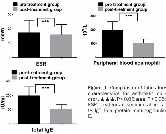
A total of 33 children with bronchial asthma were selected. All the children were treated with low-dose budesonide powder for a long time and then divided into two groups: the pre-treatment group and the post-pre-treatment group, as shown in Table 1 and Figure 1. These obser -Table 1. Comparisons of clinical and laboratory characteristics
be-tween the pre-treatment and post-treatment groups (_x ± SD)
Items Pre-treatment group Post-treatment group P-value
Age (year, mean ± SD) 8.7 ± 1.2 8.9 ± 1.0 0.563
Male gender 16 (48.5%) 17 (51.5%) 0.976
Duration of asthma (years) 2.3 ± 1.2 2.4 ± 1.3 0.863
Allergic history 17 (51.5%) 15 (45.4) 0.772
Total IgE (IU/ml) 323.8 ± 136.9 179.0 ± 79.3 < 0.001
ESR (mm/h) 17.3 ± 8.7 15.9 ± 7.1 0.067
Peripheral blood eosinophil (109/L) 0.32 ± 0.15 0.15 ± 0.07 0.010
IgE: immunoglobulin E; ESR: erythrocyte sedimentation rate.
PAGE). After electrophores- is, the proteins were
trans-ferred to PVDF membrane filters (Millipore Biotechno-logy, Billerica, MA, USA). The
membranes were incubated overnight at 4°C with the appropriate primary antibo-
dies. The membranes were
then incubated with horse-radish peroxidase-conjugat-ed secondary antibodies in
a blocking solution for 1 h at
room temperature, and the immunoreactive bands were visualized with a
chemilumi-nescence reagent (ECL, Mi-llipore Biotechnology,
Bille-rica, MA, USA) and
quanti-fied using a Bio-Rad imag-ing system (Bio-Rad
Labo-ratories, Inc, Hertfordshire, UK).
Statistical analysis
The data was analyzed
us-ing GraphPad Prism (version 6.01; GraphPad Software, Inc.) and expressed with (_x ± SD). The expression of
β2-AR mRNA, the concentra- tions of the inflammatory
factors, the parameters of lung function and airway re- modeling, age, duration of
[image:3.612.92.384.98.218.2] [image:3.612.91.383.245.481.2]vations in age, sex, duration of asthma, ESR, or allergic history did not differ between the
pre-treatment and post-pre-treatment groups (all P > 0.05). Nevertheless, the significant differences
in total IgE and peripheral blood eosinophil were determined between the two groups of asthmatic children (all P < 0.05).
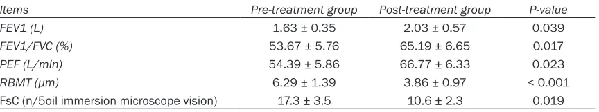
Results of pulmonary function and airway remodeling
Pulmonary function in all subjects was
evaluat-ed, including FEV1, FEV1/FVC, and PEF. The outcomes in FEV1, FEV1/FVC, and PEF for chil -dren with bronchial asthma were shown to prominently increase after treatment compar- ed with those before treatment (all P < 0.05).
All subjects underwent fiberoptic bronchosco -pies to assess their airway remodeling during
asthma remission. The observations of these
airway remodeling parameters showed that the
results of RBMT and FsC after treatment were significantly lower than those before treatment
(both P < 0.05), as shown in Table 2 and Figure 2.
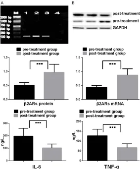
Results of β2ARs and inflammatory factors
The expressions of mRNA and protein for β2ARs in the BALF of asthmatic children were detect
-ed by RT-PCR and Western blot, respectively. The serum concentrations of IL-6 and TNF-α were determined by ELISA, as shown in Table 3, Figures 1, 3. The augmented expressions of
mRNA and protein for the β2-ARs were observ- ed for the asthmatic children after treatment (both P < 0.05). However, these lower serum
concentrations of IL-6 and TNF-α were revealed
in the post-treatment group (both P < 0.05). Correlation analysis
Pearson’s method was applied to the correla-tive analysis of the research data, as shown in Figure 4. The expression of β2Ars mRNA was
shown to be negatively correlated with IL-6, TNF-α, RBMT, and FsC in asthmatic children (P
< 0.05).
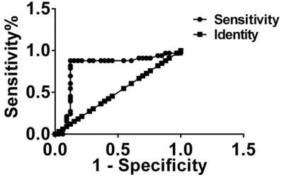
ROC curve analysis
The data in the pre-treatment group and the
post-treatment group served as the dependent
variable. The area under the curve (AUC) of the β2Ars mRNA expression was 0.823 (95% CI:0.682~0.936, P < 0.05). The sensitivity and specificity of diagnosing asthma at the best critical value were 87.9% and 89.2%. These findings suggested that β2ARs is useful in assessing the remission of asthma. The ROC curves of β2Ars mRNA expression are shown in
Figure 5. Discussion
In this study, the expression of mRNA and pro-tein for β2ARs in BALF prominently increased after treatment with budesonide in children with asthma. However, these decreased
out-comes of RBMT, FsC, IL-6, as well as TNF-α
were also observed in the post-treatment gr-
oup. These negative correlations between the expression of β2Ars mRNA and the concen-trations of IL-6 and TNF-α, RBMT, and FsC,
respectively, were observed both before and after treating the asthmatic children. We also
demonstrated the increased results of FEV1, PEF, and FVC in children with bronchial asthma
after treatment. Moreover, information from
the ROC curve confirmed the positive effect of β2ARs on assessing the remission of bron -chial asthma.
Increasing evidence suggests that the receptor theory could respond intensely to the
patho-genesis of asthma. The surface receptors found in airway epithelial cells, such as toll-like
receptors and other pattern recognition re-
[image:4.612.93.525.97.178.2]ceptors, could distinguish specific patterns on
Table 2. Comparisons of the parameters of pulmonary function and airway remodeling between the pre-treatment and post-treatment groups (_x ± SD)
Items Pre-treatment group Post-treatment group P-value
FEV1 (L) 1.63 ± 0.35 2.03 ± 0.57 0.039
FEV1/FVC (%) 53.67 ± 5.76 65.19 ± 6.65 0.017
PEF (L/min) 54.39 ± 5.86 66.77 ± 6.33 0.023
RBMT (μm) 6.29 ± 1.39 3.86 ± 0.97 < 0.001
FsC (n/5oil immersion microscope vision) 17.3 ± 3.5 10.6 ± 2.3 0.019
FEV1: forced expiratory volume in 1 sec; FVC: forced vital capacity; PEF: peak expiratory flow; RBMT: reticular basement mem
pathogen molecules (pathogen-associated mo-
lecular patterns) and lead to increased infla-mmation, Th2 cell activation, and alternative
macrophage activation for asthma patients.
These and alternative macrophages were con
-The exogenous catecholamines, such as the β2-adrenergic agonists inhaled in asthma
tr-eatment, can cause airway dilation by acting
on membrane-bound β2ARs on airway epitheli
[image:5.612.90.520.70.426.2]-al and smooth muscle cells [8]. This may be
Figure 2. Comparison of the parameters of pulmonary function and airway remodeling. ■■■, P < 0.05; (A) shows the thickened basement membrane in pre-treatment group (The black arrows indicates this); (B) shows the thinned basement membrane in the post-treatment group (The black arrows indicate this). Furthermore, these reduced outcomes of FEV1, FEV1/FVC and PEF in the post treatment group compared with those in the pre-treatment group (P < 0.05). Nevertheless, RBMT and FsC in the same condition give an enhanced result (P < 0.05). FEV1: forced expiratory volume in 1 sec; FVC: forced vital capacity; PEF: peak expiratory flow; RBMT: reticulation basement mem-brane thickness; FC: fibroblast count.
Table 3. Comparison of the expressions of mRNA and
protein for β2ARs, and inflammatory factors (_x ± SD)
Items Pre-treatment group Post-treatment group P-value β2ARs mRNA 0.43 ± 0.07 0.86 ± 0.23 < 0.001
β2ARs protein 0.51 ± 0.09 0.95 ± 0.27 < 0.001
IL-6 (ng/L) 197.23 ± 63.70 103.29 ± 31.67 < 0.001
TNF-α (ng/L) 127.60 ± 33.22 67.58 ± 19.26 < 0.001
β2ARs: beta 2 adrenergic receptors; IL-6: Interleukin-4; TNF-α: tumor necrosis factor-α.
sidered to regulate the production of
growth factors, including TGF-β and the
vascular endothelial growth factor, which could lead to airway remodeling [6]. In
addition, data from the IL-4 receptors sug
-gest that the expressions of the IL-4 receptors may impair the inhibition of Treg
cells from the lungs and participate in respiratory syncytial virus infections in
[image:5.612.91.324.550.628.2]related to the properties, density and
confor-mations of the molecular structure of β2ARs
on the cell surface. Several single nucleotide polymorphisms (SNP) within the promoter af-
fect the expression/regulation of β2ARs cod-ing 39UTR domains [9-11]. Highly resistant β2AR+79*G (Glu27) may contribute to
agonist-stimulated receptor downregulation [12]. Clini-
cal studies have confirmed that genetic varia -tion might result in a differential clinical res-
ponse to mild asthma. Furthermore, bronchiec
-tasis is more likely to be caused by salbutamol
TNF-α was confirmed to have a very
impor-tant effect on asthma pathogenesis. It was a
multifunctional proinflammatory cytokine in re-sponse to inflammation, infection,
macropha-ges, eosinophils, epithelial cells, and
neutro-phils [22]. In addition, an increased TNF-α
release in monocytes can be stimulated th-
rough β2-ARs downregulation induced by endo-genous catecholamines in septic shock [23]. This presented sufficiently the correlation be-tween TNF-α and β2-ARs, in spite of this not
arising in asthmatic patients.
Figure 3. Comparison of the expression of mRNA and protein for β2ARs in BALF and the concentrations of the inflammatory factors in serum for asthmatic children. ■■■, P < 0.05; (A, B) show the expression of mRNA and protein for β2ARs in BALF on the basis of the electrophoretic analysis of PCR amplification products and the gray level analysis of Western blot, respec-tively. In Figure a, M shows the expression of the marker (DL 2000), and 1, 2, 3 and 4 show the expression of β2ARs mRNA in the pre-treatment group, the post- treatment group, the β-actin group, and the negative control group, respectively. (B) shows the protein expression before and after treatment for β2ARs. These histograms describe the upregulation of β2ARs mRNA as well as the downregulation of IL-6 and TNF-α in the post treatment group compared with the upregulation and downregulation in the pre-treatment group (P < 0.05). β2ARs: beta 2-adrenergic receptors; BALF: bronchoalveo-lar lavage fluid; IL-6: Interleukin-6; TNF-α: tumor necrosis factor-α.
responding to β2AR+46 (a
ho-mozygote) compared to ho- mozygous individuals with the
G allele [13, 14]. Data from
larger pharmacogenetic stud-ies reveals that mild or moder-ate asthma disease almost
exclusively involved β2ARs
po-lymorphisms, but severe asth-ma reasth-mained unclear [15-18].
We confirmed that β2Ars mR-NA expression in BALF was si-gnificantly upregulated after
tr-eatment with budesonide for asthmatic children,
suggest-ing that β2ARs might be relat -ed to the pathological process of asthma, especially in the treatment surveillance of as- thma.
Increasing evidence has re-
vealed inflammation involving
asthma. It has been
suggest-ed that T regulatory (TREG; Foxp3+CD4+CD25+) cells can prevent chronic
inflammato-ry and autoimmune diseases due to the homeostasis of cel-lular immune responses [19]. Previous studies revealed th-
at increased TREG suppres -sive function was responsible
for TREG cells from aerobical-ly exercised mice in Th: TREG
cell co-cultures, and both ef- fectively relieved chronic
air-way inflammation and
[image:6.612.91.374.71.412.2]bronchial epithelial cells, smooth muscle cells,
and fibroblasts. In this process, an increased
β2-ARs release can be presented [29]. In the
present study, a similar point of view is
present-ed. The concentrations of IL-6 and TNF-α in
serum presented discrepant changes before and after treatment in asthmatic children, whi- ch were apparently correlated with the
expres-sion of β2-ARs in BALF. These observations pro
-vided evidence of β2-ARs participating in the
pathological mechanism of asthma in children as a monitoring effect on asthmatic treatment.
Airway remodeling is defined as a variation
of airway construction, including subepithelial
fibrosis, smooth muscle hyperplasia, and gob
[image:7.612.87.377.73.434.2]-let cell hyperplasia. However, little is known
Figure 4. Evidence of correlation analysis. These negative correlations be- tween β2ARs mRNA and IL-6, TNF-α, RBMT, and FsC are indicated (P < 0.05). β2ARs: beta 2-adrenergic receptors; IL-6: Interleukin-6; TNF-α: tumor necro- IL-6: Interleukin-6; TNF-α: tumor necro-sis factor α; RBMT: reticulation basement membrane thickness; FsC: fibro-blasts count.
Figure 5. Evidence of ROC curve analysis. The AUC of β2ARs mRNA expression is 0.823 (95% CI: 0.682~0.936, P < 0.05). The sensitivity and specific-ity at the best critical value in the evaluation of the therapeutic effect on asthma are 87.9% and 89.2%, respectively.
IL-6 has been described as
being involved in systemic in-
flammation and metabolic
dy-sfunction, and a lower lung function was found in
high-IL6-level asthma patients with frequent aggravating attacks [24]. The effect of salmeterol as a long-acting β2-agonist
decreases the concentration
of proinflammatory cytokines such as IL-6 and TNF-α in a
model of allergen-challenged mice [25], which presents a potent bronchodilating effect on moderate treatment for se- vere asthma [26]. In this
pro-cess, the activation of β2-ARs
were induced by salmeterol that can go through the lipid bilayer on cell membranes wh-
ere β2-ARs exist [27].
Notably, the enhanced plasma
concentrations of TNF-α were
revealed to respond to the development of childhood as- thma [28]. Interestingly, the
analysis of inflammatory cyto
-kines displayed that the
se-verity of airway diseases such as asthma is associated with
plasma concentrations of IL-4, IL-8, IL-10, and TNF-α [2]. Me-anwhile, β2-AR agonists have been disclosed to
[image:7.612.89.288.512.637.2]about the development mechanisms of airway remodeling [30]. A previous analysis proposed that airway remodeling for patients with asth-ma might be inspired by chronic airway in-
flammation such as inflammatory mediators and cytokines [31]. Nevertheless, with the inhi -bition of airway remodeling via prophylaxis for
the inflammation of the airway [32], a novel
therapy that controlled airway remodeling for asthmatic patients required a further under-standing of its mechanism.
Most review papers indicate that airway remod-eling for asthmatics might be associated with repeated episodes of asthma accompanied by
recurrent bronchoconstriction [2]. β2AR ago
-nists in response to β2ARs on airway epithelial
and smooth muscle cells contribute to cAMP-elevating agents to result in elevated cAMP expression that presents a potent inhibiting
effect on bronchial constriction. This mecha -nism has not yet been fully elucidated [33,
34].Traditionally, the activation of cAMP brings
about a relaxation of the airway’s smooth mus-cle [35] due to this process of mediating acti-
vation of protein kinase A(PKA) that partici -pates in the phosphorylation process of
multi-ple proteins, including myosin light chain kina-se and potassium channels. Thekina-se proceskina-ses
result in a changed microstructure for airway smooth muscle cells, further resulting in a sar-comere extension to attain the rapidly reversed bronchoconstrictor effect [36-38]. Other stud-ies have shown that PKA acts independently
on β2ARs mediating the relaxation of tracheal
smooth muscle in a guinea pig model. Ne- vertheless, the mechanism behind this is un- clear [39].
It has been observed that airway smooth mus-cle cell proliferation and migration are restra-
ined by cAMP elevators such as β2ARs agonists that induce the activation of β2-ARs, especially
in chronic asthma where an increase in airway smooth muscle mass is caused by cell
prolifer-ation which is more likely to lead to airway remodeling [40]. The expansion of the blocked airway by using drugs that activate β2-ARs can relieve asthma attacks. This evidence reveals the positive effects of β2-ARs in response to
the relaxation of airway smooth muscle and suggests that a decrease in the number of
β2-ARs or hypofunction can lead to the onset
of asthma. An increased frequency of asthma
attacks further advances airway remodeling.
In the present study, these declines of RBMT and FsC in airway tissue were observed in the
post-treatment children with asthma. A similar
condition also occurred with inflammatory cyto
-kines in serum. Moreover, lung function after
treatment for children with bronchial asthma was also found to be clearly improved. Me- anwhile, these parameters of airway
remodel-ing and inflammatory cytokines negatively cor
-related with the expression of β2Ars mRNA in BALF. Furthermore, information from the ROC curve confirmed the nice AUC of β2Ars mRNA
expression between the two groups of asthma sufferers and the higher sensitivity and
speci-ficity assessing the remission of bronchial as-thma. This suggested that active treatment by regulating the number of β2ARs could relieved
airway remodeling for asthmatic children, fol-lowing the improvement of symptoms and the
remission of inflammatory reactions. It further established that β2ARs can be an important marker of assessing airway remodeling for
br-onchial asthma. Conclusions
This clinical study confirmed that these obser -vations in age, sex, duration of asthma, ESR, or allergic history did not differ between the pre-treatment group and the post-pre-treatment gr-
oup. Nevertheless, these significant differenc -es in total IgE and peripheral blood eosinophil were revealed between the two groups of as- thma sufferers. Moreover, the expression of
β2-ARs mRNA correlated with these parame
-ters of inflammation and airway remodeling in asthmatic children, suggesting that β2-AR had an influence on the pathogenesis of asthma,
especially on the treatment of asthma, even though the details of the underlying mecha-nisms remained to be elucidated.
Acknowledgements
We thank Yi Liu from the First People’s Hospital
of Xian Yang City for his critical suggestions and comments on this manuscript.
Disclosure of conflict of interest
None.
Abbreviations
recep-tors; BALF, bronchoalveolar lavage fluid; IL-6, Interleukin-6; TNF-α, tumor necrosis factor α; FEV1, forced expiratory volume in 1 sec; FVC, forced vital capacity; PEF, peak expiratory flow; RBMT, reticular basement membrane thick-ness; FsC, fibroblasts count; ROC curve, partici-pant’s work characteristic curve; AUC, area under the ROC curve; AHR, airway hyperrespon-siveness; ESR, erythrocyte sedimentation rate; IgE, total protein immunoglobulin E; SD, stan-dard deviation.
Address correspondence to: Ping Wang, Depart- ment of Pediatrics, The First People’s Hospital of Xian Yang City, Xian Yang 712000, Shaanxi Provin- ce, China. Tel: +86-15706030285; E-mail: 212368- [email protected]
References
[1] Fehrenbach H, Wagner C and Wegmann M. Air-way remodeling in asthma: what really mat-ters. Cell Tissue Res 2017; 3: 551-569. [2] Huang AX, Lu LW, Liu WJ and Huang M. Plasma
inflammatory cytokine IL-6, IL-8, IL-10, and TNF-α levels correlate with pulmonary function in patients with asthma-chronic obstructive pulmonary disease (COPD) overlap syndrome. Med Sci Monit 2016; 22: 2800-2808.
[3] Gaffin JM, Raby BA, Petty CR, Hoffman EB, Baccarelli AA, Gold DR and Phipatanakul W. β-2 adrenergic receptor gene methylation is associated with decreased asthma severity in inner-city School Children. Clin Exp Allergy 2014; 44: 681-9.
[4] Hou C, Zhu X and Chang X. Correlation of vita-min D receptor with bronchial asthma in chil-dren. Exp Ther Med 2018; 15: 2773-2776. [5] Ställberg B, Naya I, Ekelund J and Eckerwall G.
Real-life use of budesonide/formoterol in clini-cal practice: a 12-month follow-up assessment in a multi-national study of asthma patients established on single-inhaler maintenance and reliever therapy. Int J Clin Pharmacol Ther 2015; 53: 447-55.
[6] Sidhu SS, Yuan S, Innes AL, Kerr S, Woodruff PG, Hou L, Muller SJ and Fahy JV. Roles of epi-thelial cell-derived periostin in TGF-beta activa-tion, collagen producactiva-tion, and collagen gel elasticity in asthma. Proc Natl Acad Sci U S A 2010; 107: 14170-5.
[7] Krishnamoorthy N, Khare A, Oriss TB, Raund-hal M, Morse C, Yarlagadda M, Wenzel SE, Moore ML, Peebles RS Jr, Ray A and Ray P. Early infection with respiratory syncytial virus impairs regulatory T cell function and increas-es susceptibility to allergic asthma. Nat Med 2012; 18: 1525-30.
[8] Barnes PJ. Receptor heterodimerization: a new level of cross-talk. J Clin Invest 2006; 116: 1210-2.
[9] Hawkins GA, Tantisira K, Meyers DA, Ampleford EJ, Moore WC, Klanderman B, Liggett SB, Pe-ters SP, Weiss ST and Bleecker ER. Sequence, haplotype, and association analysis of ADRbe-ta2 in a multiethnic asthma case-control study. Am J Respir Crit Care Med 2006; 174: 1101-9. [10] Kobilka BK, Frielle T, Dohlman HG, Bolanowski
MA, Dixon RA, Keller P, Caron MG and Lefkow-itz RJ. Delineation of the intronless nature of the genes for the human and hamster beta 2-αdrenergic receptor and their putative pro-moter regions. J Biol Chem 1987; 262: 7321-7.
[11] Reihsaus E, Innis M, MacIntyre N and Liggett SB. Mutations in the gene encoding for the beta 2-αdrenergic receptor in normal and asth-matic subjects. Am J Respir Cell Mol Biol 1993; 8: 334-9.
[12] Green SA, Turki J, Innis M and Liggett SB. Ami-no-terminal polymorphisms of the human beta 2-αdrenergic receptor impart distinct agonist-promoted regulatory properties. Biochemistry 1994; 33: 9414-9.
[13] Martinez FD, Graves PE, Baldini M, Solomon S and Erickson R. Association between genetic polymorphisms of the beta2-αdrenoceptor and response to albuterol in children with and with-out a history of wheezing. J Clin Invest 1997; 100: 3184-8.
[14] Silverman EK, Kwiatkowski DJ, Sylvia JS, Laza-rus R, Drazen JM, Lange C, Laird NM and Weiss ST. Family-based association analysis of be- ta2-αdrenergic receptor polymorphisms in the childhood asthma management program. J Al-lergy Clin Immunol 2003; 112: 870-6.
[15] Anderson HR, Ayres JG, Sturdy PM, Bland JM, Butland BK, Peckitt C, Taylor JC and Victor CR. Bronchodilator treatment and deaths from asthma: case-control study. BMJ 2005; 330: 117.
[16] Bleecker ER, Postma DS, Lawrance RM, Mey-ers DA, Ambrose HJ and Goldman M. Effect of ADRB2 polymorphisms on response to lon-gacting beta2-αgonist therapy: a pharmacoge-netic analysis of two randomised studies. Lan-cet 2007; 370: 2118-25.
[17] Bleecker ER, Yancey SW, Baitinger LA, Edwards LD, Klotsman M, Anderson WH and Dorinsky PM. Salmeterol response is not affected by beta2-αdrenergic receptor genotype in sub-jects with persistent asthma. J Allergy Clin Im-munol 2006; 118: 809-16.
SC, Lemanske RF Jr, Markezich A, Martin RJ, Permaul P, Peters SP, Ramsdell J, Sorkness CA, Sutherland ER, Szefler SJ, Walter MJ, Wasser-man SI, Israel E; National Heart, Lung and Blood Institute’s Asthma Clinical Research Network. Effect of [beta] 2-αdrenergic re- ceptor polymorphism on response to longact-ing [beta] 2 agonist in asthma (LARGE trial): a genotype-stratified, randomised, placebo-controlled, crossover trial. Lancet 2009; 374: 1754-64.
[19] Vignali DA, Collison LW and Workman CJ. How regulatory T cells work. Nat Rev Immunol 2008; 7: 523-32.
[20] Lowder T, Dugger K, Deshane J, Estell K and Schwiebert LM. Repeated bouts of aerobic ex-ercise enhance regulatory T cell responses in a murine asthma model. Brain Behav Immun 2010; 1: 153-9.
[21] Pastva A, Estell K, Schoeb TR, Atkinson TP and Schwiebert LM. Aerobic exercise attenuates airway inflammatory responses in a mouse model of atopic asthma. J Immunol 2004; 172: 4520-6.
[22] Despotovic M, Stoimenov TJ, Stankovic I, Pav-lovic D, SokoPav-lovic D, Cvetkovic T, Kocic G, Basic J,Veljkovic A and Djordjevic B. Gene polymor-phisms of tumor necrosis factor alpha and an-tioxidant enzymes in bronchial asthma. Adv Clin Exp Med 2015; 24: 251-6.
[23] Link A, Selejan S, Maack C, Lenz M and Böhm M. Phosphodiesterase 4 inhibition but not be-ta-adrenergic stimulation suppresses tumor necrosis factor-alpha release in peripheral blood mononuclear cells in septic shock. Crit Care 2008; 12: R159.
[24] Peters MC, McGrath KW, Hawkins GA, Hastie AT, Levy BD, Israel E, Phillips BR, Mauger DT, Comhair SA, Erzurum SC, Johansson MW, Jar-jour NN, Coverstone AM, Castro M, Holguin F, Wenzel SE, Woodruff PG, Bleecker ER, Fahy JV; National Heart, Lung, and Blood Institute Se-vere Asthma Research Program. Plasma IL6 levels, metabolic dysfunction, and asthma se-verity: a cross-sectional analysis of two co-horts. Lancet Respir Med 2016; 4: 574-584. [25] Hu Z, Chen R, Cai Z, Yu L, Fei Y, Weng L, Wang
J, Ge X, Zhu T, Wang J and Bai C. Salmeterol attenuates the inflammatory response in asth-ma and decreases the pro-inflamasth-matory cyto-kine secretion of dendritic cells. Cell Mol Im-munol 2012; 9: 267-75.
[26] Nguyen LP, Lin R, Parra S, Omoluabi O, Hana-nia NA, Tuvim MJ, Knoll BJ, Dickey BF and Bond RA. Beta2-adrenoceptor signaling is re-quired for the development of an asthma phe-notype in a murine model. Proc Natl Acad Sci U S A 2009; 106: 2435-40.
[27] Johnson M. Pharmacology of long-acting beta-agonists. Ann Allergy Asthma Immunol 1995; 75: 177-9.
[28] Reyes-Gibby CC, Wang J, Spitz M, Wu X, Yen-nurajalingam S and Shete S. Genetic varia-tions in interleukin-8 and interleukin-10 are associated with pain, depressed mood, and fatigue in lung cancer patients. J Pain Symp-tom Manage 2013; 46: 161-72.
[29] Futamura K, Orihara K, Hashimoto N, Morita H, Fukuda S, Sagara H, Matsumoto K, Tomita Y, Saito H and Matsuda A. Beta2-Adrenoceptor agonists enhance cytokine-induced release of thymic stromal lymphopoietin by lung tissue cells. Int Arch Allergy Immunol 2010; 152: 353-61.
[30] Kwak HJ, Park DW, Seo JY, Moon JY, Kim TH, Sohn JW, Shin DH, Yoon HJ, Park SS and Kim SH. The Wnt/β-catenin signaling pathway regu-lates the development of airway remodeling in patients with asthma. Exp Mol Med 2015; 47: e198.
[31] ten Brinke A, Zwinderman AH, Sterk PJ, Rabe KF, Bel EH. Factors associated with persistent airflow limitation in severe asthma. Am J Respir Crit Care Med 2001; 164: 744-8.
[32] Berair R and Brightling CE. Asthma therapy and its effect on airway remodelling. Drugs 2014; 74: 1345-69.
[33] Giembycz MA and Newton R. Beyond the dog-ma: novel beta2-αdrenoceptor signalling in the airways. Eur Respir J 2006; 27: 1286-306. [34] Torphy TJ. Beta-αdrenoceptors, cAMP and
air-way smooth muscle relaxation: challenges to the dogma. Trends Pharmacol Sci 1994; 15: 370-4.
[35] Taylor DR, Drazen JM, Herbison GP, Yandava CN, Hancox RJ and Town GI. Asthma exacerba-tions during long term beta agonist use: influ-ence of beta (2) adrenoceptor polymorphism. Thorax 2000; 55: 762-7.
[36] Billington CK and Penn RB. Signaling and regu-lation of G protein-coupled receptors in airway smooth muscle. Respir Res 2003; 4: 22-24. [37] Kume H, Hall IP, Washabau RJ, Takagi K and
Kotlikoff MI. Beta-αdrenergic agonists regulate Kca channels in airway smooth muscle by cAMP-dependent and -independent mecha-nisms. J Clin Invest 1994; 93: 371-9.
[38] Scheid CR, Honeyman TW and Fay FS. Mecha-nism of beta-αdrenergic relaxation of smooth muscle. Nature 1979; 277: 32-6.
[39] Spicuzza L, Belvisi MG, Birrell MA, Barnes PJ, Hele DJ and Giembycz MA. Evidence that the anti-spasmogenic effect of the beta-αdrenoceptor agonist, isoprenaline, on guinea-pig trachealis is not mediated by cyclic AMP-dependent protein kinase. Br J Pharmacol 2001; 133: 1201-12.