Functional studies on human γδ T cells and their
interactions with dendritic cells and B cells
A dissertation submitted to Trinity College Dublin, University of Dublin,
as requirement for the degree of Doctor in Philosophy (PhD)
in Clinical Medicine
25/09/2015
Andreea Petrasca, B.Sc (Hons), M.Sc
Student number 10264277
Work carried out between September 2011 and September 2015
Supervisor: Dr. Derek Doherty
Head of Department of Immunology
ii Declaration
I, Andreea Petrasca, the undersigned declare that this thesis is the result of my own
work. It has not previously been submitted to this university or any other, and any
assistance received by way of borrowing from the work of others has been cited and
acknowledged. I have read and understood the college’s regulations on plagiarism and
I make this declaration in the knowledge that a breach of the rules pertaining to
submission may carry serious consequences.
I agree to deposit this thesis in the University’s open access institutional repository or
allow the library to do so on my behalf, subject to Irish Copyright Legislation and
Trinity College Library conditions of use and acknowledgement.
iii Acknowledgements
First and foremost I would like to thank the best and most wonderful parents anyone
could have, Ivan and Angela, without whom, I would have never gotten this far. For
their endless love and support and providing me with the best education I could have
ever dreamed of, hvala!
I would also like to express my deepest gratitude to my supervisor, Derek Doherty, for
offering me the opportunity to carry out such exciting research. For all his help,
encouragement and wisdom throughout the past 4 years and his complete dedication
and constant support which were key in completing this PhD. I would also like to thank
Derek for allowing me to be independent. His positivity has been truly motivating.
Moreover, I am very grateful for all the collaborations he has allowed me to get
involved in.
To my fiancé James, who fully understands first-handedly the pressure and dedication
that is required in doing a PhD and for forgiving me for all those late nights spent on
tissue culture and flow cytometry.
My sincerest thanks also go out to some key mentors that have helped shape the
scientist I am today. To Vinny O’Reilly, for introducing me to tissue culture and flow
cytometry and for setting a great example. To Ann Atzberger for her invaluable
teachings, and whose passion for flow cytometry sparked an interest in me for flow
cytometry. To Padraic Dunne, for all his guidance, and for allowing me to ask lots of
“silly” questions. To professors John Jackson, Con Feighery and Jacinta Kelly for being
an integral part in our lab meetings and whose wisdom, insight and enthusiasm
inspired me to excel at immunology.
I would also like to express my sincerest thanks to my younger sister, Alina, for being
understanding and putting up with me after coming home following long and tiring
days. Also, to my friend Ciara for all the fun activities we enjoyed after many
productive days. Last, but not least I would like to thank my partners in crime and
wonderful friends Yasmeen Ghnewa, Éilis Dockry and Serena Arduini for lifting my
iv Academic Outputs
Publications
“Human Vδ2+ γδ T cells differentially induce maturation, cytokine production,
and alloreactive T cell stimulation by dendritic cells and B cells”. Andreea
Petrasca, Derek G Doherty. Research Article, Frontiers in Immunology, 5: 650;
December 19, 2014.
“Vδ2 T cells differentially stimulate the production of proinflammatory cytokines by dendritic cells and B cells”. Andreea Petrasca, Derek G Doherty. In:
Proceedings of the 15th International Congress of Immunology (22-27 August
2013, Milan, Italy), Medimond, Bologna, Italy pp 21-24, 2014.
“HIV-1 Tat clade-specific cytokine induction in monocytes/macrophages is not evidenced in total or Vγ9Vδ2 T lymphocytes”. Alan Kennedy, Andreea Petrasca,
Derek G Doherty, Martina Hennessy, Paul J Spiers. Research letter, AIDS,
28:131-133, January 2, 2014.
“Synthesis and immunostimulatory activity of two α-S-galactosyl phenyl-capped ceramides”. Niamh Murphy, Andreea Petrasca, Niamh M. Murphy,
Vincent O’Reilly, Paul Evans, Derek G. Doherty, Xiangming Zhu. Research
article, Arkivoc, 2013 (ii) 363-377, February 8, 2013
Manuscripts in submission
“Human Natural Killer cell expression of ULBP2 is associated with a mature functional phenotype” Kiva Brennan, Sinéad Keating, Andreea Petrasca,
Vincent P O'Reilly, Joseph Keane, Derek G. Doherty, Clair M. Gardiner.
Velasco-v Torrijos, Carol Buggy, Andreea Petrasca, Niamh M. Murphy, Vincent O’Reilly,
Derek G. Doherty.
Serum Adipokines and Vascular Phenotype in Juvenile-onset Systemic Lupus Erythematosus. Catherine Quinlan, Andreea Petrasca, John Deanfield, Michael
Riordan, Rukshana Shroff, Pilkington Clarissa, Stephen Marks, Kjell Tullus.
Collaborations
“The role of Vγ9Vδ2 T cells in immunity against Clostridium difficile”. Micheál MacAogáin, Andreea Petrasca, Derek Doherty, Thomas Rogers.
“Investigating the potential of humanised mice as a pre-clinical model for Vδ2 T cell based immunotherapy”. Eoin O’Brien, Vincent O’Reilly, Andreea Petrasca,
Margaret Dunne, Cathal Harmon, Fionnuala Hickey, Derek Doherty, Cliona
O’Farrelly, Mark Little.
“The effect of physical activity on cells of the immune system”. Grace Lavelle, Andreea Petrasca, Derek Doherty.
“The role of Staphylococcus aureus in activating Vγ9Vδ2 T cells“. Stephen Lalor, Andreea Petrasca, Derek Doherty, Rachel McLoughlin.
Oral presentations
“Human Vγ9Vδ2 T cells induce B cell and DC maturation - applications for cancer
immunotherapy”. Institute of Molecular Medicine Annual Meeting, Trinity College
Dublin, Nov 2014.
“Human Vγ9Vδ2 T cells can selectively promote TH1 or TH2 responses via interactions
with dendritic cells or B cells in vitro”. Institute of Molecular Medicine Annual Meeting,
vi “Reciprocal activating interactions between Vγ9Vδ2 T cells, B cells and dendritic cells”.
School of Medicine Postgraduate Research Day, Trinity College Dublin, Sept 2012.
Poster presentations
“Human Vδ2+ γδ T cells promote differentiation of B cells into effector cells that
release antibody and IL-4 and stimulate alloreactive T cell proliferation”. British Society
for Immunology Congress, Brighton, UK, December 2014.
“Human Vδ2+ γδ T cells promote differentiation of B cells into effector cells that
release antibody and IL-4 and stimulate alloreactive T cell proliferation”. Irish Society
of Immunology Meeting, Dublin, Ireland, September 2014.
“Human Vδ3 T cells induce expression of co-stimulatory molecules by B cells in vitro”.
International Gamma Delta T-cell conference, Chicago, USA, May 2014.
“Human Vγ9Vδ2 T cells can selectively promote TH1 or TH2 responses via interactions
with dendritic cells or B cells in vitro”. Andreea Petrasca and Derek G. Doherty, Irish
Cytometry Society Conference, Dublin, February 2014.
“Human Vγ9Vδ2 T cells can selectively promote TH1 or TH2 responses via interactions
with dendritic cells or B cells in vitro”. Andreea Petrasca and Derek G. Doherty. British
Society for Immunology Annual Meeting, Liverpool, United Kingdom, December 2013.
“Human Vγ9Vδ2 T cells can selectively promote TH1 or TH2 responses via interactions
with dendritic cells or B cells in vitro”. International Congress of Immunology, Milan,
Italy, August 2013.
“Vγ9Vδ2 T cells induce maturation of dendritic cells but not B cells into
antigen-presenting cells”. International Gamma-delta T-cell conference, Freiburg, Germany,
vii Table of Contents
Chapter 1 Introduction ... 7
1.1 Introduction ... 8
1.2 The immune system ... 8
1.2.1 Innate immunity ... 8
1.2.2 Adaptive immunity ... 8
1.2.3 Chemical messengers of immunity ... 9
1.3 Cells of the immune system ... 11
1.3.1 Myeloid cells ... 13
1.3.1.1 Dendritic cells ... 14
1.3.1.2 Antigen presentation ... 15
1.3.2 Lymphoid cells ... 17
1.3.2.1 NK cells ... 17
1.3.2.2 B cells ... 17
1.3.3 T cells ... 19
1.3.4 NKT cells ... 20
1.3.5 MAIT cells ... 21
1.4 γδ T cells ... 22
1.4.1 The human γδ T cell receptor ... 22
1.4.2 Human γδ T cells subsets ... 23
1.4.2.1 Non-Vδ2 T cells ... 23
1.4.2.2 Vγ9Vδ2 T cells ... 24
1.4.3 Vγ9Vδ2 T cell activation ... 30
1.4.4 Phenotypes of γδ T cells ... 32
1.4.4.1 CD4 and CD8 expression ... 32
1.4.4.2 Differentiation status ... 32
1.4.4.3 Chemokine receptors ... 32
1.4.4.4 Other stimulating markers ... 33
1.4.5 Cytokine and chemokine secretion by γδ T cells ... 34
1.4.6 γδ T cells in disease ... 36
1.4.6.1 Cytotoxicity by γδ T cells ... 36
1.4.6.2 γδ T cells in anti-tumour immunity ... 36
viii
1.4.6.4 γδ T cells in bacterial infection ... 38
1.4.7 APC function by γδ T cells ... 38
1.4.8 Interaction between Vγ9Vδ2 and other immune cells ... 39
1.5 Aims & Objectives ... 40
Chapter 2 Materials and Methods ... 41
2.1 Materials & Methods ... 42
2.2 Tissue culture ... 49
2.3 Cell viability and enumeration ... 49
2.4 Cryopreservation of cells ... 50
2.5 Flow cytometry ... 51
2.5.1 Principles of flow cytometry ... 51
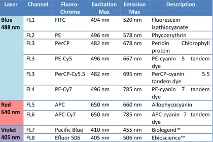
2.5.2 Labeling of cells for flow cytometry ... 52
2.5.3 Antibody -fluorochrome conjugation ... 53
2.5.4 Cell sorting... 54
2.6 Isolation and expansion of human cells ... 54
2.6.1 Isolation of human PBMC ... 54
2.6.2 Vδ2 T cell expansion ... 55
2.6.3 Vδ3 T cell expansion ... 57
2.6.4 B cell isolation ... 58
2.6.5 Dendritic cell preparation ... 59
2.6.6 Preparation of non-γδ T cell lines ... 60
2.6.7 iNKT cell expansion ... 61
2.6.8 HeLa cervical cancer cells ... 62
2.7 Analysis of cell surface markers and secreted factors from cell cultures ... 63
2.7.1 Analysis of co-stimulatory marker expression ... 63
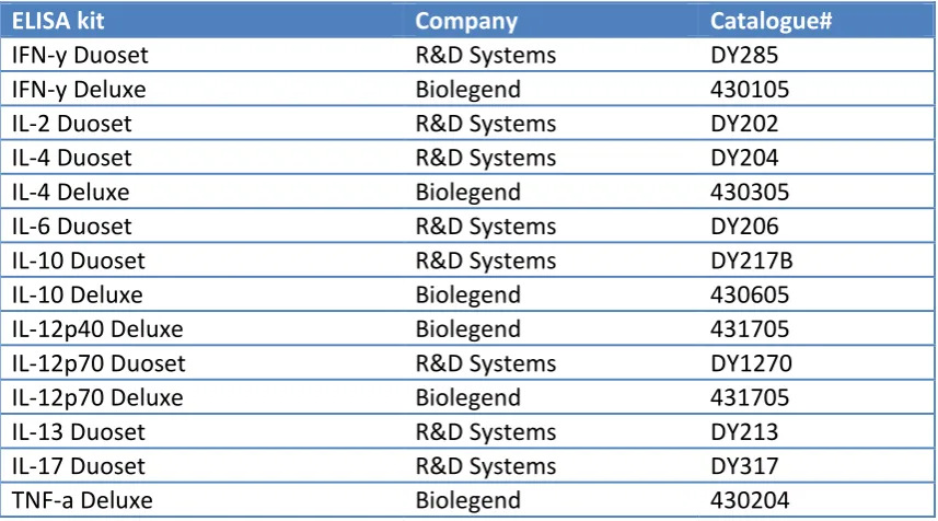
2.7.2 Analysis of cytokine release from cell cultures ... 64
2.7.3 Analysis of intracellular cytokine production ... 66
2.7.4 Measurement of antibody production by B cells ... 67
2.7.5 Blocking experiments ... 68
2.7.6 Examination of T cell proliferation and resulting cytokine release ... 69
2.7.7 Cytotoxicity assay ... 70
2.8 Vδ3 and iNKT cell stimulation using glycolipids ... 70
2.9 Statistical Analysis ... 71
ix
3.1 Introduction ... 73
3.2 Objectives ... 76
To phenotypically characterise ... 76
To phenotypically and functionally characterise the human ... 76
To phenotypically and functionally characterise V ... 76
3.3 Methods ... 77
3.3.1 Phenotypes of peripheral blood γδ T cell subsets ... 77
3.3.2 Expansion and phenotyping of Vδ2 T cell lines ... 77
3.3.3 Expansion and phenotyping of Vδ3 T cell lines ... 77
3.4 Results ... 78
3.4.1 Phenotypes of peripheral blood γδ T cell subsets ... 78
3.4.2 Frequency of CD4 and CD8 T cells in peripheral blood γδ T cell subsets ... 83
3.4.3 Memory phenotypes of peripheral blood γδ T cell subsets... 83
3.4.4. Expression of stimulatory surface markers in peripheral blood γδ T cell subsets .... 83
3.4.5. Assessing purity of HMB-PP expanded Vγ9Vδ2 T cell lines ... 91
3.4.6. Expanded Vδ2 T cell lines are CD8 positive or double negative and exhibit an effector memory phenotype. ... 91
3.4.7. Expanded Vδ2 T cells express markers of T cell activation and TLR ... 91
3.4.8 Expanded Vδ2 T cells are capable of producing IFN-γ, IL-4 and TNF-α ... 91
3.4.9 Aminobisphosphonate stimulation is more efficient than phosphoantigen stimulation in expanding Vδ2 T cells and they induce different cytokine profiles ... 101
3.4.10 Assessing purity of expanded Vδ3 T cell lines ... 101
3.4.11 Resting Vδ3 T cells produce very low cytokine levels ... 102
3.5 Discussion ... 110
Chapter 4 Reciprocal activating interactions between human Vδ2+ γδ T cells, dendritic cells and B cells ... 117
4.1 Introduction ... 118
4.2 Objectives ... 119
4.3 Methods ... 120
4.3.1 Cell line generation and co-cultures ... 120
4.3.2 Analysis of co-stimulatory marker expression by DC and B cells ... 121
4.3.3 Analysis of cytokine production ... 121
4.3.4 Measurement of antibody production by B cells ... 121
4.3.5 Blocking experiments ... 121
x
4.3.7 The effect of altering the ratio of DC to Vδ2 T cells on cytokine production ... 123
4.3.8 The effect of culturing DC, B cells and Vδ2 T cells simultaneously ... 123
4.3.9 The effect of LPS on DC and B cell activation ... 123
4.4 Results ... 125
4.4.1 Vδ2 T cells induce APC marker expression by DC and B cells ... 125
4.4.2 Vδ2 T cells induce production of distinct cytokines by DC and B cells ... 125
4.4.3 Vδ2 T cells induce pro- and anti-inflammatory cytokine secretion from DC and B cell co-cultures ... 126
4.4.4 Vδ2 T cells induce antibody production by B cells ... 132
4.4.5 Allogeneic and autologous Vδ2 equally activate DC and B cells ... 132
4.4.6 Vδ2-matured DC and B cells stimulate proliferation of resting allogeneic T cells ... 132
4.4.7 Vδ2-matured DC but not B cells stimulate cytokine production by resting allogeneic T cells. ... 133
4.4.8 Vδ2-matured DC induce proliferation of autologous resting T cells irrespective of presence of antigen... 133
4.4.9 Increasing the ratio of DC to Vδ2 T cells increases cytokine production by DC ... 134
4.4.10 The addition of DC to Vδ2-B cell cultures increases co-stimulatory marker and cytokine expression by Vδ2 and B cells ... 143
4.4.11 LPS induces co-stimulatory marker expression by DC and B cells ... 146
4.4.12 LPS induces cytokine secretion by Vδ2-DC co-cultures and synergises with Vδ2 T cells to induce IL-12 production ... 146
4.4.13 LPS induces an increase in IgG and IgA production by B cells ... 147
4.5 Discussion ... 151
Chapter 5 Analysis of antigen recognition and helper function of human Vδ3 T cells ... 158
5.1 Introduction ... 159
5.2 Objectives ... 160
5.3 Methods ... 161
5.3.1 Generation of Vδ3 T cells, iNKT cells and B cells ... 161
5.3.2 Analysis of co-stimulatory marker expression by B cells and Vδ3 T cells in co-cultures ... 161
5.3.3 Analysis of cytokine production from co-cultures of Vδ3 T cells and B cells ... 162
5.3.4 Measurement of antibody production by B cells ... 162
5.3.5 Vδ3 T cell responses to CD1 molecules in the absence of glycolipids or in the presence of cardiolipin, sulfatide and ganglioside ... 162
xi
5.4 Results ... 165
5.4.1 Vδ3 T cells induce co-stimulatory marker expression by B cells... 165
5.4.2 Vδ3 T cells do not induce cytokine production by B cells ... 165
5.4.3 B cells induce IL-17 and IL-4 production by Vδ3 T cells in the presence of PMA and ionomycin ... 166
5.4.4 Vδ3 T cells do not induce antibody production by B cells ... 166
5.4.5 Vδ3 T cells do not recognise CD1a, CD1b, CD1c or CD1d ... 173
5.4.6 Vδ3 T cells do not produce cytokines upon culture with HeLa transfectants ... 173
5.5 Discussion ... 180
Chapter 6 Preliminary studies investigating the role of Vδ2 T cells in mediating immunity against Clostridium difficile ... 184
6.1 Introduction ... 185
6.2 Objectives ... 187
6.3 Methods ... 187
6.3.1 Method for deriving C. difficile supernatant and lysate ... 187
6.3.2 Expansion of Vδ2 T cells from PBMC using C. difficile ... 188
6.3.3 Cytokine production by Vδ2 T cells expanded using C. difficile or HMB-PP ... 188
6.3.4 Investigation of the roles of B cells, monocytes and αβ T cells in Vδ2 T cell expansion in response to C. difficile ... 189
6.4 Results ... 191
6.4.1 C. difficile induces Vδ2 T cell proliferation ... 191
6.4.2 C. difficile supernatant from 3 different strains induces Vδ2 T cell expansion... 191
6.4.3 C. difficile induces moderate levels of cytokine production by Vδ2 T cells ... 192
6.4.4 C. difficile appears to be toxic to fresh γδ T cells, but this inhibition is prevented by addition of monocytes, B cells and αβ T cells. ... 192
6.4.5 C. difficile induces IFN-γ and TNF-α production by C. difficile-expanded Vδ2 T cells ... 200
6.4.6 C. difficile supernatant induces proliferation of both CD4 and CD8 subsets of Vδ2 T cells ... 200
6.4.7 C. difficile-induced activation of Vδ2 T cells is donor specific ... 200
6.5. Discussion ... 205
Chapter 7 Discussion ... 209
7.1 Discussion ... 209
7.2 Future directions ... 210
1 List of abbreviations used
ADCC antibody-dependent cell-mediated cytotoxicity
αGalCer alpha galactosylceramide
ANOVA analysis of variance
APC antigen presenting cells
APC allophycocyanin fluorophore, used in flow cytometry
βGluCer beta glucosyl ceramide
BHIS brain heart infusion medium
BSA bovine serum albumin
BTN Butyrophilin
CD cluster of differentiation
cDMEM complete Dulbecco’s modified Eagle’s medium
CDT cytolethal distending toxins
cRPMI complete Roswell Park Memorial Institute medium
Cy5/7 CyChrome 5/ CyChrome 7
DC dendritic cells
DMAPP dimethylallyl diphosphate
DMSO dimethylsulphoxide
DN double negative
EBAO ethidium bromide and acridine orange
EDTA ethylenediaminetetra-acetic acid
2 FACS fluorescence-activated cell sorting
FBS fetal bovine serum
FITC fluorescein isothiocyanate
FMO fluorescence minus one control
FSC forward scatter
γδ-T-APC γδ T cell with APC function
GM-CSF granulocyte monocyte-colony stimulating factor
HeLa Henrietta Lacks cervical cell line
HIV human immunodeficiency virus
HLA human leukocyte antigen
HMB-PP (E)-4-hydroxy-3-methyl-but-2-enyl pyrophosphate
HMG-CoA 3-hydroxy-3-methyl-glutaryl-CoA
iDC immature dendritic cells
IEL intraepithelial lymphocytes
IFN interferon
IL interleukin
iNKT invariant natural killer T
IPP isopentenyl pyrophosphate
IU international units
LF limes of flocculation
LPS lipopolysaccharide
3 MACS magnetic-activated cell sorting
MAIT mucosal-associated invariant T
MEP methylerythritol 4-phosphate
MFI mean fluorescence intensity
MHC major histocompatibility complex
MICA/MICB MHC class I-related chains A or B
NK natural killer
NKT natural killer T
NKG2 natural killer group 2
pAg phosphoantigen (phosphorylated pyrophosphate)
PAMP pathogen associated molecular pattern
PBA PBS supplemented with BSA and azide
PBMC peripheral blood mononuclear cells
PBS phosphate buffered saline
PE phycoerythrin
PFA paraformaldehyde
PerCP peridinin chlorophyll protein complex
PHA phytohaemagglutinin
PMA phorbol myristate acetate
PPD purified protein derivative
PRR pattern recognition receptors
4 SLP surface layer protein
SSC side scatter
TC cytotoxic T cell
TCM central memory T cell
TCR T cell receptor
TEM effector memory T cell
TEMRA terminally differentiated effector memory T cell
TH T helper
TFH follicular helper T
TGF transforming growth factor
TLR toll-like receptor
TMB 3,3,5,5-tetramethylbenzidine
TNF tumour necrosis factor
Treg regulatory T
TT tetanus toxoid
5 Abstract
γδ T cells are innate T cells that play central roles in protection against microorganisms
and cellular stress. There are three main subsets in humans: Vδ1, Vδ2 and Vδ3 T cells.
The most abundant of these, Vδ2 T cells, recognises phosphoantigens produced in one
of two pathways of isoprenoid synthesis, a cellular metabolic pathway employed by all
eukaryotes and many bacteria. This allows Vδ2 T cells to monitor for infection and
tumour transformation which result in altered cellular concentration of
phosphoantigens. Vδ1 and Vδ3 T cells are predominantly found in epithelial tissues
and play roles in homeostasis, tissue integrity and lipid surveillance and are found at
increased frequencies in some patients with tumours and viral infections. Upon
activation, γδ T cells can kill target cells and rapidly promote adaptive immune
responses through physical interactions with other immune cells, and rapid and
selective secretion of T helper type 1 (TH1), TH2, TH17 and regulatory cytokines.
In the present study we carried out a phenotypic and functional comparison of the
three γδ T cell subsets in human peripheral blood and examined methods for
generating lines of Vδ2 and Vδ3 T cell for functional characterisation. We found that
stimulation with the phosphoantigen (E)-4-Hydroxy-3-methyl-but-2-enyl
pyrophosphate (HMB-PP) and interleukin-2 yielded highly pure populations of Vδ2 T
cells capable of producing TH1 and TH2 cytokines upon re-stimulation. In contrast,
treatment with the aminobisphosphonate zoledronate, which promotes isoprenoid
synthesis, resulted in expansion of Vδ2 T cells that produced TH2 cytokines only. In the
absence of a known ligand for Vδ3 T cells, we used the T cell mitogen
phytohaemagglutinin to stimulate sorted Vδ3+ cells which resulted in up to 1,000-fold
expansion within 3-4 weeks.
We next examined the reciprocal activating interactions between Vδ2 T cells, dendritic
cells (DC) and B cells in co-culture experiments and defined the resulting cytokine
profiles, cell phenotypes and antibody responses and the molecular interactions
involved. We found that Vδ2 T cells promoted maturation of DC into
antigen-presenting cells capable of stimulating TH1 cell responses. In contrast, Vδ2 T cells
6 phenotypes of antigen-presenting cells, but which produced TH2 cytokines. While
co-stimulatory molecules, TH1 cytokines and cell contact were required for DC maturation
by Vδ2 T cells, they did not play major roles in B cell maturation.
The present study investigated, for the first time, the relationship between Vδ3 T cells
and B cells and revealed that while Vδ3 T cells induced co-stimulatory marker
expression by B cells, they failed to induce significant cytokine or antibody secretion.
However, activated B cells were able to induce IL-17 secretion by Vδ3 T cells. We also
assessed the ability of Vδ3 T cells to recognise CD1 molecules, but found that
freshly-isolated or expanded Vδ3 T cells showed no reactivity against CD1a, CD1b, CD1c or
CD1d molecules in the presence or absence of a number of glycolipids.
Since Clostridium difficile appears to utilise the non-mevalonate pathway of isoprenoid
biosynthesis, suggesting that it can produce PP, we assessed the ability of
HMB-PP to stimulate proliferation and cytokine secretion by Vδ2 T cells. We found, in spite
of great inter-donor variability, C. difficile secreted a Vδ2-stimulating agent which
induced T cell proliferation and cytokine production in most donors and was
comparable to the stimulating capabilities of HMB-PP. However, the identity of this
secreted factor remains to be elucidated.
These findings highlight the role of γδ T cells in immunosurveillance, innate immunity,
antigen presentation and activation of adaptive immunity. Their ability to act as a
bridge between innate and adaptive immune responses places these cells as attractive
candidates for immunotherapy for infectious and immune-mediated diseases and
7
Chapter 1
8 1.1 Introduction
1.2 The immune system
The role of the immune system is to provide protection against infectious agents.
There are two main branches of the immune system: the innate and the adaptive
immune systems.
1.2.1 Innate immunity
The innate immune system, which provides the first line of defense, is equipped with
the ability to rapidly respond to invading pathogens, and is triggered when
pathogen-associated molecular patterns (PAMP) are recognised through pattern recognition
receptors (PRR), which can also recognise damaged or injured cells. The innate system
is non-specific and short-lived and does not result in immunological memory. It also
includes physical, chemical and microbiological barriers to prevent pathogen entry.
There are many components that play roles including complement, clotting factors,
antimicrobial protein and secreted molecules which induce highly regulated cascade
pathways developed for pathogen elimination (Janeway, 2010).
The main cellular components of the innate system are monocytes, macrophages,
granulocytes, natural killer (NK) cells and innate, unconventional T cells, which display
immediate effector function upon pathogen detection. Phagocytes such as
macrophages and neutrophils engulf and eliminate pathogens, but can also release
cytokines, prostaglandins and other factors that drive innate immune responses. The
functions of each of these cell types are described in detail in section 1.3.
1.2.2 Adaptive immunity
Adaptive or acquired immunity refers to pathogen-specific defense mechanisms,
which can take several days to mount. These responses improve upon pathogen
re-encounter due to immune memory, a feature unique to adaptive immunity. This form
of immunity develops throughout life and is shaped by the host’s exposure to
pathogens. Adaptive immunity is mediated by T and B lymphocytes which bear unique
9 specificity. This diversity is generated by random genetic rearrangements and
recombinations that take place between gene segments that encode adaptive immune
receptors. This process is exclusive to cells of the adaptive immune system and allows
for generation of a diverse repertoire of antigen-specific receptors. B cells can further
undergo somatic mutation to increase affinity for antigen binding. It is estimated that
as many as 1011 specificities may be generated in this manner (Tonegawa, 1983).
Although the adaptive and innate immune systems comprise two distinct arms of the
body’s defense, interaction between them is key to successfully defending against
harmful pathogens.
1.2.3 Chemical messengers of immunity
Cytokines are chemical mediators of immune signalling, secreted mainly by cells of the
immune system, but can also be secreted from epithelial cells, endothelial cells and
fibroblasts. They include interleukins, interferons, lymphokines, chemokines and
tumour necrosis factors. Cytokines are involved in orchestrating complex immune
process and play key roles in infection, inflammation and cancer. Cytokines can also be
subdivided into pro-inflammatory or anti-inflammatory cytokines. 1, IFN-γ, 12,
IL-18 and TNF-α are characterised as pro-inflammatory cytokines, whereas IL-4, IL-10
IFN-α and TGF-β are often recognised as anti-inflammatory cytokines and IL-6 exhibits
features of both (Cavaillon, 2001). Some of the main cytokines and their functions in
the immune system are summarised in table 1.
Chemokines are chemoattractant cytokines which can stimulate the activation and
migration of cells to other sites. They also play roles in cell adhesion, proliferation,
differentiation, apoptosis and malignant transformation of cells (Rossi and Zlotnik,
2000). Chemokines are subdivided into CXC or CC subgroups, and their receptors are
10 Table 1.1. The effects and cellular source of some major cytokines discussed in
this study
Cytokine Main source Acts on Function
IFN-γ CD4 and CD8 T cells; NK cells
macrophages NK cells
Immunity against viruses, bacteria, parasites and tumours IL-1 Monocytes
macrophages B cells, DC
T cells macrophages
Inflammation T cell co-stimulation
chemoattractant IL-2 CD4 T cells T cells
B cells
Anti-microbial infection Discrimination between foreign and
self antigens; T cell development, survival & maintenance IL-4 CD4 T cells
Basophils Mast cells
T cells, B cells mast cells basophils
Humoral immunity Allergy
Differentiation into TH2
IL-6 Monocytes Fibroblasts Eosinophils
T cells
T cells Regulating immune responses IL-1 and TNFα inhibition and IL-10
activation
IL-10 CD4 T cells monocytes macrophages
DC
Inhibition of TH1 cells,
monocytes, NK cells
Regulating immune responses Tolerance
Antibody production
IL-12 DC macrophages monocytes neutrophils
NK cells T cells
Differentiation into TH1
Angiogenic
IL-13 TH2 cells Mast cells NK cells DC B cells monocytes Antibody production
Inhibits pro-inflammatory cytokines IL-1, TNF, IL-6
IL-17 TH17 cells neutrophils Allergic responses
Immune-mediated inflammatory diseases; Immunity against extracellular bacteria and fungi
IL-21 Activated CD4 T cells;
NKT cells
NK cells
T cells
B cells
Immunity against virus-infected cells and tumour cells
TGF-β T cells Monocytes
11 B cells
neutrophils
Differentiation into TH17
TNF-α Macrophages NK cells
Neutrophils
Monocytes
CD4 T cells
DC
hepatocytes
Inflammation
DC activation
Cytokine release
1.3 Cells of the immune system
The innate and adaptive immune responses rely on the actions of leukocytes. These
originate in the bone marrow as haematopoietic stem cells, which give rise to myeloid
and lymphoid progenitor cells. Cells of myeloid origin include macrophages, dendritic
cells (DC) and granulocytes, while cells of lymphoid origins include T lymphocytes, B
lymphocytes and NK cells (Janeway, 2001). A summary of these cells and their
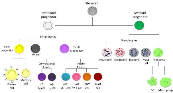
12 Figure 1.1. Cells of the immune system with lymphoid or myeloid origins. Lymphoid progenitor cells branch into B cell progenitor cells (which differentiate into plasma or memory cells), T cell progenitor cells and NK cells. T cell progenitor cells can develop into conventional T cells which include helper T (TH) and cytotoxic T (TC) cells, or innate T cells which include γδ T cells, NKT cells and MAIT cells. Myeloid progenitor cells include
[image:23.842.130.737.100.427.2]13 1.3.1 Myeloid cells
Myeloid cells originate in the bone marrow and they give rise to monocytes,
granulocytes, erythrocytes and platelets.
Monocytes, which constitute 2-10% of human peripheral blood leukocytes, are
normally found in the periphery, where they maintain numbers of resident
macrophages, but during inflammation, they migrate to sites of infection where they
enter the tissues and differentiate into macrophages or DC. They can directly provide
defense against microbial pathogens by secreting antimicrobial factors. Other
functions include phagocytosis, antigen presentation and cytokine production (Leon,
et al., 2005, Ziegler-Heitbrock, 2014).
Macrophages, which are monocyte-derived tissue resident cells, play important roles
in immune surveillance and defense. They express PRR such as toll-like receptors (TLR),
receptor kinases, C-type lectin receptors and NOD-like receptors and are major
sensors of pathogens in the tissues (Taylor, et al., 2005). Macrophages are highly
specialised in removing dead cellular debris via phagocytosis. They are also capable of
antigen presentation to T cells and thus are able to stimulate adaptive immune
responses.
Granulocytes include neutrophils, eosinophils, basophils and mast cells. Neutrophils
are the most abundant phagocyte and comprise 50-60% of circulating leukocytes and
are involved in defense against microbes which they mediate via phagocytosis,
neutrophil extracellular traps and secretion of granules. Eosinophils are involved in
killing parasites via toxic granules. Basophils, which are one of the rarest subtypes in
blood, are involved in immunity against parasites and allergens and thus their granules
contain various substances including histamine, heparin and prostaglandins. Mast cells
are also particularly rich in histamine and heparin, and thus play important roles in
14 1.3.1.1 Dendritic cells
DC are the most potent professional antigen presenting cells (APC), specialised for
recognising foreign organisms through antigen uptake and processing which results in
migration to lymphoid organs and presentation of antigens to T cells (Steinman, 1991).
They express PRR such as TLR, NLR and RLR and are well equipped to internalise
antigens by phagocytosis, pinocytosis and endocytosis (Doherty, 2015). DC can
potently activate naïve T cells and promote expansion and effector T cell
differentiation needed for protection, but also play key roles in self-tolerance (Tisch,
2010). The ability of monocytes to differentiate into DC was first described by Sallusto
and Lanzavecchia and since then this method of generating myeloid DC has been
widely implemented in studies on human DC (Leon, et al., 2005, Sallusto and
Lanzavecchia, 1994). DC are distributed throughout tissues to maximise antigen
capture. They can be found in two distinct functional states. They can be immature,
whereby they induce tolerance to self, or mature DC, which confer protection against
foreign antigens. Progression from immature DC (iDC) to mature DC upon activation
induces changes in cytokine profiles, morphology, phenotype, cell surface markers and
adhesion molecules (Banchereau, et al., 2000). DC maturation results in upregulation
of major histocompatibility complex (MHC) and co-stimulatory molecules and an
increase in IL-12 secretion, which is critical in development of helper T 1 (TH1)
responses. In addition, they can also secrete IL-18, IL-21 and IL-23 which promote
differentiation of naïve T cells. Lipopolysaccharide (LPS) and proinflammatory
cytokines are capable of activating DC, as are certain T cells. DC can also be classed as
tolerogenic or immunogenic owing to the capacity of DC which are not fully mature to
induce T cell tolerance in vivo. Antigens can be delivered to specific DC populations
without the need for maturation stimuli (Steinman, et al., 2003b). In contrast, mature
DC are thought to be immunogenic, a role which was first described in the context of
transplantation and were termed “nature’s adjuvants”. This was demonstrated by
exposing immature DC to antigens and injecting these into mice, which resulted in T
cells restricted to the antigens the DC were exposed to (Steinman, 2007).
Plasmacytoid DC, which derive their name from their resemblance to plasma cells,
15 mediating antiviral immunity by producing high amounts of IFN-α in response to viral
infection. In contrast to myeloid DC, they respond to IL-13 rather than GM-CSF and
although they are capable of endocytosis and antigen presentation they are weak at
capturing soluble and particulate antigens (Steinman, 2003).
1.3.1.2 Antigen presentation
Antigen presentation is an essential step in initiating adaptive immune responses, and
it involves MHC molecules class I (MHC-I) and class II (MHC-II), which are involved in
binding to peptide fragments and displaying them on the cell surface for recognition
by T cells. Presentation of foreign peptides to CD4 T cells via MHC-II mediates
adaptive immune responses, while presentation of self peptides to CD8 T cells via
MHC-I is involved in destruction of malignant or infected host cells.
APC, which include DC, B cells or macrophages present peptide fragments within MHC
molecules to T cells. Recognition of a peptide fragment by the T cell receptor (TCR)
constitutes the first signal in T cell activation. Recognition of costimulatory molecules
such as CD80 and CD86 by the T cell, as well as interaction between CD40 and CD40L
provides the second signal in T cell activation. Receipt of these two signals by CD4+ T
cells results in proliferation of naïve T cells which can differentiate into TH cells or
regulatory T (Treg) cells. Cytokine secretion by the APC determines the fate of the naïve
T cell which in turn determines the nature of the immune response. Depending on the
cytokines secreted, naïve CD4 T cells can differentiate into TH1, TH2, TH17, Treg or
follicular helper T cells (TFH) cells (Fig. 1.2). In contrast, naïve CD8 T cells, which
recognise intracellular peptides coupled to MHC-I molecules, differentiate into
17 1.3.2 Lymphoid cells
1.3.2.1 NK cells
NK cells are cytotoxic lymphocytes with critical roles in the innate immune system.
They express CD56 and NKp46 on the cell surface and are CD3- (Walzer, et al., 2007).
They also express activating receptors, NKG2C, NKG2D, natural cytotoxicity receptors
(NCR) and CD16 and the inhibitory receptors killer-cell immunoglobulin-like receptors
(KIR), which are found only in humans and leukocyte inhibitor receptors (LIR). NK cells
can effectively kill tumour cells via perforin and granzyme secretion or
receptor-mediated cytotoxicity (Kim, et al., 2000), and they can also secrete cytokines and
chemokines (Poli, et al., 2009). They are a major source of IFN-γ (Yao, et al., 1999) but
they can also secrete TNF-α and IL-10. NK activation results in activation of
macrophages, neutrophils and DC, which subsequently induces antigen-specific T and
B cell responses. Furthermore, NK cells are also implicated in virus-infected cell
clearance (Guo, et al., 2011) and in antimicrobial immune responses.
The role of lymphocytes is to distinguish self from non-self antigens and promote
inflammatory responses against foreign invaders. This is mediated by receptors of
structural similarity, pertaining to two distinct classes of lymphocytes: the T cell
receptor (TCR) found on T lymphocytes and the B cell receptor (BCR) on B
lymphocytes. Receptor diversity is achieved through recombination of variable region
genes (Appleman 2003).
1.3.2.2 B cells
B lymphocytes are an essential part of the humoral immune system. They are derived
from the bone marrow where they differentiate from lymphoid progenitor cells. They
then migrate to the spleen and secondary lymphoid tissues where they mature and
differentiate. B cell activation is triggered by antigen binding to the BCR.
The principal and unique function of B cells is antibody production in response to
antigens, but they can also secrete cytokines and act as APC, presenting antigen to T
18 and cell surface presentation on MHC molecules. These are presented to T cells, which
in turn upregulate the co-stimulatory marker CD40L, which is required for B cell
activation. CD40L then engages CD40 on the B cells, which results in differentiation of
naïve B cells into short-lived plasmablasts, long-lived antibody-secreting plasma cells,
germinal centre B cells or memory B cells (Lanzavecchia, et al., 2006, MacLennan, et
al., 2003). Protective antibodies secreted by long-lived plasma cells provide a first line
of defense against re-infection. If antibody levels are not sufficiently high, a second
line of defense consisting of pathogen-experienced memory B cells is triggered to
produce antibodies. Memory B cells have a wider repertoire of antigen specificity than
long-lived plasma cells, thus playing a crucial role in host immunity upon infection with
a novel pathogen (Kurosaki, et al., 2015).
TFH, also known as follicular B helper T cells, are antigen-experienced CD4 T cells
expressing the B cell follicle homing receptor CXCR5 and are found in B cell follicles or
secondary lymphoid tissues. Through expression of CD40L and IL-4 and IL-21 secretion,
they trigger formation and maintenance of germinal centres where they play crucial
roles in mediating selection and survival of B cells (Glatman Zaretsky, et al., 2009, Seo,
et al., 2009).
There are 5 different immunoglobulin isotypes secreted by B cells: IgM, IgA, IgD, IgG
and IgE, and they can be distinguished by their C regions. They have the same antigen
specificity, but activate different effector mechanisms. IgM antibody, which is
expressed as a monomer on the B cell surface, or secreted as a pentamer, is involved
in early stages of humoral immunity. IgA antibodies, which are found as dimers, are
found mainly at mucosal sites and are involved in preventing colonisation by
pathogens (Underdown and Schiff, 1986). The monomeric IgD plays a role as an
antigen receptor on B cells that have not been exposed to antigens, and can activate
basophils, mast cells and secrete antimicrobial factors (Chen, et al., 2009, Geisberger,
et al., 2006). IgE, which is also a monomer, is involved in allergic responses as it
triggers histamine release by mast cells and basophils upon allergen encounter.
Furthermore, it plays a protective role against parasitic worms. IgG, which is
subcategorised into 4 different monomers, provides the main role in antibody-based
19 passive immunity to the growing foetus. Immature B cells express only IgM prior to
antigen exposure. Mature (naïve) B cells express both IgM and IgD, thus allowing them
to respond to antigens (Goding, 1978). B cell activation results in differentiation into
plasma cells thus facilitating antibody secretion. Furthermore, a B cell can undergo
isotype switching, thus altering the class of antibody it produces over the course of an
immune response and thus can secrete IgE, IgA or IgG (Janeway, 2010).
1.3.3 T cells
T lymphocytes, referred to as αβ T cells, as they express a TCR composed of α and β
chains, are central orchestrators of immune responses, and play a vital role in the
adaptive immune system. They comprise the majority of peripheral blood T
lymphocytes. T cell development begins in the thymus where bone marrow-derived
progenitor cells undergo stringent positive and negative selection resulting in naïve T
cell populations exhibiting unique TCR. This diverse antigen specificity allows T cells to
recognise a broad array of complex protein antigens presented on the surface of APC
by MHC molecules (den Haan, et al., 2014).
CD4 T cells, also known as TH cells release cytokines and make contact-dependent
interactions with other cells, thereby stimulating and regulating adaptive immune
responses. They are capable of promoting B cell maturation, activation of
macrophages and cytotoxic T cells, to name but a few. CD4 T cells are activated when
presented with peptide antigens by APC via MHC-II molecules. Once activated, they
proliferate and differentiate into effector T cells that can secrete TH1, TH2, TH17, Treg or
TFH cytokines. These are summarised in Fig.1.2.
CD8 T cells, also known as TC, are involved in killing tumour and virus-infected cells.
CD8 T cells recognise antigens presented within MHC-I molecules and are tightly
regulated by Treg cells to prevent autoimmunity. Treg, which represent up to 10% of
CD4+ T cells are known to mediate antigen-specific suppression and prevention of
autoreactive T cells, which they mediate through direct contact, as suggested by in
vitro studies, and through secretion of regulatory cytokines IL-10 and TGF-β (Longhi, et
20 Memory T cells, which usually express CD45RO, can persist long after an infection has
resolved.They quickly expand into effector T cells upon re-exposure to their cognate
antigen, and thus providing immune “memory” against previously encountered
pathogens. Memory T cells can be further subdivided depending on homing potential
and effector functions into central memory (TCM), effector memory (TEM), and
terminally differentiated effector memory (TEMRA) T cells. In contrast to naïve T cells,
antigen-specific memory T cells do not require TCR signals, but require IL-7 and IL-15
for their long-term survival (den Haan, et al., 2014).
Regulatory T cells can undergo T cell anergy, which is a tolerance mechanism that can
occur whereby the T cell is functionally inactivated following antigen encounter but is
maintained for an extended period of time in an “unresponsive state”. This serves to
preserve immune responses from premature inactivation by Treg. Clonal anergy
describes unresponsiveness at the cellular level, where T cells do not proliferate or
secrete IL-2 upon antigen stimulation. Exogenous IL-2 can however reverse anergy by
stimulating the IL-2 receptor on these anergic T cells (Appleman and Boussiotis, 2003,
Schwartz, 2003).
A small proportion of αβ T cells express TCR that have low variation. These
semi-invariant populations include semi-invariant Natural Killer T cells (iNKT) and Mucosal
Associated Invariant T (MAIT) cells (Adams, et al., 2015) and these will be described in
further detail below.
1.3.4 NKT cells
NKT cells comprise T cells with properties of both NK cells (including NKG2C and
NKG2D) and T lymphocytes (Bendelac, et al., 2007, Brigl and Brenner, 2004). They are
categorised into type I, also known as invariant NKT (iNKT), or type II NKT cells.
Type I NKT cells are characterised by expression of a semi-invariant TCR consisting of
Vα24Jα18 paired with Vβ11 (Bendelac, et al., 2007) and a similar subset is found in
mice. They are strongly reactive to marine sponge-derived glycolipid alpha
galactosylceramide (αGalCer) and can also recognise bacterial-derived lipids and
21 isoglobotrihexosyl ceramide (Arrenberg, et al., 2010). They express high levels of the
activation markers CD69, CD44, CD122 and low expression of CD62L, a marker
expressed by LN-homing naïve T cells (Matsuda, et al., 2000, Bendelac, et al., 1992).
Type I NKT cells play important roles in antimicrobial and local and systemic immune
responses and in controlling tumour development. However, they can also have
negative effects in the pathogenesis of autoimmune and allergic disorders
(Macho-Fernandez and Brigl, 2015). They can generate large amounts of IFN-γ, IL-2, IL-4, IL-9,
IL-10, IL-13, IL-17, IL-21 and GM-CSF (Coquet, et al., 2008, Gumperz, et al., 2002,
Stetson, et al., 2003) and cytotoxic molecules such as perforin and granzyme
(Gumperz, et al., 2002, Lee, et al., 2002) and can interact with various other immune
cells (Brigl and Brenner, 2004, Brennan, et al., 2013). Greater proportions of CD4+ type
I NKT cells produce TH2 cytokines, while all NKT cells can produce both TH1- and TH
2-type cytokines (O'Reilly, et al., 2011).
Human type II NKT cells, also termed non-invariant NKT cells (Godfrey, et al., 2000),
express a diverse range of TCR (Behar, et al., 1999) and are CD1d-restricted cells but
do not recognise αGalCer and do not express the invariant Vα14-Jα18 TCR. Although
they have distinct antigen specificities, they share many features with type I NKT cells
such as rapid secretion of IFN-γ, IL-2, IL-4, IL-10, IL-17, GM-CSF and perforin (Weng, et
al., 2014, Zhao, et al., 2014) and high autoreactivity (Gumperz, et al., 2000). Some type
II NKT cells have been shown to recognise the self-glycolipid sulfatide and upon
sulfatide stimulation they result in inhibition of iNKT cell function, while mediating
protection against liver disease and type I diabetes in mouse models (Arrenberg, et al.,
2011, Halder, et al., 2007, Subramanian, et al., 2012).
Cytokine production by NKT cells can result in activation of T cells, NK cells (Lin, et al.,
2006, Carnaud, et al., 1999) and macrophages (Zeng, et al., 2013) but can also
suppress functions of neutrophils (De Santo, et al., 2008, Hwang, et al., 2006).
1.3.5 MAIT cells
Mucosal associated invariant T (MAIT) cells express a semi-invariant TCR composed of
an α chain (Vα7.2/Jα33) paired with an oligoclonal β chain (Porcelli, et al., 1993, Tilloy,
22 The TCR of MAIT cells recognises riboflavin and folic acid metabolites bound to MR1 in
a conserved docking mode, thus acting like a PRR (Birkinshaw, et al., 2014, Lepore, et
al., 2014, Treiner, et al., 2003). These cells are abundant in humans and are
predominantly found in the gut lamina propria (Kjer-Nielsen, et al., 2012). MAIT cells
are activated by bacteria and are capable of producing IL-2, IFN-γ and IL-17 and
granzymes (Dusseaux, et al., 2011, Guo, et al., 2015). MAIT cells have been shown to
be involved in defense against various pathogens such as Escherichia coli,
enterobacteria, staphylococci and mycobacterium, and yeasts that contain the
riboflavin synthetic pathway, but not viruses (Gold, et al., 2013, Le Bourhis, et al.,
2010). This indicates that they recognise an antigen that is conserved among a range
of intracellular microbes.
1.4 γδ T cells
In contrast to αβ T cells, γδ T cells, which account for 0.5-5% of total blood T cells and
higher proportions in tissues (Morita, et al., 2000), recognise unconventional antigens
such as phosphorylated microbial metabolites and lipid antigens in an
MHC-unrestricted manner. γδ T cells are rarely found in the spleen, lymph nodes, the
thymus or Peyer’s patches but they are localised in tissues (Hein and Mackay, 1991,
Bucy, et al., 1988) such as the gut and liver (Rajoriya, et al., 2014, Brandes, et al., 2003,
Li, et al., 1996, McCarthy, et al., 2013). In the intestine, γδ T cells are found as
intraepithelial lymphocytes (IEL) (Hein and Mackay, 1991, Bucy, et al., 1988, Goodman
and Lefrancois, 1988, Deusch, et al., 1991). There are 5 times as many αβ as γδ T cells
among intestinal IEL, while in blood αβ T cells outnumber γδ T cells 50 to 1 (Hayday,
2000).
1.4.1 The human γδ T cell receptor
The αβ TCR has a diverse repertoire due to the presence of over 50 Vα and Vβ gene
segments available for TCR gene rearrangement (Kabelitz and He, 2012) and although
γδ T cells have a rather small repertoire of Vγ and Vδ segments to select from, their
diversity is just as large as that of the αβ TCR. This is owing to non-germline encoded
mechanisms such as insertion of N-nucleotides during gene rearrangement which
23 the δ chain rearrangement allows for the incorporation of multiple Dδ segments, thus
further increasing diversity (Elliott, et al., 1988, Hata, et al., 1988).
In γδ TCR rearrangement, the three most frequently used δ chains are Vδ1, Vδ2 and
Vδ3, while the less common δ segments include Vδ4, Vδ5, Vδ5 and Vδ7, named
according to their locations on the δ locus (Thedrez, et al., 2007). The δ chains can pair
with one of seven functional Vγ gene segments, namely Vγ2, Vγ3, Vγ4, Vγ5, Vγ8, Vγ9
and Vγ11 (Adams, et al., 2015, Porcelli, et al., 1991, Hinz, et al., 1997).
It is hypothesised that γδ T cells have developed to respond to unique stress antigens
that are markers of cell infection or transformation, rather than directly recognising a
diversity of microbial antigens and thus serving as a “first line of defense” (Hayday,
2000, Janeway, et al., 1988).
1.4.2 Human γδ T cells subsets
Human γδ T cells fall into one of two categories: Vγ9Vδ2 T cells or non-Vδ2 T cells with
the former being the predominant subset in blood, while the latter is more common in
tissues. The two groups show distinct functions, migratory patterns and homing
capabilities (Zheng, et al., 2013).
1.4.2.1 Non-Vδ2 T cells
Non-Vδ2 T cells, which are functionally distinct from Vγ9Vδ2 T cells, include Vδ1, Vδ3
and Vδ5 T cells and these are found to be expanded in cytomegalovirus infection in
kidney transplant patients and are thought to play protective roles against tumours
and infection (Halary, et al., 2005).
Vδ1+ γδ T cells are mainly tissue resident and are the most frequent subset among
human intraepithelial cells in the skin (Ebert, et al., 2006) and small intestine
(Holtmeier, et al., 2001, Hayday, et al., 2001) and they represent over 50% of foetal
blood γδ T cells at birth (Dimova, et al., 2015). They can also be found in lymph nodes
(Brandes, et al., 2003) and certain tumours (Maeurer, et al., 1996). Vδ1 T cells have
been reported to recognise stress-inducible MHC class I-related molecules MICA and
24 al., 2007, Bai, et al., 2012, Hayday and Vantourout, 2013, Luoma, et al., 2013) and
UL-16 binding proteins (ULBP) which bind to cytomegalovirus-infected cells (Cosman, et
al., 2001). They have also been reported to respond to epithelial tumours (Maeurer, et
al., 1996, Groh, et al., 1998, Coscas, et al., 2004) and lymphomas (Catellani, et al.,
2007, Hacker, et al., 1992) and can recognise different members of the MHC
superfamily (Vantourout and Hayday, 2013). Vδ1 T cells have also been reported to be
expanded in HIV patients (Poles, et al., 2003, Rossol, et al., 1998, Wesch and Kabelitz,
2003), cytomegalovirus (Dechanet, et al., 1999) and Candida (Fenoglio, et al., 2009,
Maher, et al., 2015) infection and B cell chronic lymphocytic leukaemia (Siegers and
Lamb, 2014) and have been shown to produce IFN-y and IL-17.
Vδ3 T cells represent the third most common subset in peripheral blood, and little is
known about this subset. Its ligand specificities are unknown, but they are reported to
be expanded in renal and stem cell transplant patients and patients with leukaemia
and chronic viral infection (Halary, et al., 2005, Dechanet, et al., 1999, Knight, et al.,
2010, Couzi, et al., 2010) and have been shown in one study to recognise CD1d+ cells
(Mangan, et al., 2013).
1.4.2.2 Vγ9Vδ2 T cells
γδ T cells consisting of Vγ9 paired with Vδ2 account for 50-95% of γδ T cells in healthy
individuals (Porcelli, et al., 1991, Hinz, et al., 1997, Kabelitz, et al., 1999) and are
unique to humans and primates. These are found in very small numbers in newborns,
and exposure to environmental microorganisms stimulates their expansion (Parker, et
al., 1990). Furthermore, they change with age and differ between genders (Caccamo,
et al., 2006b). Vγ9Vδ2 recognise organic-based pyrophosphate molecules termed
phosphoantigens (pAg) which are intermediates of isoprenoid metabolism (Fig. 1.3), a
pathway essential for cell survival (Morita, et al., 2000, Constant, et al., 1994, Hintz, et
al., 2001, Puan, et al., 2007, Tanaka, et al., 1995). There are two distinct pathways of
isoprenoid biosynthesis, with the methylerythritol 4 phosphate (MEP) pathway being
used by most bacteria and protozoa (Chen and Letvin, 2003) but not eukaryotes. In
contrast, all eukaryotic and some prokaryotic cells use the mevalonate pathway. Both
25 cellular components. A table of pathways employed by common pathogens is
illustrated below (Table 2).
The most potent pAg, (E)-4-hydroxy-3-methyl-but-2-enyl pyrophosphate (HMB-PP),
which is an intermediate of the non-mevalonate MEP pathway (Fig. 1.3, Fig. 1.4)
specifically activates Vγ9Vδ2 T cells at pico- to nanomolar concentrations (Altincicek,
et al., 2001) and thus allow Vγ9Vδ2 T cells to recognise foreign pathogens that use this
pathway. The mevalonate pathway does not produce HMB-PP but results in
production of IPP, which requires up to 10,000-fold higher concentrations for
activating Vγ9Vδ2 T cells (Fig. 1.4) than intermediates of the microbial
non-mevalonate pathway (Puan, et al., 2007). However, the IPP levels normally found in
healthy cells are not sufficient to trigger Vγ9Vδ2 T cell activation, but cell stress and
other triggers can result in dysregulated metabolism which could be indicative of
infection or cancerous cells, overproduce the metabolite IPP, and therefore allow
Vγ9Vδ2 T cells to recognise IPP as a self-antigen in conditions of disease.
Aminobisphosphonates, which are compounds in clinical use for the treatment of
osteoporosis and bone metastasis can also activate Vδ2 T cells in vivo and in vitro.
They have been shown to result in accumulation of IPP through inhibition of the
IPP-processing enzyme farnesyl pyrophosphate synthase (Fig. 1.3) (Gober, et al., 2003,
Kunzmann, et al., 1999, Thompson, et al., 2006). Aminobisphosphonates have also
been shown to activate Vγ9Vδ2 T cells in vitro (Das, et al., 2001, Kabelitz, et al., 2004).
As Vγ9Vδ2 T cells frequencies are found to be decreased in many diseases, therapies
involving in vivo expansion of Vγ9Vδ2 T cell using aminobisphosphonates are currently
in place to increase the numbers of circulating Vγ9Vδ2 T cells. However, Wang and
colleagues (Wang, et al., 2011) have discovered toxicity when various
aminobisphosphonates were used continuously to expand Vγ9Vδ2 T cells from
peripheral blood mononuclear cells (PBMC), thus suggesting that prolonged exposure
to aminobisphosphonates inhibits their proliferation due to inhibition of the
isoprenoid pathway. To overcome this, they suggest pulsing them to limit toxicity
26 aminobisphosphonates through fluid phase endocytosis, which simulates the rapid
clearance through renal excretion in vivo (Wang, et al., 2011).
A class of mycobacterial nonphosphorylated alkyl amines are also known to stimulate
Vγ9Vδ2 T cells, but require much higher (milimolar) concentrations which are 106 -108
fold higher than those of HMB-PP (Bukowski, et al., 1999). These include
isobutylamine and isoamylamine, which when used to stimulate PBMC, in the
presence of IL-2 were able to induce more than 10-fold expansion of Vγ9Vδ2 T cells
29 Table 1.2. Distribution of the MEP and mevalonate pathways amongst Gram-positive and Gram-negative pathogens (adapted from Heuston et al., 2012).
Gram + pathogen MEP Mevalo-nate
Gram - pathogen MEP Mevalo-nate
Bacillus anthracis + - B. abortus + -
Bacillus subtilis + - Borrelia burgdorferi - + Clostridium difficile + - Chlamydia trachomatis + - Clostridium botulinum + - Chlamydia pneumonia + - Clostridium perfringens + - S. enterica + - Entercoccus faecalis - + Escherichia coli + -
L. monocytogenes + + F. tularensis + -
L. innocua - + Legionella pneumophila - +
Listeria seeligeri - + P. aeruginosa + -
Nocardia terpenica + - V. cholera + -
Stahylococcus aureus - + K. pneumonia + -
Streptomyces pyogenes - + Bordetella pertussis + -
S. pneumonia - + Haemophilus influenza + -
Helicobacter pylori + - Shigella dysenteriae + - Neisseria gonorrhoeae + - Neisseria meningitides + -
C. jejuni + -
30 1.4.3 Vγ9Vδ2 T cell activation
The recent discovery of butyrophilin 3A1 (BTN3A1) and its role in Vγ9Vδ2 T cell
activation has provided a major breakthrough towards understanding the process of
pAg-induced Vγ9Vδ2 T cell activation (Adams, et al., 2015). BTN3A1 belongs to the
butyrophilin family, also known as CD277, which encompasses proteins with diverse
roles in host homeostasis (Abeler-Dorner, et al., 2012, Arnett and Viney, 2014). The
three members found in humans are BTN3A1, BTN3A2 and BTN3A3 (Rhodes, et al.,
2001) which are structurally homologous to the B7 superfamily of proteins. BTN3A1
and BTN3A3 also possess an intracellular “B30.2” domain which is thought to be
involved in pAg-mediated activation of Vγ9Vδ2 T cells (Adams, et al., 2015). The
precise mechanism by which BTN3A1 contributes to Vγ9Vδ2 T cell activation has been
controversial. One model proposes that the BTN3A molecule acts as an
antigen-presenting molecule which captures and presents pAg on the cell surface to Vγ9Vδ2 T
cells which recognise it directly through their TCR (Vavassori, et al., 2013). However,
while one study demonstrated the requirement of all three isoforms (Rhodes, et al.,
2015), a different study suggested that only BTN3A1 is capable of mediating
pAg-induced activation of Vγ9Vδ2 T cells and that this activity was pinpointed to the B30.2
domain of BTN3A1 (Harly, et al., 2012). In support of this, direct binding between
HMB-PP and the B30.2 domain was shown via nuclear magnetic resonance studies
(Hsiao, et al., 2014) and affinity studies (Altincicek, et al., 2001, Sandstrom, et al.,
2014). Furthermore, it was revealed that the difference between BTN3A1 and BTN3A3
was confined to a single amino acid difference within the binding pocket of B30.2 at
position 351 where BTN3A1 contained a histidine, while BTN3A3, an arginine.
Swapping of this histidine residue onto BTN3A3 provided BTN3A3 with the ability to
bind pAg and mediate Vγ9Vδ2 T cell activation, while introduction of arginine onto
BTN3A1 abrogated its activity (Sandstrom, et al., 2014), thus confirming that the
32 A model has been proposed whereby binding of the pAg intracellularly is transitioned
to the extracellular side of the membrane. The pAg binding to B30.2 would induce a
conformational change which could modulate the structure of the extracellular
domains of BTN3A. It is not known however whether Vγ9Vδ2 T cells can directly
recognise BTN3A (Adams, et al., 2015, Sandstrom, et al., 2014, Wang, et al., 2013). The
potential mechanisms of activation are illustrated in Fig. 1.5 (Harly, et al., 2014).
1.4.4 Phenotypes of γδ T cells
1.4.4.1 CD4 and CD8 expression
Most peripheral γδ T cells lack CD4 and CD8 expression, which is not surprising since
CD4 and CD8 are components of MHC recognition, and as mentioned above, γδ T cells
do not rely on MHC molecules (Hayday, 2000, Kabelitz, et al., 2000).
1.4.4.2 Differentiation status
γδ T cells express molecules associated with different stages of differentiation which
can be classified through CD27 and CD45RA expression, as shown by Dieli et al (2003),
while αβ T cells classification also requires CCR7 expression (Okada, et al., 2008). Naïve
cells are defined as CD45RA+CD27+, T
CM are CD45RA-CD27+, while TEMRA are
CD45RA+CD27- and T
EM are negative for both markers (CD45RA-CD27-). These are
summarised in Fig. 1.6.
1.4.4.3 Chemokine receptors
Vγ9Vδ2 T cells have distinct migration properties compared to αβ T cells (Brandes, et
al., 2003). Since Vγ9Vδ2 T cells recognise antigens independently of MHC molecules,
therefore they do not migrate to secondary lymphoid tissues where antigen
presentation by professional APC takes place (Moser and Brandes, 2006). In line with
this, most Vγ9Vδ2 T cells (>80%) lack expression of the lymph node (LN)-homing
receptor CCR7, whereas most αβ T cells express the receptor. Instead, Vγ9Vδ2 T cells
express receptors for a range of inflammatory chemokines (Chen and Letvin, 2003).
Some γδ T cells express chemokine receptor CCR5, which binds inflammatory