Original Article
Loss of epithelial cell adhesion molecule
(EpCAM) in infiltrative basal cell carcinoma
Maria Rita Gaiser1,2, Daniela Hirsch3, Timo Gaiser3
1Department of Dermatology, Venereology and Allergology, University Medical Center Mannheim, Ruprecht-Karl
University of Heidelberg, Mannheim, Germany; 2Skin Cancer Unit, German Cancer Research Center (DKFZ),
Heidelberg, Germany; 3Institute of Pathology, University Medical Center Mannheim, Ruprecht-Karl University of
Heidelberg, Mannheim, Germany
Received October 27, 2017; Accepted November 9, 2017; Epub January 1, 2018; Published January 15, 2018
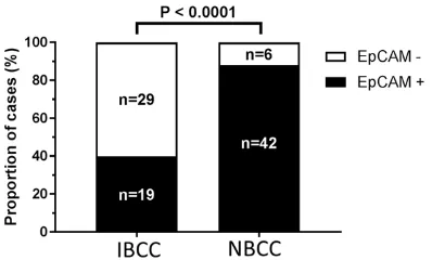
Abstract: Basal cell carcinoma (BCC) is the most common type of skin cancer and expresses high protein levels of the epithelial cell adhesion molecule (EpCAM, syn. CD326). Though BCCs only rarely metastasize, infiltrative and destructive growth do occur. EpCAM has been studied extensively in the context of adhesion and carcinogenesis but results of studies relating EpCAM expression to invasive potential or patient prognosis have been inconsistent. In an attempt to link EpCAM expression with infiltrative potential, we retrospectively stained paraffin embedded tissue samples of nodular and infiltrative BCCs. A total of 96 samples comprising 48 nodular and 48 infiltrative BCC cases were immuhistochemically stained with anti-EpCAM clone BerEP4. Loss of EpCAM expression along the tumor inva-sive front was detected in 6 of 48 (12.5%) of the nodular BCC as compared to 29 of 48 (60.4%) of the infiltrative BCC cases (P < 0.0001). These results exemplify the important role of EpCAM for cell adhesion. BCC infiltration seems to be promoted by down-regulation of EpCAM along the tumor invasion front.
Keywords: Basal cell carcinoma, EpCAM, epithelial cell adhesion molecule, CD326
Introduction
Basal cell carcinoma (BCC) is the most com-mon type of skin cancer accounting for appro- ximately 70% of all skin malignancies [1]. Th- ough BCCs are usually slow-growing, non-aggressive tumors, mostly cured by surgical treatment, a minority of cases shows an aggres- sive, rarely even metastatic behavior [2]. Ackermann classified BCC as trichoblastic car-cinoma [3] but the cell of origin is still matter of debate. Most likely, the majority of BCCs arises from the lowermost layers of the epidermis but there has also been evidence that some BCCs may originate from the outer root sheath of the pilosebaceous unit [4, 5]. Interestingly, BCC is strictly stroma dependent, thus, an xeno-trans-plantation into mice is unsuccessful if the stro-ma is not included [6]. This strostro-mal dependen-cy is the most likely reason for the low incidence of metastasis of these tumors. The morphology of BCC is quite variable. Consequently, various histopathological subtypes have been defined
including nodular (solid), micronodular, pig-mented, keratotic, superficial (multifocal), cys-tic, adenoid, fibroepitheliomatous, infiltrating, sclerosing, infundibulocystic, metatypical, and basosquamous [7]. The non-aggressive nodu-lar type accounts for approximately 70% of all cases whereas only approx. 5% represent the infiltrating type, characterized by invasive growth pattern with clinically indistinct borders [4]. Mixed patterns are quite common. The vast majority of BCC are closely attached to the basal layer of the epidermis while longer exist-ing lesions usually extend into the lower dermis. Further growth usually occurs diffusely or along the cutaneous adnexae [8]. Perineural invasion is present in nearly 1% of all BCC cases with an increasing incidence in aggressive variants [9-11].
the most primitive follicular tumor [13]. EpCAM is a transmembrane cell surface glycoprotein that is expressed by developing and differenti-ated epithelia [14-16], carcinomas, tumor-initi-ating cells, circultumor-initi-ating tumor cells, and stem cells [17, 18]. EpCAM has many faces and the literature regarding its function is extensive (for review [15, 19, 20]). Among the activities attributed to EpCAM, it has been reported that it mediates adhesion [21], that it reduces adhe-sion [22], and that it functions as an outside- in signaling molecule [23]. In humans, EpCAM
mutations induce congenital tufting enteropa-thy, a rare diarrheal syndrome that is caused by severe intestinal epithelial dysplasia and loss of epithelial integrity [24]. The role of EpCAM for tumorigenesis is also ambiguous. EpCAM has been intensively studied as a tumor antigen that may represent a suitable therapeutical target [25], and because it may play a role in cancer pathogenesis [17]. In some settings, EpCAM may facilitate cancer cell invasion and metastasis [23], and its expression in tumors may indicate poor prognosis [15, 19, 20]. In contrast, other studies could demonstrate that in some tumors EpCAM expression appears to be beneficial [20, 26]. It seems likely that because EpCAM is a molecule that interacts with surrounding cells, tissue context and microenvironment are important.
In BCC it has been demonstrated that islands of tumor cells along the tumor front are sur-rounded by a stroma that is different form the adjacent dermis and that BCC cells express decreased protein levels of basement mem-brane components (e.g. bullous pemphigoid antigens 1 and 2, integrins alpha6 and beta4, and beta3 chain of laminin), which may
facili-tate the capability of tumor cells to invade [6, 8]. Furthermore, a loss of expression of epi- thelial markers and junctional proteins, such as E-cadherin, beta-catenin, and desmoglein among the invasive tumor front has been shown in canine oral and cutaneous squamous cell carcinomas [27]. It is further known that the classical cadherins (primarily E-cadherin) and EpCAM are co-expressed in some tissues, and previously it has been reported that EpCAM can modulate cadherin-mediated adhe-sion [22].
In an attempt to link immunohistochemical EpCAM expression and infiltrative potential we retrospectively stained paraffin embedded tis-sue samples of nodular and infiltrative BCCs.
Material and methods
Formalin fixed paraffin-embedded (FFPE) BCC samples, that had been surgically removed between 2011 and 2012, were obtained from the Department of Dermatology and the Ins- titute of Pathology of the University Medical Center Mannheim, University of Heidelberg. Clinical data sets included age, sex of the patients and histopathological features. Dia- gnosis of BCC was verified histopathologically. Additionally, selected cases were subjected to immunohistochemical staining with GATA3, EMA, Vimentin, and S100. All procedures were performed according to the principle of the Declaration of Helsinki and approved by the local medical ethics committee (2014-835R- MA).
A total of 121 BCC cases were included into the study. Of those, 25 cases had to be omitt- ed because due to a mixed growth pattern, a clear classification into nodular or infiltrating BCC subtype was not possible. The remaining cases included 48 nodular BCC and 48 infil- trating BCC. Loss of EpCAM was defined as an obvious decrease (less than 50% staining intensity as compared to the rest of the tu- mor) of EpCAM staining intensity occurring on tumor borders or tumor islands infiltrating the dermis.
Immunohistochemistry
Tissue sections were stained for EpCAM (cl- one Ber-EP4, 1:50; cat # M0804, Dako, Agilent, Santa Clara, CA, USA), EMA (clone E29, 1:200;
Figure 1. EpCAM loss (EpCAM-) is associated with
[image:2.612.91.290.73.193.2]cat # M0613, Dako), GATA3 (clone L50-823, 1:100; cat # 390M-16, Medac, Wedel, Ger- many), S100 (polyclonal, 1:4000; cat # Z0311, Dako), and vimentin (clone SP20, 1:400; cat # RM-9120-s, Thermo Fisher Scientific, Waltham, MA, USA). Sections were subjected to heat-induced citrate-based (for EpCAM and vimen-tin) or EDTA-based (for EMA and GATA3) anti- gen retrieval. Antibody binding was visualized using the EnVision Detection System, Pero- xidase/DAB, Rabbit/Mouse (cat # K5007, Dako) according to the manufacturer’s ins- tructions.
Statistics
Statistical analysis was performed using Gra- phPad Prism software version 7.03 (GraphPad Software, La Jolla, CA, USA, www.graphpad. com). Differences between groups were esti-mated by Fisher’s exact test. P < 0.05 was considered significant.
Results
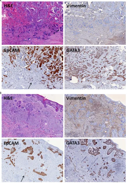
All BCC cases tested immunohistochemically positive for the expression of EpCAM. Loss of EpCAM along the tumor front/infiltrating islands was observed in 29 of 48 (60.4%) in- filtrative BCC and in 6 of 48 (12.5%) nodular BCC (P < 0.0001) (Figure 1). EpCAM loss was mainly restricted to the invasive front. EpCAM staining demonstrated a heterogeneous ex- pression pattern with strong EpCAM expres-sion in superficial and central tumor parts and weak to total EpCAM loss at the deeper infil- trating tumor fingers including the invasive front (Figure 2). Perineural invasion as demon-strated by tumor cells being adjacent to S100 positive neural structures was observed in
(100%) infiltrative BCC and 10 of 10 (100%) nodular BCC. EMA expression was absent in seven of seven (100%) infiltrative BCC and 10 of 10 (100%) nodular BCC (Table 1). Vimentin counter staining of the stroma was used for a better identification of small BCC tumor islets (Figure 2).
Discussion
To our knowledge, the loss of EpCAM along the invasive front of infiltrative BCC has not been described so far. We found a statistically relevant loss of EpCAM expression in infiltrat- ing BCC as compared to nodular BCC. EpCAM loss was mainly restricted to the tumor inva- sion front as well as deeper infiltrating tumor islands. Immunhistochemical stainings for GATA3 and EMA showed an universal positivity and negativity in 10 cases, respectively. Thus, especially in infiltrative BCC cases the use of GATA3 can facilitate detecting single tumor islands that have lost their EpCAM expression and are otherwise too small in order to be detected in conventional H&E staining.
[image:4.612.90.381.137.217.2]Loss of other intercellular adhesion molecules and junctional proteins, such as E-cadherin, beta-catenin, and desmoglein among the tu- mor invasion front has been described in squamous cell carcinoma [27] but not in BCC. It is known that loss of those molecules is associ-ated with an increase of tumor cell invasion; thus, they are considered useful prognostic markers in human carcinomas [28-31]. At least one of those cell adhesion markers, E- cadherin, is known to be co-expressed with EpCAM and it has been illustrated that EpCAM can modulate cadherin-mediated adhe-sion [22]. Akin et al. could demonstrate that would have otherwise been missed by H&E or EpCAM-staining. (A) Scale bar: 500 µm; (B) Scale bar: 200 µm, arrow depicting the infiltrating tumor islands with detected EpCAM loss (H&E, anti-vimentin, anti-EpCAM, anti-GATA3).
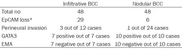
Table 1. Immunohistochemical features of infiltrative and nodular basal cell carcinoma
Infiltrative BCC Nodular BCC
Total no 48 48
EpCAM loss* 29 6
Perineural invasion 3 out of 12 cases 1 out of 24 cases GATA3 7 positive out of 7 cases 10 positive out of 10 cases EMA 7 negative out of 7 cases 10 negative out of 10 cases
*Fisher’s exact test, P < 0.0001.
EpCAM modulates adhesion and tight junction function by regulating intracellular localization and degradation of selected claudins, especial-ly claudin-7 and claudin-1 [32]. Our results underline the positive role of EpCAM expres-sion for cell adheexpres-sion in BCC while its loss at the tumor front presumably facilitates tumor invasion.
Besides, the downregulation of EpCAM has been associated with epithelial-mesenchymal transition (EMT) [33, 34], a process, that plays a key role in carcinoma progression and is necessary for invasion and metastasis [35-37]. Interestingly, in contrast to other highly malig-nant EpCAM expressing tumors, such as Merkel cell carcinoma [38], even though the majority of BCC strongly expresses EpCAM [12], BCC only rarely metastasize.
It has been demonstrated that for BCC tumori-genesis a histological continuum exists that moves from low-risk superficial and nodular BCC subtypes via less frequent transitional, mixed types toward the high-risk micronodular, morpheic, and infiltrating types [39]. The host immune response and stromal alterations ac- company this progression.
Thus, when considered in conjunction with the results reported herein, our data suggests that dynamic changes of EpCAM expression facilitate infiltration along the tumor invasion front and may be accompanied by histological changes towards more aggressive BCC sub- types.
Detection of EpCAM loss along the invasive BCC front could therefore serve as prediction marker for a destructive BCC growth pattern leading to substantial tissue damage. Ten- tatively and still theoretical, in cases where EpCAM loss is detected it might be reasona- ble to choose a larger safety margin.
Further studies on adhesion/junctional mark-ers known to be associated with EpCAM (e.g. claudins and cadherins) are needed to shed further light on the mechanisms of EpCAM loss along the tumor invasion front.
Acknowledgements
This work was supported in part by grants of the German research foundation (Deutsche Forschungsgemeinschaft) GRK2099 (M.R.G.).
Disclosure of conflict of interest
None.
Address correspondence to: Dr. Timo Gaiser, In- stitue of Pathology, University Medical Centre Ma- nnheim, Ruprecht-Karl University of Heidelberg, Theodor-Kutzer-Ufer 1-3, 68135, Mannheim, Ger- many. Tel: +49 621 383 2876; Fax: +49 621 383 2005; E-mail: [email protected]
References
[1] Mertens RB, de Peralta-Venturina MN, Balzer BL and Frishberg DP. GATA3 expression in nor-mal skin and in benign and nor-malignant epider-mal and cutaneous adnexal neoplasms. Am J Dermatopathol 2015; 37: 885-891.
[2] Tjarks BJ, Pownell BR, Evans C, Thompson PA, Kerkvliet AM, Koch MR and Jassim AD. Evalua-tion and comparison of staining patterns of factor XIIIa (AC-1A1), adipophilin, and GATA3 in sebaceous neoplasia. J Cutan Pathol 2017; [Epub ahead of print].
[3] Ackermann AB, Reddy VB and Soyer HP. Neo-plasms with follicular differentiation. Ardor Scribendi New York 2001, 2001.
[4] Wrone DA, Swetter SM, Egbert BM, Smoller BR and Khavari PA. Increased proportion of ag-gressive-growth basal cell carcinoma in the veterans affairs population of Palo Alto, califor-nia. J Am Acad Dermatol 1996; 35: 907-910. [5] Miller SJ. Biology of basal cell carcinoma (Part
II). J Am Acad Dermatol 1991; 24: 161-175. [6] Hales SA, Stamp G, Evans M and Fleming KA.
Identification of the origin of cells in human basal cell carcinoma xenografts in mice using in situ hybridization. Br J Dermatol 1989; 120: 351-357.
[7] Strutton GM. Pathological variants of basal cell carcinoma. Australas J Dermatol 1997; 38 Suppl 1: S31-35.
[8] Mehregan AH. Aggressive basal cell epithelio-ma on sunlight-protected skin. Report of eight cases, one with pulmonary and bone metasta-ses. Am J Dermatopathol 1983; 5: 221-229. [9] Mark GJ. Basal cell carcinoma with intraneural
invasion. Cancer 1977; 40: 2181-2187. [10] Ratner D, Lowe L, Johnson TM and Fader DJ.
Perineural spread of basal cell carcinomas treated with Mohs micrographic surgery. Can-cer 2000; 88: 1605-1613.
[11] Brown CI and Perry AE. Incidence of perineural invasion in histologically aggressive types of basal cell carcinoma. Am J Dermatopathol 2000; 22: 123-125.
and immunocytochemical findings in fine-nee-dle aspirates. Diagn Cytopathol 1998; 18: 403-408.
[13] Sellheyer K and Krahl D. Basal cell (trichoblas-tic) carcinoma common expression pattern for epithelial cell adhesion molecule links basal cell carcinoma to early follicular embryogene-sis, secondary hair germ, and outer root sheath of the vellus hair follicle: a clue to the adnexal nature of basal cell carcinoma? J Am Acad Der-matol 2008; 58: 158-167.
[14] Nagao K, Ginhoux F, Leitner WW, Motegi S, Bennett CL, Clausen BE, Merad M and Udey MC. Murine epidermal Langerhans cells and langerin-expressing dermal dendritic cells are unrelated and exhibit distinct functions. Proc Natl Acad Sci U S A 2009; 106: 3312-3317. [15] Trzpis M, McLaughlin PM, de Leij LM and
Harmsen MC. Epithelial cell adhesion mole-cule: more than a carcinoma marker and adhe-sion molecule. Am J Pathol 2007; 171: 386-395.
[16] Schiechl H and Dohr G. Immunohistochemical studies of the distribution of a basolateral-membrane protein in intestinal epithelial cells (GZ1-Ag) in rats using monoclonal antibodies. Histochemistry 1987; 87: 491-498.
[17] Baeuerle PA and Gires O. EpCAM (CD326) find-ing its role in cancer. Br J Cancer 2007; 96: 417-423.
[18] Martowicz A, Seeber A and Untergasser G. The role of EpCAM in physiology and pathology of the epithelium. Histol Histopathol 2016; 31: 349-355.
[19] Trzpis M, Bremer E, McLaughlin PM, de Leij LF and Harmsen MC. EpCAM in morphogenesis. Front Biosci 2008; 13: 5050-5055.
[20] van der Gun BT, Melchers LJ, Ruiters MH, de Leij LF, McLaughlin PM and Rots MG. EpCAM in carcinogenesis: the good, the bad or the ugly. Carcinogenesis 2010; 31: 1913-1921. [21] Litvinov SV, Velders MP, Bakker HA, Fleuren GJ
and Warnaar SO. Ep-CAM: a human epithelial antigen is a homophilic cell-cell adhesion mol-ecule. J Cell Biol 1994; 125: 437-446.
[22] Litvinov SV, Balzar M, Winter MJ, Bakker HA, Briaire-de Bruijn IH, Prins F, Fleuren GJ and Warnaar SO. Epithelial cell adhesion molecule (Ep-CAM) modulates cell-cell interactions me-diated by classic cadherins. J Cell Biol 1997; 139: 1337-1348.
[23] Maetzel D, Denzel S, Mack B, Canis M, Went P, Benk M, Kieu C, Papior P, Baeuerle PA, Munz M and Gires O. Nuclear signalling by tumour-as-sociated antigen EpCAM. Nat Cell Biol 2009; 11: 162-171.
[24] Sivagnanam M, Mueller JL, Lee H, Chen Z, Nel-son SF, Turner D, Zlotkin SH, Pencharz PB,
Ngan BY, Libiger O, Schork NJ, Lavine JE, Taylor S, Newbury RO, Kolodner RD and Hoffman HM. Identification of EpCAM as the gene for con-genital tufting enteropathy. Gastroenterology 2008; 135: 429-437.
[25] Sears HF, Atkinson B, Mattis J, Ernst C, Herlyn D, Steplewski Z, Hayry P and Koprowski H. Phase-I clinical trial of monoclonal antibody in treatment of gastrointestinal tumours. Lancet 1982; 1: 762-765.
[26] Akita H, Nagano H, Takeda Y, Eguchi H, Wada H, Kobayashi S, Marubashi S, Tanemura M, Takahashi H, Ohigashi H, Tomita Y, Ishikawa O, Mori M and Doki Y. Ep-CAM is a significant prognostic factor in pancreatic cancer patients by suppressing cell activity. Oncogene 2011; 30: 3468-3476.
[27] Nagamine E, Hirayama K, Matsuda K, Okamo-to M, Ohmachi T, Uchida K, Kadosawa T and Taniyama H. Invasive front grading and epithe-lial-mesenchymal transition in canine oral and cutaneous squamous cell carcinomas. Vet Pathol 2017; 54: 783-791.
[28] Bankfalvi A, Krassort M, Buchwalow IB, Vegh A, Felszeghy E and Piffko J. Gains and losses of adhesion molecules (CD44, E-cadherin, and beta-catenin) during oral carcinogenesis and tumour progression. J Pathol 2002; 198: 343-351.
[29] Mattijssen V, Peters HM, Schalkwijk L, Manni JJ, van’t Hof-Grootenboer B, de Mulder PH and Ruiter DJ. E-cadherin expression in head and neck squamous-cell carcinoma is associated with clinical outcome. Int J Cancer 1993; 55: 580-585.
[30] Schipper JH, Frixen UH, Behrens J, Unger A, Jahnke K and Birchmeier W. E-cadherin ex-pression in squamous cell carcinomas of head and neck: inverse correlation with tumor dedif-ferentiation and lymph node metastasis. Can-cer Res 1991; 51: 6328-6337.
[31] Wang Q, Sun ZX, Allgayer H and Yang HS. Downregulation of E-cadherin is an essential event in activating beta-catenin/Tcf-dependent transcription and expression of its target genes in Pdcd4 knockdown cells. Oncogene 2010; 29: 128-138.
[32] Wu CJ, Mannan P, Lu M and Udey MC. Epithe-lial cell adhesion molecule (EpCAM) regulates claudin dynamics and tight junctions. J Biol Chem 2013; 288: 12253-12268.
[33] Frederick BA, Helfrich BA, Coldren CD, Zheng D, Chan D, Bunn PA Jr and Raben D. Epithelial to mesenchymal transition predicts gefitinib resistance in cell lines of head and neck squa-mous cell carcinoma and non-small cell lung carcinoma. Mol Cancer Ther 2007; 6: 1683-1691.
JN, Hartmann LC, Manjili MH, Radisky DC, Fer-rone S and Knutson KL. Immune-induced epi-thelial to mesenchymal transition in vivo gen-erates breast cancer stem cells. Cancer Res 2009; 69: 2887-2895.
[35] Guarino M. Epithelial-mesenchymal transition and tumour invasion. Int J Biochem Cell Biol 2007; 39: 2153-2160.
[36] Kalluri R and Weinberg RA. The basics of epi-thelial-mesenchymal transition. J Clin Invest 2009; 119: 1420-1428.
[37] Thiery JP. Epithelial-mesenchymal transitions in tumour progression. Nat Rev Cancer 2002; 2: 442-454.
[38] Gaiser MR, Daily K, Hoffmann J, Brune M, Enk A and Brownell I. Evaluating blood levels of neuron specific enolase, chromogranin A, and circulating tumor cells as Merkel cell carcino-ma biocarcino-markers. Oncotarget 2015; 6: 26472-26482.
![A STUDY ON NETWORK ON CHIP [NOC]](data:image/gif;base64,R0lGODlhAQABAIAAAP///wAAACH5BAEAAAAALAAAAAABAAEAAAICRAEAOw==)