IJSRR, 8(1) Jan. – Mar., 2019 Page 2841
Research article Available online www.ijsrr.org
ISSN: 2279–0543
International Journal of Scientific Research and Reviews
Validation of LC-MS/MS Electrospray Ionisation method for the
Estimation of
Binimetinib
in Human Plasma
*K. Poorna chandra rao, G. Vidya sagar
1and K.R.S.Sambasiva rao
2*JNTUK,Kakinada-533003, Andhra Pradesh, India.
1Srinivasa rao College of Pharmacy, Visakhapattanam – 530041, Andhra Pradesh, India.
Mizoram University(Central university) Tanhril, Aizawl –796009, Mizoram, India.
ABSTRACT
A rapid, specific and robust bioanalytical method for quantification of Binimetinib was developed and validated in micro volumes (300 μL) of human plasma by liquid chromatography -electrospray ionization tandem mass spectrometry in positive ion mode. Binimetinib-13C2-D4 was used as an internal standard. Precursor to product ion transitions of m/z442.2 (parent ion) to 363.9 m/z (product ion) and m/z448.5 m/z (parent ion) to 363.9 m/z (product ion) were used to measure the analyte and the internal standard (ISTD), respectively. Chromatographic separation was carried out in reverse phase conditions using XTerra MS C18 Column, 125Å, 3.5 µm, 1 mm X 150 mm
with an isocratic mobile phase consisting of Methanol: 20mM Ammonium acetate (pH: 4.5, Adjusted with diluted aceticacid) (90: 10, v/v) at a flow rate of 0.5 mL min−1. The extraction procedure yielded a recovery of 94.12 and 85.92% for Binimetinib and the internal standard, respectively. The assay exhibited a linear dynamic range of 20.00–200.00pg/mL. The RSD% of intra- and inter-day assay was ≤15%. For its sensitivity, reliability and lower plasma volume
requirement, the proposed method is suitable for pharmacokinetic studies.
*Corresponding author
K. Poorna chandra rao
Research scholar, School of Pharmacy,
JNTUK,Kakinada-533003
Andhra Pradesh, India.
IJSRR, 8(1) Jan. – Mar., 2019 Page 2842
INTRODUCTION
Binimetinib (BT) is an orally available inhibitor of mitogen-activated protein kinase kinase 1
and 2 (MEK1/2) with potential antineoplastic activity. Binimetinib, noncompetitive with ATP, binds
to and inhibits the activity of MEK1/2. Inhibition of MEK1/2 prevents the activation of MEK1/2
dependent effector proteins and transcription factors, which may result in the inhibition of growth
factor-mediated cell signalling 1,2, 3, 4, 5, 6.
This may eventually lead to an inhibition of tumor cell proliferation and an inhibition in
production of various inflammatory cytokines including interleukin-1, -6 and tumor necrosis factor.
MEK1/2 are dual-specificity threonine/tyrosine kinases that play key roles in the activation of the
RAS/RAF/MEK/ERK pathway and are often up regulated in a variety of tumor cell types 4, 5, 6, 7, 8, 9,
10, 11 .
MEK proteins are upstream regulators of the extracellular signal-related kinase (ERK)
pathway. In vitro, binimetinib inhibited extracellular signal-related kinase (ERK) phosphorylation in
cellfree assays as well as viability and MEK-dependent phosphorylation of BRAF-mutant human
melanoma cell lines. Binimetinib also inhibited in vivo ERK phosphorylation and tumor growth in
BRAF-mutant murine xenograft models 7, 8, 9, 10, 12, 16.
The chemical name is 5-[(4-bromo-2-fluorophenyl)amino]-4-fluoro-N-(2
hydroxyethoxy)-1-methyl-1H-benzimidazole-6-carboxamide. The molecular formula is C17H15BrF2N4O3 and the
molecular weight is 441.2 daltons. Binimetinib is a white to slightly yellow powder. In aqueous
media, binimetinib is slightly soluble at pH 1, very slightly soluble at pH 2, and practically insoluble
at pH 4.5 and higher. The chemical structure of binimetinib is shown in Figure-1.0 6, 7, 8, 9.
A B
IJSRR, 8(1) Jan. – Mar., 2019 Page 2843
Literature survey reveals, conventional HPLC methods are not utilizing by the bioanalytical
scientists due to limitations in its rapidity, resolution and sensitivity. Hence there is a need for fast or
ultra–fast methods such as LC–MS/MS without compromising on the sensitivity and efficiency.
LC–MS/MS methods are widely adopted in bioanalytical applications due to its specificity
and high sensitivity. For a bioavailability and bioequivalence studies, it is necessary to quantify the
Binimetinib (BT) concentrations in in–vivo samples. Till date, no LC–MS/MS method has been
reported for the determination of Binimetinib in any of the biological matrices.
With the above, we made an attempt to develop a specific, sensitive and rapid LC–MS/MS
method for simultaneous determination of Binimetinib (BT) in 300 µL of human plasma using
Binimetinib-13C-D4 (BTIS) as internal standard and simple Liquid-Liquid extraction method using
dichloromethane as extraction solvent shows high-throughput tool for bioanalysis. The developed
method was found to be significantly free from the possible matrix interferences and finally validated
as per FDA guidelines 18, 19, 20.
MATERIALS AND METHODS
Materials:
Chemical Resources
Binimetinib (BT) was obtained from Arbro Pharmaceuticals, India. Binimetinib-13C2-D4
(BTIS) was procured from Alsachim, France. Water (HPLC Grade), Ammonium acetate (analytical
grade) were purchased from Merck, Mumbai, India. Methanol (HPLC Grade) and dichloromethane
(HPLC grade) were obtained from J.T. Baker, USA. Human plasma was procured from Navjeevan
Blood Blank, Hyderabad. Milli Q water was taken from the in-house Milli-Q system.
Instrument Resources
An API 4000 HPLC-ESI-MS/MS system (Applied Biosystems), 1200 Series HPLC system
(Agilent Technologies, Waldbronn, Germany), data acquisition and processing were accomplished
using Analyst® Software 1.4.1.
Methods:
Chromatographic conditions and Internal standard selection
After a sequence of trials, chromatographic separation was achieved with Methanol: 20mM
Ammonium acetate (pH: 4.5, Adjusted with diluted aceticacid) (90: 10, v/v), gave the best peak
shape and low baseline noise was observed using the XTerra MS C18 Column, 125Å, 3.5 µm, 1 mm
IJSRR, 8(1) Jan. – Mar., 2019 Page 2844
was set to 40°C for the column oven. The sample volume for the injection into mass spectrometry
was adjusted to 10 µL for better ionization and chromatography.
For selection of internal standard; Osimertinib and Sunitinib were tried with optimized
mobile phase and column conditions. Finally Binimetinib-13C2-D4 (BTIS) was selected as IS
(internal standard) due to its compatibility with analyte chromatographic conditions. The peak
elution times for the BT and BTIS were found at 4.49 min and 4.42 ± 0.05min respectively.
Detection
The pure drug (Binimetinib) and Internal standard (Binimetinib-13C2-D4) were prepared in
acetontrile (100.00 pg/mL) and injected with a flow rate of 5 µL/min into positive ion mode mass
spectrometer for optimization of mass parameters like source temperature, IS, heater gas, nebulizer
gas, curtain gas, CAD gas (all gas channels were purged with ultra high pure nitrogen gas), EP, DP,
CE, FP and CXP were optimized. Analysis was performed using MRM positive ion mode with mass
transitions of 442.2 m/z (parent ion) to 363.9 m/z (product ion) for BT. Similarly, BTIS mass
transitions were obtained from 448.5 m/z (parent ion) to 363.9 m/z (product ion). The mass
spectrums of parent, product ions were depicted in Figure-2&3.
IJSRR, 8(1) Jan. – Mar., 2019 Page 2845
Figure.3: Parent ion mass spectra (Q1) and (Q3) of Binimetinib-13C2-D4
Standard calibration and quality control samples preparation
Standard stock solutions of BT (10.0mg/mL) and BTIS (10.0 mg/mL) were prepared in
Methanol. The IS spiking solution (100.0 pg/mL) was prepared in mobile phase solution (Methanol:
20mM Ammonium acetate (pH: 4.5, Adjusted with diluted aceticacid) (90: 10, v/v)) from BTIS
stock solution. Standard stock solutions and IS spiking solutions were stored in refrigerator
conditions of 2–8ºC until analysis. Standard stock solutions of LT (10.0 mg/mL) were added to
drug-free screened human plasma to obtain concentration levels of 20, 40, 60, 80, 100, 120, 140, 160, 180
and 200 pg/mL for analytical standards and 20 (LLOQ), 65 (LQC), 110 (MQC) and 190 pg/mL
(HQC) for quality control (QC) standards, and stored in the freezer at -30ºC until analysis. The
aqueous standards were prepared in a mobile phase solution (Methanol: 20mM Ammonium
acetate (pH: 4.5, Adjusted with diluted aceticacid) (90: 10, v/v) and stored in the refrigerator at
2–8ºC until analysis.
Sample extraction
The LLE method was used to isolate BT and BTIS from human plasma. For this purpose, 50
µL of BTIS (10 pg/mL) and 300 µL of plasma sample were added to the labelled polypropylene
IJSRR, 8(1) Jan. – Mar., 2019 Page 2846
dichloromethane was added and vortexed for about 10 min. Next, the samples were centrifuged at
6000 rpm for approximately 10 min at ambient temperature. From each, a supernatant sample was
transferred into labelled polypropylene tubes and evaporated to a dryness of 55ºC briefly, and then
reconstituted with a mobile phase solution (Methanol: 20mM Ammonium acetate (pH: 4.5,
Adjusted with diluted aceticacid) (90: 10, v/v), and the sample was transferred into autosampler
vials and injected into the LC-MS/MS for study.
Method validation
The developed method was validated over a linear concentration range of 20.0–200.0 pg/ml.
The validation parameters include selectivity and specificity, LOQ, Linearity, precision and
accuracy, matrix effect, recovery, stability (freeze–thaw, auto sampler, bench top, long term) was
evaluated under validation section.
Selectivity and Specificity
Ten lots of blank plasma samples were analyzed out of which six lots free from
interference were selected for assessing the selectivity and specificity. The endogenous/potential
interfering peak areas for blank samples must be less than 20% of the LLOQ peak area of drug
(Binimetinib) retention time and less than 5% for Internal standard (Binimetinib-13C2-D4) retention
time.
Limit of Quantification (LOQ)
Six LLOQ standards were prepared in screened plasma lot along with IS
(100.00 pg/ml) and signal to noise ratio (S/N) was calculated using analyst software.
Linearity
Calibration standards were prepared to obtain linearity range of 20, 40, 60, 80, 100, 140, 180
and 200 pg/mL pg/ml and assayed in five replicates on five different days.
Precision & Accuracy
One set of calibration standards and one set contains four different concentrations of quality
control standards of Lower limit QC (20.00 pg/ml), Low QC (65.00 pg/ml), Mid QC (110.00 pg/ml)
and High QC (190.00 pg/ml) concentrations were prepared in screened plasma and analyzed each
quality control (QC) standards in six replicates on the same day (Intra day) and five different days
IJSRR, 8(1) Jan. – Mar., 2019 Page 2847
Matrix Effect
Six extracted blank plasma samples in three replicates were spiked with the un-extracted
concentration of mid QC (110.00 pg/ml) and compared with un-extracted standards of the same
concentration.
Recovery
The recovery of samples was performed by protein precipitation method.The extraction
recovery was determined in sextuplicate by comparing the extracted QC standards with un-extracted
QC standards at three different concentrations of Low QC (65.00 pg/ml), Mid QC (110.00 pg/ml)
and High QC (190.00 pg/ml).
Stability studies
Bench top Stability (Room Temperature Stability, 24 h)
Six replicates of spiked low and high concentrations (BT stability samples) were set aside at
ambient temperature up to 24 h. Samples were processed and compared with newly prepared low and
high concentrations (comparison samples).
Freeze and thaw stability (after 3
rdcycle at -30°C)
Six replicates of low and high concentrations (FT stability samples) were frozen at -30°C and
subjected to three freeze-thaw cycles of 24, 36 and 48 h (-30°C to room temperature) and compared
with newly prepared low and high concentrations (comparison samples).
Autosampler stability (2-8°C, 55 h)
Six replicates of low and high concentrations (AS stability samples) were stored in
auto-sampler up to 55 h at 2-8°C. Stability samples were compared with newly prepared low and high
concentrations (comparison samples).
Long-term Stability (-30°C, 40 Days)
After completion of the stability period stored at -30 °C (40 days) six replicates of low and
high concentrations (LT stability samples) were compared with newly prepared low and high
concentrations (comparison samples).
RESULTS AND DISCUSSION
Method development
On the way to develop a simple and easy applicable method for determination of Binimetinib
in human plasma, HPLC-MS/MS was selected as the method of choice. During method development
IJSRR, 8(1) Jan. – Mar., 2019 Page 2848
volume), mass spectrometric, sample extraction and internal standard parameters were optimized in
logical and sequential manner to achieve the best results.
After a series of trials a mobile phase consisting of 10mM ammonium acetate and methanol:
acetonitrile in varying combinations were tried. Using a mobile phase containing Methanol: 20mM
Ammonium acetate (pH: 4.5, Adjusted with diluted aceticacid) (90: 10, v/v), gave the best
signal along with a marked improvement in the peak shape and low baseline noise was observed
using the XTerra MS C18 Column, 125Å, 3.5 µm, 1 mm X 150 mm analytical column with a flow
rate of 0.5 ml/min and reduced runtime to 7 min. The column oven temperature was kept at a
constant temperature of about 38 °C and temperature of auto sampler was maintained at 4°C.
Injection volume of 10 µl sample was adjusted for better ionization and chromatography.
Binimetinib-13C2-D4 (BTIS) was selected as IS (internal standard) due to its compatibility
with analyte chromatographic conditions in terms of better extractability.
The peak elution times for the Binimetinib and Binimetinib-13C2-D4 were found at 4.49 min
and 4.42 ± 0.05min, respectively with runtime 7 min. Various organic solvents and buffers were
optimized to extract BT and BTIS from the plasma sample. After a series of trials, dichloromethane
was selected as appropriate due to high recovery efficiency and matrix free interference.
The predominant peaks in the primary ESI spectra of Binimetinib and Binimetinib-13C2-D4
were obtained using MRM positive ion mode with mass transitions of 442.2 m/z (parent ion) to
363.9 m/z (product ion) and 448.5 m/z (parent ion) to 363.9 m/z (product ion). respectively.
Method validation
Selectivity and Specificity, Limit of Quantification (LOQ)
No significant response was observed at retention times of Binimetinib and
Binimetinib-13C2-D4 in blank plasma as compared to LLOQ and blank with IS samples. The limit of
quantification for this method was proven as the lowest concentration of the calibration curve which
IJSRR, 8(1) Jan. – Mar., 2019 Page 2849
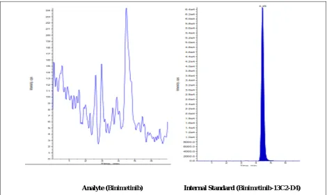
Analyte (Binimetinib) Internal Standard (Binimetinib-13C2-D4)
Fig.4.0 - Representative chromatograms of interference free blank plasma sample
IJSRR, 8(1) Jan. – Mar., 2019 Page 2850
Analyte (Binimetinib) Internal Standard (Binimetinib-13C2-D4) Fig. 6- Chromatogram of LLOQ sample (Binimetinib and Binimetinib-13C2-D4)
Linearity
Linearity was plotted as a peak area ratio (Binimetinib peak area / Binimetinib-13C2-D4 peak
area) on the y-axis against Talazoparib concentration (pg/ml) on the x-axis. Calibration curves were
found to be consistently accurate and precise for Binimetinib over a linearity range of 20 to 200.00
pg/ml. The correlation coefficient was greater than 0.9990for Talazoparib. The %CV was less than
15% and mean %accuracy was ranged between 99.63 – 101.45%. Results were presented in Table 1.
Table. 1 - Calibration curve details of Binimetinib
Spiked plasma Concentration
(pg/ml)
Concentration measured (pg/ml)
(Mean±S.D)
%CV (n=5) %Accuracy
20.00 20.13±0.56 2.78 100.63
40.00 40.38±0.66 1.62 100.96
60.00 59.78±0.33 0.56 99.63
80.00 80.79±1.04 1.29 100.98
100.00 100.38±0.84 0.83 100.38
140.00 142.03±1.03 0.72 101.45
180.00 179.67±1.67 0.93 99.81
200.00 201.20±0.71 0.35 100.60
Precision & Accuracy
Intra and inter batch %accuracy for Binimetinib was ranged between 98.41-100.14 and
IJSRR, 8(1) Jan. – Mar., 2019 Page 2851
Table.2-Precision and accuracy (Analysis with spiked samples at three different concentrations) of Binimetinib
Spiked Plasma Concentration
(pg/ml)
Within-run (Intra-day) Between-run (Inter-Day)
Concentration measured (n=6;pg/ml;mean±S.D)
%CV %Accuracy
Concentration measured (n=6;pg/ml;mean±S.D)
%CV %Accuracy
65.0 63.97±2.17 3.40 98.41 63.00±2.04 3.23 99.93
110.00 109.76±0.45 0.41 99.78 110.36±1.75 1.58 100.33
190.00 190.27±0.47 0.25 100.14 191.83±2.54 1.32 100.96
Recovery
The mean %recovery for LQC, MQC, HQC samples of Binimetinib were 97.64%, 97.30%
and 87.42% respectively.
The overall mean %recovery and %CV of Binimetinib across QC levels is 94.12% and
2.30%. For the Binimetinib-13C2-D4 (internal standard) the mean % recovery and %CV is 85.92%
and 4.82%.
Matrix Effect
No significant matrix effect found in different sources of rat plasma tested for Binimetinib,
Binimetinib-13C2-D4. The %CV was found to be 7.72.
Stability (freeze–thaw, auto sampler, bench top, long term)
Quantification of the Binimetinib in plasma subjected to three freeze–thaw cycles (−30°C to
room temperature), autosampler (processed), room temperature (Benchtop), long-term stability
details were shown in Table 3.
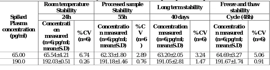
Table. 3 - Stability studies of Binimetinib in spiked plasma samples
Spiked Plasma concentration (pg/ml) Room temperature Stability Processed sample
Stability Long term stability
Freeze and thaw stability
24h 55h 40 days Cycle (48h)
Concentrati on measured (n=6;pg/ml; mean±S.D) %CV (n=6) Concentratio n measured (n=6;pg/ml; mean±S.D) %C V (n=6 ) Concentration measured (n=6;pg/ml; mean±S.D) %CV (n=6) Concentratio n measured (n=6;pg/ml; mean±S.D) %CV (n=6)
65.00 65.54±4.21 6.74 62.33±1.80 2.89 63.20±2.05 3.24 64.69±3.27 5.06
190.0 192.03±0.51 0.26 191.18±1.46 0.76 191.05±2.81 1.47 191.67±1.74 0.91
CONCLUSION
The method described in this manuscript has been developed and validated over the
concentration range of 20–200.0 pg/ml in human plasma. The intra and inter-batch precision (%CV)
was less than 15% and %accuracy ranged from 96.93-104.39%. The overall %recovery for
Binimetinib, Binimetinib-13C2-D4 was greater than 85%. The selectivity, sensitivity, precision and
accuracy obtained with this method make it suitable for the purpose of the present study. In
IJSRR, 8(1) Jan. – Mar., 2019 Page 2852
with an adequate accuracy, precision, selectivity and stability. The simplicity of the method, and
using rapid liquid-liquid extraction with run time of 7.0 min per sample, make it an attractive
procedure in high-throughput bioanalysis of Binimetinib.
ACKNOWLEDGEMENTS
The authors wish to thank the support received from Azidus clinical research laboratories, pvt
LTd, chennai, India for providing literature survey and carrying out this research work.
CONFLICT OF INTEREST:
Authors declare that, there is no conflict of interest.REFERENCES:
1. Sebolt-Leopold JS, Dudley DT, Herrera R, et al. Blockade of the MAP kinase pathway
suppresses growth of colon tumors in vivo: Nat Med 1999; 5(7):810-816.
2. Trachet E, Przybranowski S, Howard C. In vivo evaluation of MEK inhibitor, CI-1040 (PD
0184352), against a panel of human pancreatic tumor xenografts. Proc Am Assoc Cancer Res
2002; 43: 2096.
3. Yeh TC, Marsh V, Bernat BA, et al. Biological characterization of ARRY-142886
(AZD6244), a potent, highly selective mitogen-activated protein kinase kinase 1/2 inhibitor.
Clin Cancer Res, 2007;13(5):1576–1583.
4. Huynh H, Soo KC, Chow PK, et al. Targeted inhibition of the extracellular signal-regulated
kinase kinase pathway with AZD6244 (ARRY-142886) in the treatment of hepatocellular
carcinoma. Mol Cancer Ther. 2007; 6(1):138–146.
5. Davies B, Logie A, McKay J, et al. AZD6244 (ARRY-142886), a potent inhibitor of
mitogen-activated protein kinase/extracellular signalregulated kinase kinase 1/2 kinases:
Mechanism of action in vivo, pharmacokinetic/pharmacodynamic relationship, and potential
for combination in preclinical models. Mol Cancer Ther. 2007;6 (8):2209–2219.
6. Haass NK, Sproessor K, Nguyen TK, et al. The mitogen-activated protein/extracellular signal
regulated kinase kinase inhibitor AZD6244 (ARRY-142886) induces growth arrest in
melanoma cells and tumor regression when combined with docetaxel. Clin Cancer Res. 2008;
14(1):230–239.
7. Lorusso P, Krishnamurthi S, Rinehart JR, et al. A phase 1–2 clinical study of a second
generation oral MEK inhibitor, PD 0325901, in patients with advanced cancer. J Clin Oncol
2005;23(16):3011-3011.
8. Pfizer Inc. Pfizer pipeline as of July 31, 2007.
IJSRR, 8(1) Jan. – Mar., 2019 Page 2853
9. Ratain MJ, Mick R, Schilsky RL, et al. Statistical and ethical issues in the design and conduct
of phase I and II clinical trials of new anticancer agents. J Natl Cancer Inst 1993;85(20):
1637–1643.
10. Therasse P, Arbuck SG, Eisenhauer EA, et al. New guidelines to evaluate the response to
treatment in solid tumors: European Organization for Research and Treatment of Cancer,
National Cancer Institute of the United States, National Cancer Institute of Canada. J Natl
Cancer Inst 2000;92(3):205-216.
11. Waterhouse D, Rinehart J, Adjei AA, et al. A phase 2 study of an oral MEK inhibitor,
CI-1040, in patients with advanced non-small-cell lung, breast, colon, or pancreatic cancer. Proc
Am Soc Clin Oncol 2003;22(22): 4456-62.
12. Solit DB, Garraway LA, Pratilas CA, et al. BRAF mutation predicts sensitivity to MEK
inhibition.Nature. 2006; 439(7074): 358–362.
13.Adjei AA, Cohen RB, Franklin W, Morris C, Wilson D, Molina JR, Hanson LJ, Gore L,
Chow L, Leong S, Maloney L, Gordon G, Simmons H, Marlow A, Litwiler K, Brown S,
Poch G, Kane K, Haney J, Eckhardt SG. Phase I pharmacokinetic and pharmacodynamic
study of the oral, small-molecule mitogen-activated protein kinase kinase 1/2 inhibitor
AZD6244 (ARRY142886) in patients with advanced cancers. J Clin Oncol.
2008;26(13):2139–2146.
14.Delord J, Houede N, Awada A, Taamma A, Faivre SJ, Besse-Hammer T, Italiano A, Vignaud
C, Donica M, Raymond E. First-in-human phase I safety, pharmacokinetic (PK), and
pharmacodynamic (PD) analysis of the oral MEK-inhibitor AS703026 (two regimens [R]) in
patients (pts) with advanced solid tumors [abstract]. J Clin Oncol 28. 2010; 15: 2504.
15.Larkin J, Ascierto PA, Dreno B, Atkinson V, Liszkay G, Maio M, Mandala M, Demidov L,
Stroyakovskiy D, Thomas L, de la Cruz-Merino L, Dutriaux C, Garbe C, Sovak MA, Chang
I, Choong N, Hack SP, McArthur GA, Ribas A. Combined vemurafenib and cobimetinib in
BRAF-mutated melanoma. N Engl J Med 2014; 371: 1867–1876.
16. Lee J, Galloway R, Grandjean G, Jacob J, Humphries J, Bartholomeusz C,Goodstal S, Lim B,
Bartholomeusz G, Ueno NT, Rao A. Comprehensive two- and three-dimensional RNAi
screening identifies PI3K inhibition as a complement to MEK inhibitor AS703026 for
combination treatment of triple-negative breast cancer. J Cancer. 2015; 6(12): 1306–1319.
17.Long GV, Stroyakovskiy D, Gogas H, Levchenko E, de Braud F, Larkin J, Garbe C, Jouary
T, Hauschild A, Grob JJ, Chiarion Sileni V, Lebbe C, Mandala M, Millward M, Arance A,
IJSRR, 8(1) Jan. – Mar., 2019 Page 2854
V, Schadendorf D, Nathan P, Robert C, Ribas A, DeMarini DJ, Irani JG, Casey M, Ouellet D,
Martin AM, Le N, Patel K, Flaherty K. Combined BRAF and MEK inhibition versus BRAF
inhibition alone in melanoma. N Engl J Med. 2014; 371: 1877–1888.
18. Guidance for Industry, Bioanalytical Method Validation, US Department of Health and
Human Services, Food and Drug Administration Centre for Drug Evaluation and Research
(CDER), Centre for Veterinary Medicine (CVM), 2001.
19. FDA guideline, 2013. Guidance for industry: Bioanalytical method validation, US, FDA.
Rockville, MD Nix DJ, Pien C, LaButti J. Clinical pharmacology of the proteasome inhibitor
PS 341.
20. Shah, V.P.; Midha, K.K.;Dighe, S.; McGilveray, I.J.; Skelly, J.P.; and Yacobi, A. Analytical
methods validation. Bioavailability, bioequivalence and pharmacokinetic studies. Conference