https://doi.org/10.5194/fr-20-279-2017
© Author(s) 2017. This work is distributed under the Creative Commons Attribution 4.0 License.
Foramina in plesiosaur cervical centra indicate a specialized
vascular system
Tanja Wintrich1, Martin Scaal2, and P. Martin Sander1
1Bereich Paläontologie, Steinmann-Institut für Geologie, Mineralogie und Paläontologie, Universität Bonn, 53115 Bonn, Germany
2Institut für Anatomie II, Universität zu Köln, Joseph-Stelzmann-Str. 9, 50937 Cologne, Germany
Correspondence:Tanja Wintrich ([email protected])
Received: 16 August 2017 – Revised: 13 November 2017 – Accepted: 14 November 2017 – Published: 19 December 2017
Abstract.The sauropterygian clade Plesiosauria arose in the Late Triassic and survived to the very end of the Cretaceous. A long, flexible neck with over 35 cervicals (the highest number of cervicals in any tetrapod clade) is a synapomor-phy of Pistosauroidea, the clade that contains Plesiosauria. Basal plesiosaurians retain this very long neck but greatly reduce neck flexibility. In addition, plesiosaurian cervicals have large, paired, and highly symmetrical foramina on the ventral side of the centrum, traditionally termed “subcentral foramina”, and on the floor of the neural canal. We found that these dorsal and the ventral foramina are connected by a canal that extends across the center of ossification of the vertebral centrum. We posit that these foramina are not for nutrient transfer to the vertebral centrum but that they are the osteological correlates of a highly paedomorphic vascu-lar system in the neck of plesiosaurs. This is the retention of intersegmental arteries within the vertebral centrum that are usually obliterated during sclerotome re-segmentation in early embryonic development. The foramina and canals are a rare osteological correlate of the non-cranial vascular (arte-rial) system in fossil reptiles. The adaptive value of the reten-tion of the intersegmental arteries may be improved oxygen transport during deep diving and thermoregulation. These features may have been important in the global dispersal of plesiosaurians.
1 Introduction
1.1 Sauropterygian evolution and plesiosaur origins Plesiosauria are Mesozoic marine reptiles that had a global distribution almost from their origin in the Late Triassic (Benson et al., 2012) to their extinction at the end of the Cretaceous (Ketchum and Benson, 2010; Fischer et al., 2017). Plesiosauria belong to the clade Sauropterygia and are its most derived and only post-Triassic representatives, being among the most taxonomically diverse of all Meso-zoic marine reptiles (Motani, 2009). Sauropterygia orig-inated in the Early Triassic, diversifying into Placodon-tia and Eosauropterygia. Eosauropterygia include the Pis-tosauroidea, which in turn include Plesiosauria and non-plesiosaurian pistosauroids (Benson et al., 2012), most no-tably the genera Yunguisaurus, Pistosaurus, and Augus-tasaurus(sometimes grouped in the “Pistosauridae”) and Bo-bosaurus, the taxon closest to Plesiosauria. All these stem representatives are Middle Triassic and early Late Triassic (Carnian) in age, meaning that a gap of around 30 million years separates them from the plesiosaurs (Benson et al., 2012; Wintrich et al., 2017).
1.2 The plesiosaur bauplan
verte-brae, which is unique to “Pistosauridae” and Plesiosauria, all other amniotes having less than 30 cervical vertebrae (Müller et al., 2010). In Pistosauroidea, the increase in neck length evolves by an increase in vertebral number, not by an in-crease in centrum length as in other well-known long-necked animals, like sauropod dinosaurs (Sander et al., 2011; Taylor and Wedel, 2013). Neck elongation in plesiosaurians culmi-nates in Elasmosauridae with cervical numbers exceeding 70 (O’Keefe, 2001; Zammit et al., 2008; Müller et al., 2010; Noe et al., 2017). The plesiosaurian neck was remarkably stiff (Taylor, 1981; Massare, 1988, 1994; Noe et al., 2017), which appears counterintuitive especially in the long-necked forms.
1.3 Paired foramina in plesiosaurian cervical vertebrae All plesiosaurian cervical vertebrae show a pair of large foramina on the ventral surface of the vertebral cen-tra (Romer, 1956). These foramina are generally termed “subcentral foramina” (Storrs, 1991; Noe et al., 2017). The large, highly symmetrical subcentral foramina are an autapomorphy of plesiosaurs and are found with great reg-ularity in members of the clade (Wintrich et al., 2017; Ben-son and Druckenmiller, 2014; Storrs, 1991; O’Keefe, 2001), but smaller and less symmetrical foramina are found in some pistosaurids (non-plesiosaurian Pistosauroidea) such as Au-gustasaurus(Rieppel et al., 2002) andPistosaurus longaevus (Sues, 1987).
The usage of the descriptive term subcentral foramina has a long tradition, and such paired foramina are seen in many taxa of different lineages outside of Sauropterygia. However, the term subcentral foramen is also somewhat of a waste-basket term. In the case of Plesiosauria, foramina subcen-tralia (subcentral foramina) were defined by Storrs (1991). He described them as a uniquely derived character shared by virtually all plesiosaurs and as being unknown among other Sauropterygia like pachypleurosaurs, placodonts, and nothosaurid-grade Nothosauriformes. Furthermore, he inter-preted the foramina subcentralia as vertebral nutritive foram-ina in cervical vertebrae. Rothschild and Storrs (2003) hy-pothesized that the foramina indicated a rich blood supply to the interior of the centra (i.e., acting as nutrient foram-ina), protecting the vertebrae from decompression syndrome. However, they noted that the foramina showed a “large de-gree of variability” which is not what we observe (see be-low).
In addition to these ventral, paired foramina, plesiosaurian cervicals show a pair of large, highly symmetrical foramina on the floor of the neural canal. This character has not re-ceived much attention in the literature before (but see Martin and Parris, 2007), probably because it is harder to observe due to its location inside the neural canal. Damaged or sec-tioned vertebral centra as well as CT scans reveal that the two sets of foramina appear to be connected by two canals that pass through the center of the vertebral centrum. This
raises the question as to what occupied the canals in the liv-ing animal, with vascular tissue comliv-ing to mind.
1.4 Osteological correlates of postcranial vascular features
The vascular system in fossil vertebrates is hard to recon-struct because blood vessels such as arteries and veins as well as the heart are not preserved in fossils (see Malda-nis et al., 2016, for an exception). In addition, osteologi-cal correlates for features of the vascular systems in fossil reptiles remain little studied (Schwarz et al., 2007, p. 181), particularly outside the head. Some basic principles apply, though, that aid in possibly identifying such correlates. In addition, an understanding of the development of the vas-cular system is important. During development, some arter-ies in the embryo become remodeled or resorbed, and during growth, bone will grow around arteries but arteries will not lead to bone resorption. This is seen also in the structure of the human skull bone. Note that this is unlike the situation in postcranial skeletal pneumaticity in dinosaurs including birds, where respiratory tissue invades the interior of bone by inducing bone resorption (Wedel, 2009), a process that continues throughout ontogeny.
1.6 Development of the vertebral column and associated vessels
Development of intersegmental arteries is seen in all verte-brates at an early ontogenetic stage. At the onset of the devel-opment of the axial skeleton in vertebrate embryos, in a pro-cess called somitogenesis, primary segments form in cran-iocaudal sequence within the paraxial mesoderm (Benazeraf and Pourquie, 2013). These segments, which are formed syn-chronously on either side of the neural tube and the noto-chord, are called somites. While the newly formed somites are epithelial spheres, they subsequently undergo several steps of differentiation to form their tissue derivatives, which include axial skeleton, skeletal muscle, and connective tis-sue of the trunk. In amniotes, the ventral somite half be-comes a mesenchymal mass of cells, the sclerotome, which gives rise to all elements of the vertebral column, including the ribs. The dorsal half, in contrast, forms the dermootomal epithelium, which again differentiates into the my-otome giving rise to axial muscle and the dermatome giving rise to the connective tissue of the skin. A sub-compartment of the sclerotome, the syndetome, gives rise to vertebral liga-ments (Brent et al., 2003) and another sub-compartment, the arthrotome, gives rise to intervertebral joints (Mittapalli et al., 2005; Christ et al., 2007).
Importantly, the somites do not represent the definitive segments of the vertebral column as seen in the individual vertebral bones. In a process called re-segmentation, the ad-jacent cranial and caudal halves of neighboring sclerotomes unite to give rise to a single vertebra, whereas intervertebral muscles and ligaments arising from the myotome and syn-detome maintain the original somitic segmentation pattern. In other words, the derivative of a single somite is not a sin-gle vertebra, but a so-called motion segment, which includes two vertebral halves tethered together in a flexible fashion by muscles and ligaments. Without re-segmentation a mobile vertebral column would not be possible (Hall, 2015, chap. 16; Scaal, 2016).
Prior to re-segmentation, neighboring sclerotomes are sep-arated by a pair of embryonic blood vessels called the in-tersegmental artery and vein, which are ventrally connected to the dorsal aorta and posterior cardinal vein, respectively. In a process which is not yet well understood, these ves-sels usually disappear or undergo remodeling during the re-segmentation process. While at trunk level, the segmental ar-ray of vessels is still visible in the adult as, e.g., intercostal vessels, the cervical intersegmental vessels are lost and likely form the vertebral artery and vein.
We hypothesize that in plesiosaurians the intersegmental arteries were retained in cervical vertebrae into the postem-bryonic stage. Thus, in plesiosaurians, the process of the obliteration of the cervical intersegmental arteries did not happen, and the intersegmental arteries stayed in position and remained functional, extending (following the direction of blood flow) through the center of the cervical vertebral
centrum from the ventral surface of the centrum to the floor of the neural canal. Accordingly, we here propose a new term for the large, highly symmetrical paired foramina in plesiosaurian cervicals, i.e., intersegmental artery foramen (IAF), to obtain a more precise terminology in relation to its putative embryological origin. We differentiate between the ventral IAF (vIAF), which corresponds to the traditional subcentral foramen, and the dorsal IAF (dIAF) on the floor of the neural canal. The two foramina are connected by a canal running across the center of ossification of the verte-bral centrum, the intersegmental artery canal (IAC). Blood flow in the intersegmental artery located in the IAC would have been from ventral to dorsal, the artery entering through the vIAF and exiting through the dorsal dIAF.
2 Materials and methods 2.1 Materials
Finally, we used morphological data on cervical vertebral morphology from the literature, specifically character de-scriptions compiled for phylogenetic analysis (Benson and Druckenmiller, 2014). Therefore, we transformed the infor-mation in the phylogenetic character matrix, consisting of the character descriptions and character states, into the morpho-logical information.
2.2 Methods
To test competing hypotheses regarding vascular features, i.e., nutrient canals vs. intersegmental artery canals, the inter-nal morphology of the vertebral centra needs to be revealed, which can be done by µCt scan and by a transversal histo-logical (petrographic) thin section. Histohisto-logical sectioning (Fig. 1) of the vertebrae was possible for only one specimen: the posterior cervical (SMNS 50845) from Holzmaden. Of course, the problem with histological sectioning is that this method is destructive and thus was not allowed for the other plesiosaur vertebrae which were used in this study. For the two complete vertebrae, we used high-resolution µCt scans to obtain virtual sections and reconstruct the IAC (see be-low).
2.2.1 Histological sectioning of plesiosaur vertebra In the study of the IAC in plesiosaur vertebra, obviously an accurate plane of the section that will intersect the canal is crucial. This will be the transverse plane of the vertebral cen-trum (perpendicular to the body axis), passing through the center of ossification of the bone (Fig. 1). The proper plane can be detected easily if the floor of the neural canal and thus the dorsal IAF is visible in addition to the ventral surface of the centrum. Before sectioning, vertebra SMNS 50845 was molded and cast for reconstruction after sectioning. Next, the area of the surface trace of the plane of the section was covered by a removable epoxy putty (Technovit) to ensure a clean cut of the outer bone surface. Then, two cuts spaced about 5 mm apart were placed on either side of the plane of sectioning to obtain a thin slice of bone containing the IAC. After sectioning, the putty was removed from the bone sur-face and the gap in the bone was filled in with plaster, with the two halves of the bone being held in place by the mold.
The transverse slice of bone was then processed into a pet-rographic thin section 50 to 80 µm in thickness, following the standard procedure for fossil bone most recently outlined by Lamm (2013). The sections were then observed under a Le-ica DM2500LP polarizing microscope, and digital photomi-crographs were taken with a Leica DFC420 color camera mounted on this microscope and edited using the 2007 Le-ica Image Access EASYLAB 7 software. Overview images were obtained with an Epson V750 high-resolution scanner. Terminology follows Francillion-Vieillot et al. (1990).
C
1 cm
Figure 1.Transversal histological thin section of the posterior cer-vical vertebra SMNS 50845 from Holzmaden. The left and the right canals appear to meet in the center of ossification (C). However, CT data from other vertebrae suggest that the canals do not meet in the center of ossification, and the apparent connection in this section is probably caused by bone resorption during the formation of the medullary cavity and by damage during grinding of the section.
2.2.2 µCt scanning and 3-D reconstruction of plesiosaur vertebra
The µCt scans for the virtual sections and canal reconstruc-tion were obtained with the v|tome|x s CT scanner manufac-tured by GE Phoenix X-ray at the Division of Paleontology, Steinmann Institute, University of Bonn. On average, each scan was based on 1200 images. Kilovolt and microampere were set to 190 kV and 150 µA, respectively, with a voxel size of 79 µm.
We reconstructed a surface model of the scanned verte-brae with the program Avizo 7.1.1. In order to process the data from the µCt scan, an image stack was created from the dorsoventral plane (Fig. 2). For this, all 1200 µCt record-ings were first uploaded into VG Studio Max and then trans-formed into image stacks in a JPEG format. The images in the stack were then edited and individual structures of in-terest were marked and color-coded and became visible, a process known as segmentation. The result is a 3-D model of the vertebra with the course of the canals having been traced (Figs. 2, 3). The modeling software Polyworks was used to visualize the course of the intersegmental arteries (Fig. 3).
2.2.3 Morphology information based on the phylogenetic matrix
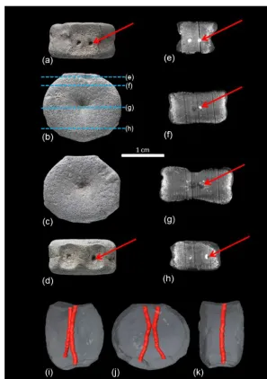
Figure 2.Fetal vertebral centrum of a plesiosaur (LWL-MFN P 64372) from the Rhaetian (latest Triassic) bone bed of Bonenburg (Germany).
(a)Dorsal view with paired intersegmental artery foramina on the floor of the neural canal (red arrow).(b)Posterior view of the fetal vertebral
centrum with the locations of the µCt virtual sections(e)to(h)indicated.(c)Anterior view of the centrum.(d)View of the ventral surface
of the centrum with paired intersegmental artery foramina (red arrow). Note that the foramina are set in a sunken area.(e–h)µCt virtual
sections through the centrum. The sections also show the orientation of the vascular spaces in the bone, which are arranged radially from the center of ossification. The high-density (white) infillings of these vascular spaces and the intersegmental artery canals are pyrite. The
darker fillings are either air or sediment. The red arrows mark the trace of the right canal.(e)Section near the dorsal surface of the vertebral
centrum. The paired intersegmental artery foramina as the entrance to the intersegmental artery canals are clearly visible.(f)This section
is ventral to(e); the intersegmental artery canal comes closer together.(g)Section through the center of ossification in the middle of the
centrum. The canals are close to each other but are still separated.(h)Section through the ventral region of the centrum, where the canals
are widely separated.(i–k)Reconstructed paired intersegmental artery canals connecting the paired dorsal and ventral intersegmental artery
foramina, with the fetal centrum rendered semitransparent. Reconstruction was performed with Avizo 7.1.1.(i)Oblique anterolateral view.
(j)Anterior view. The reconstruction shows clearly that the intersegmental artery canals approach each other one third along their course
from dorsal to ventral, close to the center of ossification of the centrum. The connection between the canals is limited, but it gives the canals
a characteristic X shape. Note the sunken areas of the ventral surface of the centrum.(k)Lateral view, showing that the canals are located in
1
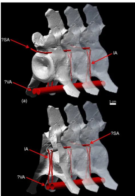
Figure 3. (a)Reconstruction of the arterial system in the plesiosaur neck based on µCT scans, segmentation of the intersegmental artery canals, and modeling of hypothetical vessels in Polyworks. The vir-tual vertebrae with the intersegmental artery canals are the same
cervical (no. 23) ofCryptoclidusIPB R 324 repeated three times.
Anterior is to the left.(b)Cutaway view of the anteriormost
cervi-cal vertebra of this row with the intersegmental arteries crossing the centrum in dorsoventral direction. Abbreviations: IA – inter-segmental artery passing through the vertebral centrum; VA – hy-pothetical vertebral artery from which the intersegmental arteries branched off; SA – hypothetical spinal artery receiving blood from intersegmental arteries.
Our analysis was based on the phylogenetic character matrix of Benson and Druckenmiller (2014), with updates for this study. The matrix consists of 80 taxa from the Late Triassic to the Late Cretaceous and of 270 characters: the character de-scription and character state dede-scription as well as the coding. Eight characters (141, 152, 156, 166, 177, 179, 187, 191) of the 270 characters deal with special aspects of vertebral mor-phology such as nutrient foramina, subcentral foramina, and the number of cervical vertebrae that have implications for the reconstruction of the plesiosaurian neck arterial system.
3 Results
3.1 External morphology
Based on the analysis of the phylogenetic character list, the cervical vertebrae of all plesiosaurian terminal taxa in the matrix have large, paired symmetrical foramina on the ventral side of the cervical vertebral centra, conforming to the definition of vIAF. In the dorsal vertebrae there is no evidence of subcentral or nutrient foramina. The ab-sence/presence and possible morphology of ventral foramina is unknown in the transitional pectoral vertebrae, which we consider as part of the trunk because of a lack of sufficiently informative material and a lack of published descriptive in-formation.
Both the ventral and the dorsal IAFs are part of character 156 of Benson and Druckenmiller (2014): “Cervical verte-brae, subcentral foramina and foramina on the dorsal surface of the centrum, within the neural canal”. This has three states: “both absent (0); both present (1); dorsal foramina present, but subcentral foramina very small or absent (2)”. Charac-ter 166 is also relevant for this study in that it captures the presence of a midline keel or rounded ventral ridge on the centrum. Beyond this, the shape of the ventral keel and the size of the pits and foramina differ depending on taxon. We observed that in all cervical vertebrae (with the exception of the atlas–axis complex) from the Rhaetic bone beds of Bo-nenburg and France and in some of the Early Jurassic ple-siosaur cervicals (e.g., Benson et al., 2012), there are paired deep ventral pits. At the bottom of the pits are the vIAFs. The cervical vertebrae of the Triassic articulated skeleton (see Wintrich et al., 2017) and the vertebrae of the indeter-minate Jurassic plesiosaur SMNS 50845 and ofCryptoclidus IPB 324 have a more even ventral surface without the large keel and deep pits.
3.2 Internal structure as revealed by µCt data, histology, and fracture surfaces
Both histological sections and segmentation of µCt data re-veal that the left and the right vIAFs and the left and the right dIAF are each connected by a canal that passes through the center of ossification of the centrum. This is also seen in frac-ture surfaces of centra. In the µCt images, the canal is visible well and can be traced easily. It is also clear that these two canals do not end in the center of ossification, as a nutrient canal would do, but pass through it. The general appearance of the two canals in transverse sections is X-shaped because the canals gradually diverge from each other towards both the ventral and dorsal intersegmental foramina. The left and the right canals appear to be connected in the central region in the thin section of SMNS 50845, and it appears that the canals merge in a medullary cavity which resorbed the origi-nal center of ossification (Fig. 1). However, in the 3-D recon-structions of the fetal vertebra (WMNM P 64372) (Fig. 2) and the adult vertebra (STIPB R 324) (Fig. 3), the canals do not appear to meet in the center of ossification, only ap-proaching each other closely. These conflicting observations may be explained by the loss of trabeculae in the ossification center during the preparation of the thin section of SMNS 50845 (Fig. 1), resulting in an apparent connection between the canals.
4 Discussion
4.1 Interpretation of the paired canals
Plesiosaur vertebral foramina have been observed and de-scribed from so many taxa and have been used as charac-ters in phylogenetic analyses that it is clear that they are a pervasive feature of plesiosaur cervicals (Storrs, 1991; O’Keefe, 2001; Benson and Druckenmiller, 2014; Wintrich et al., 2017), with the possible exception of the pliosaur-type forms (see above). Thus, we feel that our results are representative of all plesiosaur cervicals although we only investigated three specimens in detail. The course of the paired canals through the vertebral centrum would suggest that these structures originally housed continuous arteries traversing the vertebral centrum in ventrodorsal direction, which opens up the possibility that they contained persisting intersegmental arteries, not nutrient ones, which would have ended within the central region of the vertebra. Furthermore, the crossing of the vertebral centrum is a feature which we argue should originate at an early developmental stage. This is because in the case of nutritive canals, the vascular system does spread into bone tissue (as mentioned above), whereas in continuous blood vessels, the bone tissue grows around the vessels instead. It is also known that bone tissue cannot resorb or displace features of the vascular system such as ar-teries because osteoclasts only resorb mineralized surfaces
(Hall, 2015, chap. 15). This suggests that the arteries were already present in the vertebral primordium at the stage in early development when the sclerotomes were re-segmented and, subsequently, the cartilage primordia of the centra of the vertebra were formed. As the primordium of the centrum grew and ossified, the arteries and with them the canals also enlarged in size. The divergence of the canals is explained by the retention of the homologous locations inside the cen-trum and on its surface as ventral and dorsal intersegmental foramina.
While posterior caudal and fluke vertebrae of extant whales have similar canals piercing the vertebral centra and housing arteries (Slijper, 1939), their morphology and origin is rather different, as described in detail by Slijper (1939) and confirmed by a study of the tail segment of a complete adult skeleton of the bottlenose dolphinTursiops truncatus (LACM 97723; Slijper, 1939). First of all, the paired canals do not pass through the center of ossification of the cen-tra but in an arch around it. This indicates that the vessels were incorporated into vertebrae only in the juvenile, not in the embryo. Second, it can be observed that the canals form by the gradual (from anterior to posterior, not ontogeneti-cally) incorporation of an artery lateral to the centrum. The artery more anteriorly only pierces the transverse process and then more posteriorly becomes incorporated deeply into the centrum. The canals in the whale caudal vertebrae are thus not homologous to those in the plesiosaurian cervical verte-brae. A possible exception to the non-homology of the canals in plesiosaurs and whales may be the vertebra depicted by Houssaye et al. (2015, fig. 14) in which canals are seen pass-ing dorsoventrally through the center of ossification. How-ever, these canals do not show the strong symmetry that is so typical of plesiosaurs. The lack of symmetry in the canals inBasilosaurussuggest that they do not represent persisting intersegmental arteries but originated later in ontogeny. 4.2 Developmental retention of the intersegmental
arteries in plesiosaurs
In the embryo, a paired primary dorsal aorta differenti-ates by vasculogenesis and is located underneath the paraxial mesoderm. The primary dorsal aortae gradually change po-sition from lateral to medial. Eventually, both dorsal aortae fuse in the midline of the embryo ventral to the notochord, which leads to the formation of a single large median aorta (Wiegreffe et al., 2007; Garriock et al., 2010). Initially, the intersegmental arteries branch off in dorsal direction from the paired dorsal aortae, passing in between the sclerotomes before re-segmentation. Their subsequent development is not well studied. In the trunk at thoracic levels, they relocate lat-erally to form the intercostal arteries. In the neck, they seem to obliterate in their proximal part during re-segmentation, whereas their distal part outside the vertebral centra fuses with neighboring segments to form the vertebral artery (Arey, 1924, p. 212).
Importantly, we found evidence for intersegmental artery retention only in the cervical vertebral centra, not in the dor-sal vertebral centra. Thus, if plesiosaurians retained interseg-mental arteries, then the question arises as to which vessels these intersegmental arteries were connected to. As men-tioned above, in principle the intersegmental arteries in ex-tant embryos arise from the paired aortae. In the neck, how-ever, they form longitudinal anastomoses which give rise to the aorta vertebralis, while the connections to the dorsal aorta become obliterated. In snakes likeElaphe obsoleta, the Arte-ria vertebralis, in turn, emits segmental branches which reach the spinal canal where they anastomose longitudinally to give rise to the A. spinalis and associated vessels (Zippel et al., 1998). As in plesiosaurs the intersegmental vessels lead to the spinal canal. Thus, we postulate that in analogy to the anatomy of snakes, the intersegmental vessels join the longi-tudinal spinal artery in the spinal canal. As to their origin, we speculate that they branch off a longitudinal vertebral artery which has arisen from the paired dorsal aortae of earlier em-bryonic stages (Fig. 3). As no fossil correlates are preserved, this scenario remains forcibly speculative, but the vascular anatomy as postulated here would likely not be problematic in an adult plesiosaurian from a functional point of view. The extreme evolutionary neck elongation by an increase in seg-ment number (not segseg-ment elongation) in the plesiosaur line (including pistosaurids) may have required or have been fa-cilitated by a developmental retention of strong bilateral ar-teries derived from the paired aortae and with it the interseg-mental arteries arising from them.
We shall now evaluate the hypotheses explaining why ple-siosaurians did not resorb the intersegmental arteries, re-taining the embryonic vascular system. The first hypothe-sis involves developmental constraints linked to the uniquely high number of cervicals in plesiosaurs (the low number of pliosaur-type plesiosaurs being secondarily derived). This, like any other hypothesis explaining the persistence of inter-segmental arteries, has to be consistent with the lack of IAF in the dorsal and presumably pectoral vertebrae, the numbers of which are not unusually high in plesiosaurians compared
to other amniotes (Müller et al., 2010; Coffin and Poole, 1988).
The uniquely high number of cervical vertebrae means that in the plesiosaurian embryo there was also a uniquely high number of cervical somites and sclerotomes. As noted above, no other vertebrate group evolved such an enormously long neck via an increase in the number of segments, i.e., vertebrae (Müller et al., 2010). A model for understanding the development of the very high number of cervical seg-ments in plesiosaurians might be the segmentation process in snakes, that have evolved very high numbers of dorsal seg-ments. There, it has been shown that the molecular mecha-nisms of somitogenesis are principally the same as in ver-tebrates with lower segment numbers, but that somitogene-sis proceeds much faster leading to initially smaller somites which, however, later on grow to a normal size relative to the size of the snake species concerned (Gomez et al., 2008). In snakes, the extremely high number of dorsal vertebrae (“precloacal” in morphological terminology) correlates with a corresponding extension the expression of thorax-specific Hoxgenes, likeHox6, along the body axis (Cohn and Tickle, 1999). It is therefore likely that in long-necked plesiosaurian embryos, cervicalHoxgene expression was maintained over many segmentation rounds, which probably occurred rela-tively rapidly when compared to short-necked species (both ancestral to plesiosaurians and derived within plesiosaurians, i.e., in the pliosaur type). A potential link between frequency and speed of somitogenesis on the one hand and the forma-tion of intersegmental vessels on the other hand is yet un-known.
Furthermore, we do not know if plesiosaurians developed paired vertebral arteries arising from the aorta in addition to retaining the intersegmental arteries. These vertebral arteries would have extended along (?) the vertebral centrum, with nutrient arteries branching off and vascularizing the vertebral body through lateral nutrient foramina as seen in mammals (Rothman and Simeone, 1975). Such lateral nutrient foram-ina are common in marine mammals but differ from the vIAF of plesiosaurians in their smaller size, larger number, and asymmetrical irregular placement.
In the tuatara,Sphenodon punctatus, there are also paired foramina in the caudal centra, and classical embryologi-cal research (Schauinsland, 1906, fig. 323) clearly shows a paired artery extending dorsoventrally across the cartilage primordium of the caudal centrum in dolphins (Schauins-land, 1906).
4.3 Functional interpretation and adaptive value of intersegmental arteries
seemingly retain intervertebral arteries. In the tree-climbing rat snake Elaphe obsoleta, intersegmental arteries are re-tained in the neck region. This is interpreted as an adapta-tion for maintaining cerebral blood flow in spite of grav-itational stress, e.g., during climbing (Zippel et al., 1998). This observation offers an exciting parallel to the situation in plesiosaurians, where high hydrostatic pressure during deep diving (> 200 m) might have required segmental transverte-bral arterial anastomoses to provide sufficient ceretransverte-bral blood supply. If intravertebral intersegmental arteries were devel-oped as additional arteries in plesiosaurians, there would be a higher oxygen transport capacity than in a single pair of intersegmental arteries per vertebra. This capacity would en-able faster transport of oxygen for storage into muscles dur-ing deep divdur-ing. A similar hypothesis was presented by Roth-schild and Storrs (2003), suggesting that the increased blood flow to the interior of the centra protected them from avas-cular necrosis. However, this hypothesis is not consistent with the persistence of intersegmental arteries because they would not have supplied the interior of the centrum with ex-tra blood.
Other functional hypotheses explaining the retention of in-tersegmental arteries related to deep diving involve the pro-tection from compression of the blood vessels by their loca-tion in canals in the vertebrae. If there were anastomoses with the vertebral arteries, an increase in the number of segments would mean more anastomoses and again greater transport capacity. Other, as yet less easily hypothesized advantages might be related to the compression of the upper respiratory and digestive tracts. Hypotheses involving a long neck and high cervical vertebral numbers invite future tests based on the comparison with the cervical vertebral column of short-necked plesiosaurian (i.e., pliosaur-type) that evolved several times in the history of plesiosaurians, together with the seem-ing loss of ventral IAF (see phylogenetic data matrix in Ben-son and Druckenmiller, 2014).
4.4 Persistent intersegmental arteries: increasing adaptation to a pelagic lifestyle?
As discussed above, IAFs are a unique character of ple-siosaurians. However, it is not only plesiosaurs that have paired foramina on the ventral side of their cervical verte-brae. The non-plesiosaurian pistosauroids Pistosaurus lon-gaevusandAugustasaurus hagdornialso show paired foram-ina (Sues, 1987; Rieppel et al., 2002), whereasYunguisaurus liae (Sato et al., 2014) andBobosaurus forojuliensis (Dalla Vecchia, 2006) do not share the character. The reason for this could be the degree of adaptation to the pelagic habi-tat and colder waters in the pistosauroid lineage (Krahl et al., 2013). Augustasaurusis the first and only unequivocal non-plesiosaurian pistosauroid which is found outside the Tethys realm on the western coast of North America (Sander et al., 1997). Possible other non-plesiosaurian pistosauroids outside the Tethys are Corosaurusfrom Wyoming (Storrs,
1991) andAlexeyisaurusfrom Arctic Russia (Sennikov and Arkhangelsky, 2010), but the systematic position of the for-mer is unstable, and the latter is too poorly preserved for a reliable systematic assignment.
Since available evidence suggests that sauropterygians originated in the Tethys (Neenan et al., 2013) and all other Triassic pistosauroids are known from this realm (Benson et al., 2012), the ancestors ofAugustasaurus must have emi-grated from the warm equatorial waters of the Tethys and either dispersed around the polar northern or southern coast of Pangaea or across Panthalassa. Either way, an elevated metabolic rate and endothermy appear to be prerequisites for this dispersal (Krahl et al., 2013). The persistence of intersegmental arteries in the neck could have been incipi-ently present inAugustasaurusand would have been useful for improved thermoregulation in the colder pelagic waters of Panthalassa. If pistosauroids dispersed across Panthalassa to reach western North America, several other adaptations would be necessary. These include, for example, cruising adaptations in aquatic locomotion by underwater flight. Also, the ability of deep diving (see above) is an adaptation to the pelagic habitat because prey there is sparser and more evenly distributed across the water column (and not at the sea bot-tom) than in coastal habitats. Plesiosaurians show all these features of adaptation to a pelagic habitat and not surpris-ingly are globally distributed at least by the Middle Jurassic (Bardet et al., 2014).
5 Conclusions
of the persistent intersegmental arteries must be sought in deep diving and in thermoregulation in the neck.
Data availability. All data needed to evaluate the conclusions in the paper are present in the paper. Additional data related to this paper may be requested from the authors.
The specimens and thin sections on which this study is based are reposited in the following institutions, here listed with their abbre-viations: LWL-MFN – LWL- Museum für Naturkunde, Muünster, Germany; SMNS – Staatliches Museum für Naturkunde, Stuttgart, Germany; STIPB – Steinmann Institute Paleontology Collection, University of Bonn, Bonn, Germany.
Competing interests. The authors declare that they have no conflict of interest.
Special issue statement. This article is part of the special issue “Secondary adaptation of tetrapods to life in water – Proceedings of the 8th International Meeting, Berlin 2017”. It is a result of the 8th International Meeting on the Secondary Adaptation of Tetrapods to Life in Water, Berlin, Germany, 3–8 April 2017.
Acknowledgements. First and foremost we thank Michael Mertens of Schwaney (North Rhine-Westphalia, Germany) for his untiring efforts in collecting marine reptiles from the Rhaetian bone beds of Bonenburg and facilitating their transfer to the LWL-MFN collections. We thank Olaf Dülfer (Bonn) for help with specimen preparation, Georg Oleschinski (Bonn) for photography, and Rico Schellhorn (Bonn) for help with illustrations and discussion. Reviews by Alexandra Houssaye and two anonymous reviewers are gratefully acknowledged. This project was funded by the Deutsche Forschungsgemeinschaft (DFG, grant number SA 469/47-1) and by the LWL-Museum für Naturkunde (Münster, Germany) through the archeological and paleontological heritage mitigation scheme of the State of North Rhine-Westphalia.
Edited by: Florian Witzmann
Reviewed by: Alexandra Houssaye and two anonymous referees
References
Alberch, P., Gould, S. J., Oster, G. F., and Wake, D. B.: Size and shape in ontogeny and phylogeny, Paleobiology, 5, 296–317, 1979.
Andrews, C. W.: A descriptive catalogue of the marine reptiles of the Oxford Clay, The British Museum (Natural History) London, London, 202 pp., 1910.
Arey, L. B.: Developmental Anatomy. W. B. Saunders Company, Philadelphia and London, 1924.
Bardet, N., Falconnet, J., Fischer, V., Houssaye, A., Jouve, S., Pereda Suberbiola, X., Pérez-García, A., Rage, J.-C., and Vin-cent, P.: Mesozoic marine reptile palaeobiogeography in
re-sponse to drifting plates, Gondwana Research, 26, 869–887, https://doi.org/10.1016/j.gr.2014.05.005, 2014.
Bénazéraf, B. and Pourquié, O.: Formation and segmentation of the vertebrate body axis, Annu. Rev. Cell Dev. Bi., 29, 1–26, 2013. Benson, R., Evans, M., and Druckenmiller, P.: High diversity,
low disparity and small body size in plesiosaurs (Reptilia, Sauropterygia) from the Triassic – Jurassic boundary, PLoS ONE, 7, e31838, https://doi.org/10.1371/journal.pone.0031838, 2012.
Benson, R. B. J. and Druckenmiller, P. S.: Faunal turnover of marine tetrapods during the Jurassic–Cretaceous transition, Biol. Rev., 89, 1–23, https://doi.org/10.1111/brv.12038, 2014.
Böhmer, C., Rauhut, O., and Wörheide, G.: Correlation between Hox code and vertebral morphology in archosaurs, Proc. Roy. Soc. B, 282, 20150077, https://doi.org/10.1186/s40851-017-0069-4, 2015.
Brent, A. E., Schweitzer, R., and Tabin, C. J.: A somitic compart-ment of tendon progenitors, Cell, 113, 235–248, 2003.
Christ, B., Huang, R., and Scaal, M.: Amniote somite derivatives, Dev. Dynam., 236, 2382–2396, 2007.
Coffin, J. D. and Poole, T. J.: Embryonic vascular development: im-munohistochemical identification of the origin and subsequent morphogenesis of the major vessel primordia in quail embryos, Development, 102, 735–748, 1988.
Cohn, M. J. and Tickle, C.: Developmental basis of limblessness and axial patterning in snakes, Nature, 399, 474–479, 1999. Dalla Vecchia, F. M.: A new sauropterygian reptile with
ple-siosaurian affinity from the Late Triassic of Italy, Rivista Italiana di Paleontologia e Stratigrafia, 112, 207–225, 2006.
Fischer, V., Cappetta, H., Vincent, P., Garcia, G. r., Goolaerts, S., Martin, J. E., Roggero, D., and Valentin, X.: Ichthyosaurs from the French Rhaetian indicate a severe turnover across the Triassic-Jurassic boundary, Naturwissenschaften, 101, 1027– 1040, https://doi.org/10.1007/s00114-014-1242-7, 2014. Fischer, V., Benson, R. B. J., Zverkov, N. G., Soul, L. C.,
Arkhangelsky, M. S., Lambert, O., Stenshin, I. M., Uspensky, G. N., and Druckenmiller, P. S.: Plasticity and convergence in the evolution of short-necked plesiosaurs, Current Biology, 27, 1667–1676.e3, https://doi.org/10.1016/j.cub.2017.04.052, 2017. Francillon-Vieillot, H., de Buffrénil, V., Castanet, J., Geraudie, J., Meunier, F., Sire, J. Y., Zylberberg, L., and de Ricqles, A.: Mi-crostructure and mineralization of vertebrate skeletal tissues, in: Skeletal Biomineralization: Patterns, Processes and Evolutionary Trends, Vol. 1, edited by: Carter, J. G., Van Nostrand Reinhold, New York, 471–530, 1990.
Garriock, R. J., Czeisler, C., Ishii, Y., Navetta, A. M., and Mikawa, T.: An anteroposterior wave of vascular inhibitor downregulation signals aortae fusion along the embryonic midline axis, Develop-ment, 137, 3697–3706, 2010.
Gomez, C., Özbudak, E. M., Wunderlich, J., Baumann, D., Lewis, J., and Pourquié, O.: Control of segment number in vertebrate embryos, Nature, 454, 335, 2008.
Hall, B. K.: Bones and Cartilage. 2nd Edition. Developmental and Evolutionary Skeletal Biology, Academic Press, San Diego, 2015.
Ketchum, H. F. and Benson, R. B.: Global interrelationships of Plesiosauria (Reptilia, Sauropterygia) and the pivotal role of taxon sampling in determining the outcome of phylogenetic anal-yses, Biol. Rev., 85, 361–392, https://doi.org/10.1111/j.1469-185X.2009.00107.x, 2010.
Krahl, A., Klein, N., and Sander, P. M.: Evolutionary implications
of the divergent long bone histologies ofNothosaurusand
Pis-tosaurus(Sauropterygia, Triassic), BMC Evolutionary Biology, 13, 1–23, 2013.
Lamm, E.-T.: Chapter 4 – Preparation and sectioning of specimens, in: Bone Histology of Fossil Tetrapods. Advancing Methods, Analysis, and Interpretation, edited by: Padian, K. and Lamm, E.-T., University of California Press, Berkeley, 55–160, 2013. Lara Maldanis, L., Carvalho, M., Almeida, M. R., Freitas, F. I., de
Andrade, J. A. F. G., Nunes, R. S., Rochitte, C. E., Poppi, R. J., Freitas, R. O., Rodrigues F., Siljeström S., Lima, F. A., Galante, D., Carvalho, I. S., Perez, C. A., de Carvalho, M. R., Bettini, J., Fernandez, V. and Xavier-Neto, J.: Heart fossilization is possible and informs the evolution of cardiac outflow tract in vertebrates, Elife, 5, e14698, https://doi.org/10.7554/eLife.14698, 2016. Martin, J. E. and Parris, D. C. (Eds.).: The Geology and
Paleon-tology of the Late Cretaceous Marine Deposits of the Dakotas, Geological Society of America Special Paper 427, 2007. Massare, J. A.: Swimming capabilities of Mesozoic marine reptiles,
Paleobiology, 14, 187–205, 1988.
Massare, J. A. and Callaway, J. M.: Cymbospondylus
(Ichthyosauria: Shastasauridae) from the Lower Triassic
Thaynes Formation of southeastern Idaho, J. Vertebr. Paleontol., 14, 139–141, 1994.
McNamara, K. J.: Shapes of Time. The Evolution of Growth and Development, Johns Hopkins University Press, Baltimore, 342 pp., 1997.
Mittapalli, V. R., Huang, R., Patel, K., Christ, B., and Scaal, M.: Arthrotome: a specific joint forming compartment in the avian somite, Dev. Dynam., 234, 48–53, 2005
Motani, R.: The evolution of marine reptiles, Evolution: Education and Outreach, 2, 224–235, 2009.
Müller, J., Scheyer, T. M., Head, J. J., Barrett, P. M., Werneburg, I., Ericson, P. G. P., Pol, D., and Sánchez-Villagra, M. R.: Homeotic effects, somitogenesis and the evolution of vertebral numbers in recent and fossil amniotes, P. Natl. Acad. Sci. USA, 107, 2118– 2123, 2010.
Nakajima, Y., Hirayama, R., and Endo, H.: Turtle humeral mi-croanatomy and its relationship to lifestyle, Biol. J. Linn. Soc., 112, 719–734, 2014.
Neenan, J. M., Klein, N., and Scheyer, T. M.: European ori-gin of placodont marine reptiles and the evolution of crush-ing dentition in Placodontia, Nature Communications, 4, 1–7, https://doi.org/10.1038/ncomms2633, 2013.
Noè, L. F., Taylor, M. A., and Gómez-Pérez, M.: An integrated ap-proach to understanding the role of the long neck in plesiosaurs, Acta Palaeontologica Polonica, 62, 137–162, 2017.
O’Keefe, F. R.: Preliminary description and phylogenetic position of a new plesiosaur (Reptilia: Sauropterygia) from the Toarcian of Holzmaden, Germany, J. Paleontol., 78, 973–988, 2004. Rieppel, O., Sander, P. M., and Storrs, G. W.: The skull of the
pis-tosaurAugustasaurusfrom the Middle Triassic of northwestern
Nevada, J. Vertebr. Paleontol., 22, 577–592, 2002.
Romer, A. S.: Osteology of the Reptiles, The University of Chicago Press, Chicago, 772 pp., 1956.
Rothman, R. H. and Simeone, F. A: The spine. Vol. 1. WB Saunders, Philadelphia, 1975.
Rothschild, B. M. and Storrs, G. W.: Decompression syndrome in plesiosaurs (Sauropterygia: Reptilia), J. Vertebr. Paleontol., 23, 324–328, 2003.
Sander, P. M., Christian, A., Clauss, M., Fechner, R., Gee, C., Griebeler, E. M., Gunga, H.-C., Hummel, J., Mallison, H., Perry, S., Preuschoft, H., Rauhut, O., Remes, K., Tütken, T., Wings, O., and Witzel, U.: Biology of the sauropod dinosaurs: the evolution of gigantism, Biol. Rev. of the Cambridge Philosophical Society, 86, 117–155, 2011.
Sander, P. M., Rieppel, O. C., and Bucher, H.: A new pistosaurid (Reptilia: Sauropterygia) from the Middle Triassic of Nevada and its implications for the origin of plesiosaurs, J. Vertebr. Paleon-tol., 17, 526–533, 1997.
Sander, P. M., Wintrich, T., Schwermann, A. H., and Kindlimann, R.: Die paläontologische Grabung in der Rhät-Lias-Tongrube der Fa. Lücking bei Warburg-Bonenburg (Kr. Höxter) im Frühjahr 2015, Geologie und Paläontologie in Westfalen, 88, 11–37, 2016. Sato, T., Zhao, L.-J., Wu, X.-C., and Li, C.: A new specimen of
the Triassic pistosauroidYunguisaurus, with implications for the
origin of Plesiosauria (Reptilia, Sauropterygia), Palaeontology, 57, 55–76, 2014.
Scaal, M.: Early development of the vertebral column, Seminars in Cell and Developmental Biology, 49, 83–91, 2016.
Schauinsland, H. H.: Beiträge zur Entwicklungsgeschichte und Anatomie der Wirbeltiere, E. Nägele, Stuttgart, 168 pp., 1903. Schauinsland, H. H.: Die Entwicklung der Wirbelsäule nebst
Rip-pen und Brustbein, in: Handbuch der vergleichenden und exper-imentellen Entwicklungslehre der Wirbeltiere, edited by: Her-twig, O., Gustav Fischer Jena, Bd. 3 Teil 2, 339–562, 1906. Schwarz, D., Frey, E., and Meyer, C. A.: Pneumaticity and
soft-tissue reconstructions in the neck of diplodocid and dicraeosaurid sauropods, Acta Palaeontologica Polonica, 52, 167–188, 2007. Sennikov, A. G. and Arkhangelsky, M. S.: On a typical Jurassic
sauropterygian from the Upper Triassic of Wilczek Land (Franz Josef Land, Arctic Russia), Paleontol. J., 44, 567–572, 2010. Seymour, R. S., Smith, S. L., White, C. R., Henderson, D. M., and
Schwarz-Wings, D.: Blood flow to long bones indicates activity metabolism in mammals, reptiles and dinosaurs, Proc. Roy. Soc. B, 279, 451–456, 2012.
Slijper, E. J.:Pseudorca crassidens(Owen), ein Beitrag zur
ver-gleichenden Anatomie der Cetaceen, Zoologische Mededeelin-gen Rijksmuseum van Natuurlijke Historie Leiden, 21, 241–366, 1939.
Storrs, G. W.: Anatomy and relationships ofCorosaurus alcovensis
(Diapsida: Sauropterygia) and the Triassic Alcova Limestone of Wyoming, Bulletin of the Peabody Museum of Natural History, Yale University, 44, 1–151, 1991.
Storrs, G. W.: Fossil vertebrate faunas of the British Rhaetian
(latest Triassic), Zool. J. Linn. Soc., 112, 217–259,
https://doi.org/10.1111/j.1096-3642.1994.tb00319.x, 1994.
Sues, H.-D.: Postcranial skeleton ofPistosaurusand
interrelation-ships of the Sauropterygia (Diapsida), Zool. J. Linn. Soc., 90, 109–131, 1987.
Taylor, M. P. and Wedel, M. J.: Why sauropods had long necks; and why giraffes have short necks, PeerJ, 1:e36, https://doi.org/10.7717/peerj.36, 2013.
Wedel, M. J.: Evidence for bird-like air sacs in saurischian di-nosaurs, J. Exp. Zool. A, 311, 1–18, 2009.
Wiegreffe, C., Christ, B., Huang, R., and Scaal, M.: Sclerotomal origin of smooth muscle cells in the wall of the avian dorsal aorta, Dev. Dynam., 236, 2578–2585, 2007.
Wintrich, T., Hayashi, S., Houssaye, A., Nakajima, Y., and Sander, P. M.: A Triassic plesiosaurian skeleton and bone histology in-form on evolution of a unique body plan, Sci. Adv., 3, e1701144, https://doi.org/10.1126/sciadv.1701144, 2017.
Woltering, J. M., Vonk, F. J., Müller, H., Bardine, N., Tuduce, I. L., de Bakker, M. A., Knöchel, W., Sirbu, O., Durston, A. J., and Richardson, M. K.: Axial patterning in snakes and caecilians: evidence for an alternative interpretation of the Hox code, Dev. Biol., 332, 82–89, 2009.
Zammit, M., Daniels, C. B., and Kear, B. P.: Elasmosaur (Reptilia: Sauropterygia) neck flexibility: Implications for feeding strate-gies, Comp. Biochem. Physiol., Part A, 150, 124–130, 2008. Zippel, K. C., Lillywhite, H. B., and Mladinich, C. R. J.:
Contribu-tion of the vertebral artery to cerebral circulaContribu-tion in the rat snake