ISSN: 2231-5381
http://www.ijettjournal.org
Page 3049
Application of Bacterial Foraging Optimisation as
a De-noising filter
Suman Yaduwanshi#1 Jagroop Singh Sidhu*2
1
Student (M.Tech. ECE), DAV Institute of Engineering and Technology, Jalandhar 2
Associate Professor, ECE, DAV Institute of Engineering and Technology, Jalandhar
Abstract— De-noising of image still a concerned for researchers
working in this area. It is further challenging in case of medical images mainly images of the internal organs. Various digital filters have been developed and tried by researchers to provide ideal solution in the de-noising of medical images. In the present paper the authors present a Soft Computing approach to de-noise the medical images. Bacterial Foraging Optimisation which is a bio-inspired algorithm is used as filter to de-noise medical images like CT-Scan and MRI of pancreas. The performance metrics like MSE and PSNR are calculated which show that Bacterial Foraging Optimisation can act as potential tool for de-noising images.
Keywords— BFO, cost function, MSE, PSNR, De-noising,
Medical image
I. INTRODUCTION
During image acquisition process due to different processing such as A/D conversion, transmission etc. the digital images gets added with noise. Different types of noises corrupt digital images at different stages of image processing.[1][3] While dealing with medical images, the image gets corrupted by variety of noise mainly from bone, soft tissue, body movement etc. Therefore precautions are taken to reduce the noise to a larger possible extent in the medical images for better diagnosis. However, the demands for noise free image are increasing day by day so is the case for better de-noising filters. Soft Computing techniques in the recent past have been used by researchers either in isolation or in conjunction with Adaptive Median filter or Median filter to de-noise images. In this paper, the authors have presented a technique to de-noise medical images using Bacterial Foraging Optimization technique.
For removing the Gaussian noise, Bacterial Foraging Optimisation is hybridised with Average filter whereas for other noise BFO is applied by hybridising it with Adaptive Median filter. Bacterial Foraging Optimisation developed by Passino in 2002 [8] bio-inspired Optimisation technique that is derived from the food searching process of E. Coli bacteria. As the bacteria travel in slow speed, it gives the capability to search the pixel without jumping or slipping out pixels thus, improving the quality of image. To test the capability of proposed algorithm, medical images like CT-Scan and MRI of
pancreas are considered. Self image is also taken to see the performance of algorithm as a de-noising filter.
Along with several digital filtering techniques, Soft computing techniques are gaining importance in de-noising process. Several digital filtering techniques used by researchers to remove Salt &Pepper noise have been published. Progressively determining noisy pixel and removal of noisy pixel by switched median filter used in[10]. Modified Median filter used in [2]. Artificial neural network, Fuzzy logic, Genetic Algorithm, Optimisation techniques such as Particle Swarm Optimisation [4] are some important Soft Computing techniques. PSO is applied to de-noise images[11][12] by optimising cost function as structure of similarity, Bacterial Foraging Optimisation [8], Swine Influenza Model Based Optimisation [9] are some important Soft computing techniques. Researchers used various soft computing techniques and hybrid techniques to de-noise images [1]. In this paper, BFO is used to optimise the output of Adaptive Median filter when images are corrupted with Salt and Pepper noise and output of Average filter is optimized when images are corrupted with Gaussian noise. Experiment is performed on two benchmark images – Lena and Bridge and CT-Scan and MRI of pancreas and image of self.
The paper is organised in following sections. Section II. Presents BFO, Section III. explains the proposed soft filter, BFO Soft Filter for noise removal, Section IV. shows Results and Discussion and Section V. encompasses the Conclusion.
II. BACTERIAL FORAGING OPTIMIZATION
ISSN: 2231-5381
http://www.ijettjournal.org
Page 3050
(i) Chemo taxis: Single Chemo-tactic step completes in tumble, run and tumble if nutrient does not increase in the direction of swim and otherwise it is tumble, run(swim) and followed by run (as per defined limit of swim) if concentration of nutrient increases in the direction of swim. A unit walk with random direction represents a Tumble and unit walk with same direction in the last step indicates Run. Mainly foraging completes in Chemo-tactic step.
(ii) Swarming: The cells when stimulated, release an attractant aspirate, which helps them to aggregate into groups and move as concentric patterns of swarms. This helps to achieve global optimum value for cost function. Swarming helps in fast convergence in case of multidimensional cost function.
(iii) Reproduction: After calculating fitness value for each bacteria, reproduction allows the half healthy bacteria (with least cost value) to survive and reproduce. The remaining half unhealthy bacteria die. This step helps in the generation of values of variables which are closer to actual value. Fifty percentage of the population is removed in each state and rest fifty percentage reproduce.
(iv) Elimination and Dispersal: The chemo taxis provides a basis for local search and reproduction speeds the convergence. But to avoid the trap of bacteria in local minimum Elimination –Dispersal is done.
III.BFO AS SOFT FILTER
To test the proposed concept, initially ideal images are corrupted with noise. Then the noisy image is passed through Adaptive Median filter. Now the difference in terms of Mean Square Error between this filtered image and the original image is minimised using BFO. The block diagram of the process is shown Fig. 1
Original Signal d(k)
+
Noisy input Output y(k) -
Error signal E(k)
Fig. 1: Block diagram of Soft filter
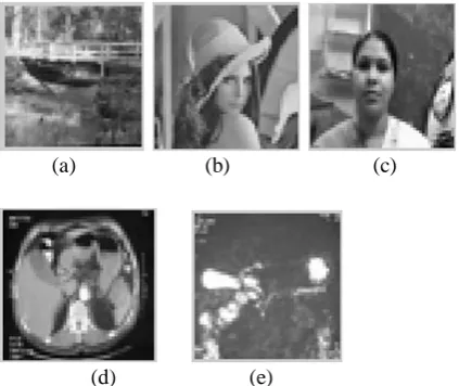
The original resized images selected for the testing are shown in Fig. 2
(a) (b) (c)
(d) (e)
Fig.2: Original resized image (a) Bridge (b)Lena (c) Self (d) CT-Scan of pancreas (e) MR Cholangiopancreatogram
BFO is used to minimize the Mean Square Error between Adaptive Median filter output and target image to restored image which is closer to actual image. Parameters selected to de noise image are-
Number of bacteria in population used for searching (S) = M×N size of image, in this case it is 50×50 (downsized) Dimension of search space (p) = 1
Number of Chemo tactic steps (Nc) = 2 Number of swimming steps (Ns) = 1 Number of reproduction steps (Nre) = 2 Number of elimination and dispersal (Ned)=2 Probability of elimination and dispersal (ped) = 0.25 Cost function used to minimize using BFO is MSE.
MSE = ∑ ∑
)
))
Where f’(x, y) = filtered output F(x, y) = target image
Due to pixel –by- pixel operation, f’(x, y) = P(i, j , k ,l) and f(x, y) = R(x, j, k, l )
J(i, j, k, l) =| P(i, j , k ,l) - R(x, j, k, l ) |2 Where
P(i, j , k ,l) = location of ith pixel (bacteria) at jth chemo tactic step, kth reproduction step and lth elimination step.
R(x, j, k, l) = location of xth target pixel at jth chemo tactic step, kth reproduction step and lth elimination step.
J(i, j, k, l) = Cost of ith pixel (bacteria) at jth chemo tactic step, kth reproduction step and lth elimination step.
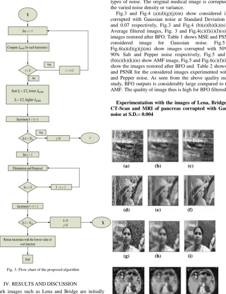
A. Algorithm used for de noising images
For xth target pixel optimization takes in following steps: Initialize parameters p, S, NC, NS, Nre, Ned, ped, and C(i), i= 1,2,3…………S.
C(i) = Step size in the random direction Step 1: Elimination –dispersal loop :l=l+1 Step 2: Reproduction loop : k=k+1 Step 3: Chemo taxis loop : j=j+1 Adaptive Median Filter
Bacterial Foraging Optimisation
ISSN: 2231-5381
http://www.ijettjournal.org
Page 3051
a) For i=1,2,…….,S, take a chemo tactic step for
bacterium i as follows.
b) Let Jlast=J(I,j,k,l)= | P(i, j , k ,l) - R(x, j, k, l ) |2
where
P(i, j , k ,l) = location of ith pixel (bacteria) at jth chemo tactic step, kth reproduction step and lth elimination step.
R(x, j, k, l) = location of xth target pixel at jth chemo tactic step, kth reproduction step and lth elimination step.
J(i, j, k, l) = Cost of ith pixel (bacteria) at jth chemo tactic step, kth reproduction step and lth elimination step..
c) Tumble: A random vector ∆m(i), m= 1,2,…p, a
random number in [-1,1].
d) Move: Let (j+1,k,l)= (j,k,l) + C(i) . results in a step
C(i) in the direction of the tumble for bacterium i.
e) Swim:
i. Let m= 0 (counter for swim length).
ii. While m< NS
• Let m= m+1
• If J(i,j+1,k,l) < Jlast ,
Let Jlast = J(i,j+1,k,l) and let Pi(j+1,k,l) + C(i)
And use this Pi(j+1,k,l) to compute the new J(j+1,k,l).
• Else, let m = NS.
f) Go to next bacteria (i+1) if i≠ S
Step 4: If j < NC, go to step 3. Step 5: Reproduction:
i) For the given k and l, and for each i= 1,2,………….S, let J(i,j,k,l)
ii) The Sr = S/2 bacteria with the highest value of cost function die and other Sr = S/2 bacteria with the best value (least value of cost function) split.
Step 6: If k < Nre, go to step 2.
Step 7: Elimination- dispersal: Eliminate and dispersal each bacterium.
Step 8: If l < Ned , go to the step 1. Otherwise end.
The flow chart is depicted in Fig. 3
start
Initialise p, S, NC, Nre, and Ned
Set i = 1
Compute Cost for i
J (i ,j, k,l) = ǀ P(i,,j,k,l)-R(x,,j,k,l)ǀ2
J last =J (i ,j, k, l)
Tumble: Update position and cost of bacterium i
J(i,j+1,k,l)=|P(i,j+1,k,l)- R(x,j+1,k,l)|2
Set m = 0 (Counter of swim)
Y
Is
J(i,j+1, k,
l ) < Jlast
m < NS
j <NC
i < S
Bacterium i moves one more step in the direction of tumble
Increment j = j + 1 Increment swim m = m+1
i =i + 1
X
Yes Yes
Yes
No
No
No No
ISSN: 2231-5381
http://www.ijettjournal.org
Page 3052
Fig. 3: Flow chart of the proposed algorithmIV.RESULTSANDDISCUSSION
Benchmark images such as Lena and Bridge are initially considered. To reduce the processing time, the images are downsized to (50×50). Salt and pepper noise with varied noise density 10% to 90% are used. In case of Gaussian noise
variance is changed from 0.002 to 0.07. Then medical images like CT-Scan and MRI of pancreas are considered. Pancreas being one of the innermost organs, hence is prone to different types of noise. The original medical image is corrupted with the varied noise density or variance.
Fig.3 and Fig.4 (a)(d)(g)(j)(m) show considered images corrupted with Gaussian noise at Standard Deviation 0.002 and 0.07 respectively, Fig.3 and Fig.4 (b)(e)(h)(k)(n) show Average filtered images, Fig. 3 and Fig.4(c)(f)(i)(l)(o) show images restored after BFO. Table 1 shows MSE and PSNR for
considered image for Gaussian noise. Fig.5 and
Fig.6(a)(d)(g)(j)(m) show images corrupted with 50% and 90% Salt and Pepper noise respectively, Fig.5 and Fig.6 (b)(e)(h)(k)(n) show AMF image, Fig.5 and Fig.6(c)(f)(i)(l)(o) show the images restored after BFO and Table 2 shows MSE and PSNR for the considered images experimented with Salt and Pepper noise. As seen from the above quality matrices study, BFO outputs is considerably large compared to that of AMF. The quality of image thus is high for BFO filtered case.
Experimentation with the images of Lena, Bridge, Self, CT-Scan and MRI of pancreas corrupted with Gaussian noise at S.D.= 0.004
(a) (b) (c)
(d) (e) (f)
(g) (h) (i)
(j) (k) (l)
X
Set i = 1
Compute Jhealth for each bacterium i
Increment k = k +1
Is l < Ned
X
End
i < S i = i+1
Sort Sr = S/2, lower Jhealth
Sr = S/2, higher Jhealth
Is k < Nre j=0 Y
Elimination and Dispersal Set i = 1
Is i < S
I =i + 1
Increment l =l + 1
k=0 j=0
Return bacterium with the lowest value of cost function
No Yes
ISSN: 2231-5381
http://www.ijettjournal.org
Page 3053
(m) (n) (o)Fig.3: (a)(d)(g)(j)(m) Image corrupted with Gaussian noise at S.D. of 0.004 (b)(e)(h)(k)(n) Average filtered Image (c)(f)(i)(l)(o) Image restored
after BFO
Experimentation with the images of Lena, Bridge, Self, CT-Scan and MRI of pancreas corrupted with Gaussian noise at S.D.= 0.07
(a) (b) (c)
(d) (e) (f)
(g) (h) (i)
(j) (k) (l)
(m) (n) (o)
Fig.4: (a)(d)(g)(j)(m) Image corrupted with Gaussian noise at S.D. of 0.07 (b)(e)(h)(k)(n) Average filtered Image (c)(f)(i)(l)(o) Image restored after BFO
TABLEI
COMPARISON OF AVERAGE FILTER AND BFO ON THE BASIS OF MSE AND
PSNR FOR THE IMAGES,BRIDGE,LENA,SELF CT-SCAN OF PANCREAS AND
MRCHOLANGIOPANCREATOGRAM
Imag e
S.D. (σ)
Average Filter BFO
Brid ge
MSE PSNR (dB)
MSE PSNR (dB)
0.002 439.5551 21.7007 0.4633 51.4721 0.004 435.7321 21.7386 0.1705 55.8140 0.006 435.3541 21.7424 0.1666 55.9141 0.008 435.5069 21.7409 0.1698 55.8302 0.01 436.1986 21.7340 0.1720 55.7763 0.03 468.9167 21.4198 0.2550 54.0654 0.05 543.4171 20.7795 0.9374 48.4116 0.07 665.4071 19.8999 01.6022 46.0836 Lena 0.002 432.6510 21.7694 0.0078 69.2283 0.004 433.2666 21.7633 0.0066 69.9214 0.006 449.5479 21.6030 0.0237 64.3797 0.008 428.4987 21.8113 0.0490 61.2274 0.01 441.1730 21.6847 0.0120 67.3315 0.03 470.4446 21.4057 0.0095 68.3485 0.05 541.5641 20.7943 0.0109 67.7562 0.07 669.1511 19.8756 0.0399 62.1166 Self 0.002 503.4207 21.1115 0.6266 50.1608 0.004 525.03111 20.9290 0.5953 50.3833 0.006 539.1716 20.8135 1.2647 47.1110 0.008 511.9681 21.0384 0.2537 54.0883 0.01 502.1488 21.1225 0.2879 53.5378 0.03 550.0545 20.7267 0.4845 51.2775 0.05 608.4642 20.2885 0.1525 56.2988 0.07 708.3690 19.6282 1.3943 46.6872
CT-Scan of pancr
eas
0.002 433.4032 21.7619 0.3397 52.8202 0.004 437.2765 21.7232 2.1233 44.2765 0.006 442.1166 21.6754 1.7204 45.7746 0.008 447.1674 21.6261 0.8296 48.9422 0.01 441.2470 21.6840 1.8203 45.5294 0.03 482.1797 21.2987 0.6371 50.0885 0.05 587.0902 20.4438 0.2155 54.7968 0.07 740.2602 19.4370 0.1581 56.1416 MRI
of pancr
eas
ISSN: 2231-5381
http://www.ijettjournal.org
Page 3054
Experimentation with the images of Lena, Bridge, MRI,CT-Scan of pancreas and image of self at 50% Salt & Pepper Noise
(a) (b) (c)
(d) (e) (f)
(g) (h) (i)
(j) (k) (l)
(m) (n) (o)
Fig.5: (a)(d)(g)(j)(m) Image corrupted with 50% Salt & Pepper noise (b)(e)(h)(k)(n) AMF Image (c)(f)(i)(l)(o) Image restored after BFO
Experimentation with the images of Lena, Bridge, MRI ,CT-Scan of pancreas and image of self at 90% Salt & Pepper Noise
.
(a) (b) (c)
(d) (e) (f)
(g) (h) (i)
(j) (k) (l)
(m) (n) (o)
Fig.6:.(a)(d)(g)(j)(m) Image corrupted with 50% Salt & Pepper noise (b)(e)(h)(k)(n) AMF Image (c)(f)(i)(l)(o) Image restored after BFO
TABLE II
COMPARISON OF ADAPTIVE MEDIAN FILTER AND BFO ON THE BASIS OF
MSE AND PSNR FOR THE IMAGES, BRIDGE, LENA, SELF CT-SCAN OF PANCREAS AND MRCHOLANGIOPANCREATOGRAM
Le na
Smax for AMF
% of sa lt & pe p pe
r
AMF BFO
MSE PSNR
(dB) MSE
PSNR (dB)
ISSN: 2231-5381
http://www.ijettjournal.org
Page 3055
43×43 90 2003.7 15.1124 0.3231 53.0372 Bri
dg e
5×5 10 93.9872 28.4001 0.0502 61.1231 9×9 20 151.0020 26.3410 0.0526 60.9203 11×11 30 230.2928 24.5080 0.0682 59.7955 17×17 40 297.5188 23.3957 0.1845 55.4713 21×21 50 391.1516 22.2074 0.2137 54.8332 27×27 60 538.8240 20.8163 0.2814 53.6382 33×33 70 700.4588 19.6770 0.5074 51.0776 37×37 80 963.8880 18.2905 0.6442 50.0409 43×43 90 1402.3 16.6623 0.6664 49.8935 CT -Sc an of pa ncr eas
5×5 10 165.6924 25.9378 0.1891 55.3634 9×9 20 227.9096 24.5532 0.2007 55.1049 17×17 30 324.1800 23.0229 0.2035 55.0447 21×21 40 424.4564 21.8525 0.2078 54.9535 27×27 50 682.2984 19.7911 0.2111 54.8855 31×31 60 840.2984 18.8869 0.2314 54.4864 35×35 70 1222.7 17.6284 0.2332 54.4535 39×39 80 1623.9 16.0252 0.3198 53.0815 43×43 90 3138.2 13.1639 0.6672 49.8882 M RI of pa ncr eas
5×5 10 68.6892 29.7619 0.1779 55.6286 9×9 20 75.5420 29.3489 0.2169 54.7686 17×17 30 155.3624 26.2173 0.2198 54.7097 21×21 40 196.5188 25.1968 0.2204 54.6987 25×25 50 368.6452 22.4647 0.2316 54.4834 31×31 60 504.5540 21.1017 0.2382 54.3607 35×35 70 658.2576 19.9468 0.2446 54.2455 39×39 80 802.7740 19.0849 0.3046 53.2929 43×43 90 1528.0 16.2958 0.5781 50.5109 Im
age of Sel f
5×5 10 98.6188 28.1912 0.2533 54.0948 9×9 20 110.3048 27.7049 0.2675 53.8572 11×11 30 227.3684 24.5635 0.3649 52.5093 13×13 40 393.2328 22.1843 0.3716 52.4300 15×15 50 507.0896 21.0800 0.3762 52.3762 21×21 60 776.4108 19.2299 0.3848 52.2785 29×29 70 1035.7 17.9785 0.4274 51.8221 31×31 80 1430.9 16.5747 0.5386 50.8185 43×43 90 2304.4 14.5052 1.4498 46.5176
Fig.7 shows the convergence plot of the BFO which
converges with 2500 iterations. Thus, computation
overloading is also low.
Fig.7: Convergence plot showing reduction of MSE with Number of iterations
V. CONCLUSION
The paper presents an application of BFO as a digital filter to de-noise medical image i.e., CT-Scan and MRI of pancreas. The experimentation in terms of quality matrices like MSE and PSNR show considerable improvement in the quality of restored images. The computational overloading is also low. Thus, this approach of using Soft Computing in conjunction with digital filter will definitely enhance the de-noising capability of digital filters which can find application in sensitive images like medical images.
ACKNOWLEDGMENT
I would like to place on record my deep sense of gratitude to Prof. (Dr.) S.S. Pattnaik, Prof. and HOD, ETV, NITTTR, Chandigarh, India and Dr. Jagroop Sigh Sidhu, Associate Prof., Department of Electronics and Communication Engineering, DAV Institute of Engineering and Technology, Jalandhar, India, for generous guidance, help and useful suggestions.
REFERENCES
[1] Choubey, Abha, Sinha, G. R., Choubey, Siddharth; "A Hybrid filtering Technique in Medical Image denoising Blending of Neural Network and Fuzzy Inference." ICECT Vol.1, pp. 170-177, April 2011 [2] Daiyan, G.M., Mottalib, M.A.; "Removal of high density Salt and
Pepper noise through modified median filter." International Conference on Informatics, Electronoics and vision, pp. 565-570, 2012
[3] Gonzalez,Rafel C. and Woods, Richard E.; "Digital Image Processing." Second edition, Publising House of Electronics Industry, Beijing, 2003 [4] Kennedy,J and Eberhart, R.C; "Particle Swarm Optimisation."
Proceedings of IEEE International Conference on Neural Network, , 1995 Piscataway, NJ, pp.1942-1948
[5] Kneey Kal Vin Toh and Nor Ashidi Mat Isa; "Noise Adaptive Fuzzy Switching Median filter for Salt & Pepper Noise Reduction." IEEE Signal Processing Letters, Vol. 17, No. 3, pp. 281-284 , March 2010 [6] Lu Zhang, Jiaming Chen, Yuein Zhu, Zianhua Luo; "Comparisions of
several New Denoising Methods for Medical Images." International Conference on Bioinfomatics and Biomedical Engineering, June 2009, pp. 1-4,
[7] Newton, T.H. and Potts, D.G.; "Technical Aspects of Computed Tomography in Radiology of skull and Brain." Vol.5 ISBN-0-8016-3662-0, pp. 3941-3956, 1981
[8] Passino, K.M.; "Biomimicry of Bacterial Foraging for Distributed Optimisation and Control." IEEE Control and System Magzene, pp. 52-67, June 2002
[9] Pattnaik, S.S; Backwad, K.M.; Sohi, B.S.; Ratho, R.K.; S.Devi; "Swine Influenza Model Based Optimisation (SIMBO)." Applied Soft Computing, Vol.1, pp.18-30, Sept. 2012
[10] Zhou Wang David Zhang; "Progressive Switching Median filter for the removal of Impulse Noise from Highly corrupted Images." IEEE Transaction on Circuits and System –II: Analog and Digital Processing, Vol.46,No.1 pp. 78-80 Jan 1999
[11] Zhu Youlin; Huang Cheng; "Image denoising algorithm based on PSO Optimising Structuring element." Control and Decision Conference, , 2012, pp.2404-2408