Role of nitric oxide in experimental obliterative
bronchiolitis (chronic rejection) in the rat.
E A Kallio, … , K Vaali, K B Lemstöm
J Clin Invest.
1997;
100(12)
:2984-2994.
https://doi.org/10.1172/JCI119852
.
The role of nitric oxide in obliterative bronchiolitis development, i.e., chronic rejection, was
investigated in the heterotopic rat tracheal allograft model. An increase in the intragraft
inducible nitric oxide synthase (iNOS) mRNA and mononuclear inflammatory cell iNOS
immunoreactivity was demonstrated during progressive loss of respiratory epithelium and
airway occlusion in nontreated allografts compared to syngeneic grafts. In nontreated
allografts, however, intragraft nitric oxide production was decreased, most likely because of
loss of iNOS epithelial expression. Treatment with aminoguanidine, a preferential inhibitor
of inducible nitric oxide synthase, was associated with enhanced proliferation of
alpha-smooth muscle actin immunoreactive cells and the intensity of obliterative bronchiolitis early
after transplantation. Aminoguanidine treatment did not affect iNOS mRNA synthesis or
intragraft nitric oxide production, but decreased iNOS immunoreactivity in smooth muscle
cells. Treatment with L-arginine, a precursor of nitric oxide, significantly reduced obliterative
changes. L-arginine supplementation enhanced intragraft iNOS mRNA synthesis and iNOS
immunoreactivity in capillary endothelial and smooth muscle cells as well as intragraft nitric
oxide production. Immunohistochemical analysis of allografts showed that neither iNOS
inhibition nor supplementation of the nitric oxide pathway affected the number of
graft-infiltrating CD4+ and CD8+ T cells, ED1+ and ED3+ macrophages, immune activation with
expression of IL-2R or MHC class II, or production of macrophage or Th1 cytokines. In
contrast, L-arginine treatment was associated with increased staining for […]
Research Article
Find the latest version:
J. Clin. Invest.
© The American Society for Clinical Investigation, Inc. 0021-9738/97/12/2984/11 $2.00
Volume 100, Number 12, December 1997, 2984–2994 http://www.jci.org
Role of Nitric Oxide in Experimental Obliterative Bronchiolitis (Chronic Rejection)
in the Rat
Erkki A. Kallio,* Petri K. Koskinen,* Einari Aavik,* Kirsi Vaali,‡ and Karl B. Lemstöm*
*Cardiopulmonary Research Group of Transplantation Laboratory, Haartman Institute, University of Helsinki and Helsinki University Central Hospital; and ‡Institute of Biomedicine, Department of Pharmacology and Toxicology, FIN-00014 University of Helsinki,
Helsinki, Finland
Abstract
The role of nitric oxide in obliterative bronchiolitis
develop-ment, i.e., chronic rejection, was investigated in the
hetero-topic rat tracheal allograft model. An increase in the
intra-graft inducible nitric oxide synthase (iNOS) mRNA and
mononuclear inflammatory cell iNOS immunoreactivity was
demonstrated during progressive loss of respiratory
epithe-lium and airway occlusion in nontreated allografts
com-pared to syngeneic grafts. In nontreated allografts, however,
intragraft nitric oxide production was decreased, most likely
because of loss of iNOS epithelial expression. Treatment with
aminoguanidine, a preferential inhibitor of inducible nitric
oxide synthase, was associated with enhanced proliferation
of
a
-smooth muscle actin immunoreactive cells and the
in-tensity of obliterative bronchiolitis early after
transplanta-tion. Aminoguanidine treatment did not affect iNOS mRNA
synthesis or intragraft nitric oxide production, but
de-creased iNOS immunoreactivity in smooth muscle cells.
Treatment with
L-arginine, a precursor of nitric oxide,
sig-nificantly reduced obliterative changes.
L-arginine
supple-mentation enhanced intragraft iNOS mRNA synthesis and
iNOS immunoreactivity in capillary endothelial and smooth
muscle cells as well as intragraft nitric oxide production.
Immunohistochemical analysis of allografts showed that
neither iNOS inhibition nor supplementation of the nitric
oxide pathway affected the number of graft-infiltrating
CD4
1
and CD8
1
T cells, ED1
1
and ED3
1
macrophages,
immune activation with expression of IL-2R or MHC class
II, or production of macrophage or Th1 cytokines. In
con-trast,
L-arginine treatment was associated with increased
staining for Th2 cytokines IL-4 and IL-10. In conclusion,
this study demonstrates that nitric oxide has a protective
role in obliterative bronchiolitis development in this model,
and suggests that nitric oxide either directly or indirectly
in-hibits smooth muscle cell proliferation and modulates
im-mune response towards Th2 cytokines. (
J. Clin. Invest.
1997.
100:2984–2994.) Key words: inducible nitric oxide synthase
• L-arginine
•aminoguanidine
•transplantation
•lung
Introduction
Despite recent advances in understanding chronic rejection, the regulatory and effector mechanisms underlying the disease process remain incompletely known (1). Observations in a het-erotopic rat tracheal allograft model demonstrate that recruit-ment of CD41 and CD81 T cells and macrophages into air-way wall induces local cytokine and growth factor production leading to epithelial damage, proliferation of smooth muscle cells, and gradual occlusion of airway lumen (2–4), i.e., histo-logical changes similar to those observed in human lung al-lografts with obliterative bronchiolitis (OB)1 (5).
Nitric oxide (NO) has a significant role in several physio-logical and pathophysio-logical conditions such as vasorelaxation, neurotransmission, platelet and leukocyte adhesion, and host defense mechanisms against bacteria and viruses (6). NO is generated from L-arginine by the enzyme NO synthase (NOS) (7). NOS can be either constitutively expressed (cNOS) or in-duced by cytokines and LPS (iNOS) (7). cNOS has been local-ized in vascular endothelium (eNOS), platelets, and neurons (nNOS) of the central nervous system (7). cNOS produces small quantities of NO with activity regulated by intracellular calcium/calmodulin, and is involved in a variety of normal physiological functions. iNOS can produce large amounts of NO, and is calcium-independent. iNOS is expressed in mac-rophages, vascular smooth muscle cells, endothelial cells, mes-angial cells, and airway epithelium (7–9). In contrast to many other organs, NO is continuously produced by iNOS in normal airway epithelium, and regulates bronchomotor tone (10, 11). In addition, NO may regulate several inflammatory responses in the lung such as development of asthma and granulomatous inflammation (12–14).
During chronic rejection, there is a major release of proin-flammatory molecules by infiltrating activated T cells and mac-rophages that participate in the immune reaction to foreign MHC and other antigens present in endothelial, epithelial, and other cells of the transplanted organ. Cytokines derived from activated macrophages, i.e., TNF-a and IL-1b, and those de-rived from Th1 cells, i.e., IL-2 and IFN-g, promote iNOS ex-pression (8). Recently induction of iNOS (15) and production of NO (16) during acute allograft rejection was demonstrated,
This work was presented as an abstract at The 2nd International Sym-posium of the Etiology and Pathobiology of Transplant Vascular Sclerosis in Southhampton, Bermuda on March 5–9, 1997.
Address correspondence to Dr. Erkki Kallio, Transplantation Laboratory, P.O. Box 21 (Haartmaninkatu 3), FIN-00014 University of Helsinki, Finland. Phone: 1358-9-1912-6596; FAX: 1 358-9-241-1227; E-mail: [email protected]
Received for publication 27 May 1997 and accepted in revised form 16 October 1997.
and it was shown that treatment with aminoguanidine, a pref-erential inhibitor of iNOS (17), significantly attenuated patho-genesis of acute cardiac and lung allograft rejection (18, 19). This study was undertaken to investigate whether iNOS is in-duced during experimental OB in rat tracheal allografts, and to investigate the biological role of NO by supplementation of NO pathway by L-arginine, or by inhibition of the iNOS path-way by aminoguanidine in this model.
Methods
Heterotopic rat tracheal transplantation model and study design. Spe-cific pathogen-free, 2–3-mo-old inbred male DA (AG-B4, RT1a) and
WF (AG-B2, RT1u) rats weighing 200–300 g (Laboratory Animal
Center, University of Helsinki, Helsinki, Finland) were used. Alloge-neic heterotopic tracheal transplantations were performed as de-scribed (3, 4) from DA to WF rats, and syngeneic controls from DA to DA rats. Four groups were formed: (a) nontreated syngeneic con-trols, (b) nontreated allogeneic controls, (c) allografts treated with aminoguanidine, a preferential inhibitor of iNOS (17), and (d) allo-grafts supplemented with an NO precursor, L-arginine (7). The grafts were removed 3, 10, and 30 d after transplantation to determine his-tological changes, relative intragraft iNOS mRNA, protein expres-sion, and nitrite/nitrate production. In addition, a detailed immuno-histochemical analysis was performed to localize iNOS production and to examine how NO may modulate inflammatory cell infiltration and cytokine production in the airway wall of tracheal transplants.
Aminoguanidine and L-arginine administration. Aminoguanidine-treated allografts received aminoguanidine hemisulfate (Sigma Chem-ical Co., St. Louis, MO) by injection of 400 mg/kg/d i.p. in three doses (100 mg/kg at 9:00 AM, 100 mg/kg at 1:00 PM, and 200 mg/kg at 5:00 PM) in 0.9% NaCl in a concentration of 100 mg/ml, previously shown to decrease serum NO concentrations to 30% of normal in rats (18).
L-arginine (L-arginine hydroxychloride; Sigma Chemical Co.) was given orally at a concentration of 2.25% dissolved in tap water (available ad li-bidum), a solution previously shown to increase plasma L-arginine levels twofold (20).
Histological evaluation. The grafted trachea was excised, embed-ded in Tissue-Tek (Miles Inc., Elkhart, IN), snap-frozen in liquid ni-trogen, and stored at 2708C until used. For histological evaluation, frozen sections were stained with Mayer’s hematoxylin-eosin. The histological changes in respiratory epithelium were evaluated as a percentage of normal respiratory, abnormal cuboidal and squamous epithelium, or loss of epithelium. Luminal occulusion was evaluated as a reduction of luminal area using an ocular grid. All the analyses were done in a blinded review by two observers, and the scores of these two observers were highly correlated (r25 0.95). The degree of
airway wall inflammation, the cell types of the fibroproliferative le-sion, and the number of proliferating inflammatory and myofibropro-liferative cells were analyzed using immunohistochemistry.
Single immunostaining. Serial frozen sections (4–6 mm) were air-dried on silane-coated slides, fixed in acetone at 2208C for 20 min, and stored at 2208C until used. Before immunostaining, the slides were refixed with chloroform and then air-dried. After incubation with appropriate 1.5% nonimmune serum (Vector Laboratories, Inc., Burlingame, CA) for mAbs or pAbs, frozen sections were incubated with a mouse mAb at room temperature for 30 to 60 min, or with a rabbit or goat pAb at 148C for 12 h. The primary Abs were diluted in PBS with 1% BSA and appropriate 3% nonimmune serum. With in-tervening washes in Tris-buffered saline, the slides were immersed in bionylated horse anti–mouse, goat anti–rabbit, or rabbit anti–goat rat-absorbed antibodies at room temperature for 30 min followed by avidin-bionylated horseradish complex (Vectastain Elite ABC Kit; Vector Laboratories) in PBS at room temperature for 30 min. The re-action was revealed by chromogen 3-amino-9-ethylcarbazole (AEC; Sigma Chemical Co.) containing 0.1% hydrogen peroxidase, yielding
a brown-red reaction product. The specimens were counterstained with hematoxylin and coverslips were aquamounted (Aquamount; BDH Chemicals Ltd., Poole, England).
Antibodies used. Details of the Abs and their use in immunohis-tology were described previously unless otherwise specified (3). Abs were directed against rat macrophages (ED1; Serotec Ltd., Oxford, United Kingdom), activated macrophages (ED3; Serotec Ltd.), CD4 (W3/25; Lab, Sussex, United Kingdom), and CD8 (OX8; Sera-Lab) subsets. Immune activaton was determined using mouse mAbs to MHC class II common determinant (OX6; Sera-Lab) and IL-2R (PharMingen, San Diego, CA); mouse mAbs to IFN-g (CY-047; In-nogenetics, Zwijndrecht, Belgium), TNF-a (CY-051; Innogenetics), and IL-4 (MRC OX-81; Serotec Ltd.); rabbit pAbs to IL-1a (IP-110) and IL-1b (LP-712; Genzyme Corp., Cambridge, MA); and goat IgG pAbs to IL-2 (SC-1786; Santa Cruz Biotechnology, Santa Cruz, CA) and IL-10 (AB-417-NA; R&D Systems, Minneapolis, MN). NOS was localized using rabbit IgG pAbs to mouse and rat iNOS (NOS2, SC-650; Santa Cruz Biotechnology) and cNOS (NOS3, SC-654; Santa Cruz Biotechnology).
Double immunostaining. To localize iNOS expression on mac-rophages, endothelial cells, and smooth muscle cells, double staining was applied on representative frozen sections. After staining for iNOS using the peroxidase ABC method described above (yielding a brown-red reaction product), tracheal segments were washed in Tris-buffered saline, and avidin–biotin complex from the first step was blocked by incubating the sections with an excess of avidin and biotin (Avidin/Biotin Blocking Kit; Vector Laboratories, Inc.). After appli-cation of antibodies to macrophages (ED1), endothelial cells (RECA 1; Serotec Ltd.), or a-smooth muscle cell actin (1A4; Biomakor, Re-hovot, Israel) at room temperature for 30 min, sections were incu-bated with bionylated horse anti–mouse rat-absorbed antibodies at room temperature for 30 min, followed by incubation with alkaline phosphatase avidin–biotin complex (Vectastain Elite ABC Kit; Vec-tor LaboraVec-tories, Inc.) and visualized by VecVec-tor blue, an alkaline phosphate substrate kit (Vector Laboratories, Inc.) that produced a blue reaction. Double-stained sections were not counterstained.
Specificity controls of immunostaining. Controls were performed using the same immunoglobulin concentrations of species and iso-type-matched antibodies: mouse monoclonal IgG1 antibody (catalog no. X931; Dako Corp., Carpinteria, CA) and rabbit polyclonal immu-noglobulin fraction (catalog no. X936; Dako Corp.) for mAbs and pAbs, respectively. Additional specificity controls for iNOS, cNOS, TNF-a, IL-1a, IL-1b, and IFN-g stainings involved use of a working dilution of the polyclonal antibody after overnight incubation with a 10 to 20-molar excess of recombinant cytokine (Genzyme Corp.) or peptide (Santa Cruz Biotechnology). None of these control stainings showed any immunoreactivity.
Quantitation of immunohistochemistry. The immunohistochemi-cal analysis was done in a blind review by two observers, and the score assigned was determined by consensus. The intensity of the staining was scored from 0 to 3 as follows: 0, no visible staining; 1, few cells with faint staining; 2, moderate intensity with multifocal staining; and 3, intense diffuse staining of the cells analyzed. Positive staining for IL was scored as the number of positive cells in cross-section.
In vivo labeling for cell proliferation. All recipients were injected with 400 ml i.v. of a concentrated solution of bromodeoxyuridine (BrdU; Zymed Laboratories, Inc., San Francisco, CA) 3 h before death. Cell proliferation in frozen sections was revealed by an IgG1 mouse mAb to BrdU 1:20 (M744; Dako A/S, Glostrup, Denmark) and a Vectastain Elite ABC Kit (Vector Laboratories) method as de-scribed above. Before staining, the frozen sections were fixed with buffered formalin for 15 min. After a 10-min wash in PBS, the sec-tions were microwave-treated with 500 W in 0.1 M citrate buffer, pH 6, for 5 min to break the double-stranded DNA, followed by a 10-min wash in PBS. Cell proliferation was measured by counting the num-ber of labeled nuclei in tracheal cross-sections.
normal-ization against levels of the reference gene glyceraldehyde-3-phos-phate dehydrogenase (GAPDH). Total RNA isolation was extracted from fresh trachea tissue using a guanidine-isothiocyanate prepara-tion as descibed (21, 22). The total RNA yields were estimated by spectrophotometry, and were confirmed by 1% agarose/TBE gel elec-trophoresis.
To identify the optimum PCR conditions for accurate measument of each gene, we established the logarithmic assay range with re-spect to cycle number and starting template concentration against dif-ferent dilutions of cDNA. 1 mg of total RNA was reverse-transcribed to cDNA in a 20-ml volume of master mix: 13 RT buffer (Promega Corp., Madison, WI), 300 mM dNTP (GeneAmp; Perkin-Elmer Corp., Norwalk, CT), 50 pmol oligo-dT primer (p[dT]15 primer;
Boehringer-Mannheim, Boehringer-Mannheim, Germany), 20 U RNasin (RNasin Ribonu-clease Inhibitor; Promega Corp.), and 100 U reverse transcriptase (M-MLV-RTase Reverse Transcriptase, RNase H Minus; Promega Corp.) at 1378C for 90 min. 1 ml of cDNA reaction mixture was added to 37.4 ml of water and supplemented with 5 ml 103 PCR buffer I (GeneAmp; Perkin Elmer Corp.), 10 nmol dNTP (GeneAmp; Perkin Elmer Corp.), 1 mCi a[32P]dATP, 3000 Ci/mmol (Amersham Intl.,
Buckinghamshire, England), 10 pmol iNOS sense primer (59-CTT TGT GCG GAG TGT CAG TGG-39[GenBank accession no. D12520]), 10 pmol iNOS antisense primer (59-TTC TTC CTG ATA GAG GTG GTCC-39), and 2.5 U AmpliTaq Gold (Perkin Elmer Corp.). The samples were heated to 958C for 10 min, and then cycled 37 times (948C for 30 s, 608C for 30 s, 728C for 1 min) using the 96-well PCR apparatus (GenAmp PCR System 9600 apparatus; Perkin Elmer Corp.). The relative amount of GAPDH mRNA was analyzed by a similar protocol using GAPDH sense (59-GTC TTC ACC ACC ATG GAG AAG GCT-39 [GenBank accession no. M17701]) and GAPDH antisense (59-TGT AGC CCA GGA TGC CCT TTA GTG-39) primers. The PCR protocol for GAPDH was as follows: 958C for 10 min, and then amplification for 33 cycles (948C, 1 min, 578C, 1 min, 728C, 2 min). Gene transcript levels for all samples for a single gene were amplified simultaneously, and each PCR analysis was completed in triplicate. For each experiment, the negative con-trols for which water was used instead of cDNA or reverse tran-scriptase was omitted during cDNA synthesis. The PCR samples were electrophoresed through 2% agarose. Subsequently, the gel was dried (LKB 2003 Slab Gel Dryer; LKB, Bromma, Sweden), exposed to an imaging plate (Fuji Photo Film Co., Tokyo, Japan), and the gels were quantified using a Fuji BAS1500 phosphoimager. The mean values of
the three determinations were used for final analysis, and the normal-ized iNOS mRNA levels were derived by dividing the mean of iNOS mRNA with the mean of GAPDH mRNA for each tissue sample.
Spectrometric determination of intragraft nitrites and nitrates. Intra-graft NO production was determined as stable metabolite nitrites and nitrates (NOx). Tracheas were snap-frozen and stored in 2708C.
Ho-mogenization was done in ultrapure water (MilliQ; Millipore Corp., Bedford, MA) with a Douche homogenizator at 148C. The crude ho-mogenate was centrifuged (4000 g). To prevent interference of pro-teins and hemegroups, the supernatant was filtrated through Centri-con filters (AmiCentri-con Inc., Beverly, MA), exclusive size 10.000 D, and the rest of the proteins in the filtrate were precipitated in 1:20 of 20% ZnSO4. The supernatants were divided for two assays: one for
mea-surement of NO221 NO32 and for the measurement of NO22. NO22
was measured with the Griess reaction, and nitrate reductase (EC 1.6.6.2, Aspergillus species; Boehringer Mannheim) was used to re-duce the NO32 to NO22 used in the Griess reaction (23, 24). All
de-terminations were performed in duplicate, and the results are given in nmol/mg of tissue. The detection limit of the assay was 0.25 mM, and absorbance in 540 nm was measured.
Statistical analyses. All data are expressed as mean6SEM. For multiple group comparisons of small sample size nonparametric, Kruskal-Wallis (Statview 5121 program; Brain Power Inc., Calaba-sas, CA) one-way analysis by ranks was used. The rank sums ob-tained with the Kruskal-Wallis test were then used for the Dunn test at significance levels of 5 and 1% (Medstat; Astra Group A/S, Copen-hagen, Denmark). In groups with normal distribution and standard variance (F-test), one-factor ANOVA and Fisher PLSD-test were used at significance levels of 5 and 1%. P, 0.05 was regarded as sta-tistically significant.
Results
Histology. The morphological changes of respiratory
[image:4.612.64.537.510.693.2]epithe-lium and the degree of luminal occlusion of tracheal grafts at different time points are summarized in Fig. 1. 3 d after trans-plantation, the respiratory epithelium of syngeneic grafts was slightly damaged, probably due to ischemic injury before re-vascularization of the graft. 30 d after transplantation, the res-piratory epithelium of syngeneic grafts was fully recovered,
and the grafts were filled with mucous, a sign of normal epithe-lium function. In syngeneic grafts, no myofibroproliferation occured, and overall histology resembled that of nontrans-planted tracheas (Fig. 2 A). In nontreated allografts, there was a progressive loss of respiratory epithelium. Airway wall in-flammation peaked 10 d after transplantation, and subsided thereafter. In nontreated allografts, the luminal surface was re-duced by myofibroproliferative lesion already at 10 d, and at
30 d intense myofibroproliferation nearly totally occluded the lumen (Fig. 2 B). Aminoguanidine treatment significantly ac-celerated myofibroproliferative lesion development, compro-mising the luminal area already at 3 and 10 d compared to non-treated allografts (P , 0.05). Thereafter, the difference leveled off as the tracheal lumen was nearly totally occluded in both groups at 30 d (Figs. 1 and 2 C). L-arginine treatment signifi-cantly delayed epithelial destruction, and signifisignifi-cantly
down-Figure 2. Photomicrographs of (A)
syngeneic graft, (B) nontreated, (C) aminoguanidine-treated, and (D)
L-arginine–treated allografts 30 d after transplantation. Syngeneic grafts were filled with mucus, and the airway was covered with normal respiratory epithelium (arrow). In nontreated and aminoguanidine-treated allografts, the airway lumen was severely compromised by dense myofibroproliferative lesions (B and
[image:5.612.58.427.61.346.2]C). L-arginine treatment signifi-cantly ameliorated development of the obliterative lesion. Mayer’s hae-matoxylin & eosin staining, 403 original.
Table I. Expression of iNOS in Normal Tracheas, Syngeneic Grafts, Nontreated, Aminoguanidine-, and
L-arginine–treated Allografts
Days after
transplantation Group n Epithelial cells
Myofibroproliferative lesion
Capillary endothelial cells
Mononuclear inflammatory cells
Smooth muscle cells
0 Normal DA trachea 6 0.560 060 0.260.1 0.160.1
3 Syngeneic graft 10 0.160.1 0.160.1 0.360.1 0.160.1
Allograft 12 0.360.3 0 0.860.1 0.160.1
Allograft 1 aminoguanidine 12 1.160.2 0.760.1 1.260.2 0.560.2 Allograft 1L-arginine 12 1.060.1 1.460.1‡ 1.260.1 0.960.1‡
10 Syngeneic graft 10 2.260.3 0.960.2 0.660.1* 0.560.1
Allograft 10 1.360.3 0.860.1 1.260.2 0.660.1
Allograft 1 aminoguanidine 15 1.060.2 0.460.1 0.860.1 0.160.0‡
Allograft 1L-arginine 15 1.960.3 0.960.2 1.460.2 0.560.1 30 Syngeneic graft 10 0.660.2 0.860.2 0.360.1 0.860.1
Allograft 14 0.260.1 0.660.1 0.560.1 0.460.1
Allograft 1 aminoguanidine 14 0.360.1 0.960.1 0.760.1 0.660.1 Allograft 1L-arginine 12 0.760.2 0.960.2 0.860.1 0.660.1
In allogeneic groups (allograft, allograft 1 L-arginine, and allograft 1 aminoguanidine) at 3 and 10 d, only some of the grafts had preserved epithe-lium where iNOS expression could be scored. Myofibroproliferative lesion was scored only at 30 d, as at earlier time-points it was not observed con-stantly for scoring. Values are given as mean6SEM. *P , 0.05, and ‡P , 0.01 when compared with nontreated allografts (Kruskal-Wallis and Dunn
[image:5.612.58.557.510.693.2]regulated the development of myofibroproliferative lesion at 30 d when compared to nontreated allografts (P , 0.01) (Figs. 1 and 2 D).
In vivo cell proliferation. Cell proliferation in the airway
wall and myofibroproliferative lesion of tracheal grafts was de-termined by BrdU immunoreactivity (Fig. 3). 3 d after trans-plantation, airway wall cell proliferation was prominent in both syngeneic grafts and allografts. In syngeneic grafts, the proliferative response ceased thereafter. In nontreated al-lografts, the airway wall cell proliferation peaked at 10 d, whereas intense fibroproliferation continued up to 30 d result-ing in total occlusion of the tracheal lumen as compared to syn-geneic grafts. In aminoguanidine-treated allografts, the prolif-eration of airway wall cells, mainly smooth muscle cells and mononuclear inflammatory cells, was significantly enhanced at days 10 and 30 compared to other allogeneic groups. Ami-noguanidine treatment was further associated with upregula-tion of fibroproliferaupregula-tion 10 d after transplantaupregula-tion, but not thereafter. L-arginine treatment reduced fibroproliferation at 10 and 30 d after transplantation, resulting in markedly dimin-ished luminal occlusion, but had no effect on airway wall cell proliferation.
NOS protein expression. Immunohistochemistry and
dou-ble stainings revealed iNOS expression in macrophages, smooth muscle cells, capillary endothelial cells, and epithelium (Fig. 4). Immunohistochemical analysis of normal nontransplanted tracheas localized weak iNOS expression in respiratory epithe-lium (Table I, Fig. 4 A). In syngeneic grafts, induction of epi-thelial iNOS expression was observed at 10 d, coinciding with recovery of respiratory epithelium of the grafts (Fig. 4 B). In nontreated allografts, intensity of epithelial iNOS expression was downregulated compared to syngeneic grafts, reflecting ongoing damage of epithelial cells during the alloimmune re-sponse (Fig. 4 C). Inflammatory cell iNOS expression was some-what upregulated (Fig. 4 F). Aminoguanidine treatment did not affect the intensity of epithelial iNOS expression, but re-duced the expression of capillary endothelial, ED11 mononu-clear inflammatory cell and smooth muscle cell (P , 0.01) iNOS expression at 10 d compared to nontreated allografts (Table I). L-arginine treatment significantly enhanced capillary endothelial and smooth muscle cell iNOS expression early af-ter transplantation (Table I). In normal tracheas, weak cNOS expression was recorded in capillary endothelium, and in a few inflammatory cells. Neither transplantation, aminoguanidine, or L-arginine treatment had any effect on cNOS expression (data not shown).
Production of nitrites and nitrates. Spectrometric analysis
and Griess-reaction were used to determine the intragaft NO production as nitrites and nitrates (NOx) (Table II). In a
[image:6.612.58.297.35.591.2]syn-geneic group, intragraft NOx production was comparable to
Figure 3. Myofibroproliferation (A) and airway wall cell
prolifera-tion (B) in syngeneic grafts, nontreated, aminoguanidine-treated, and
L-arginine–treated allografts at various time points. The number of BrdU-positive cells per cross section was counted. Data is expressed as mean6SEM. One-factor ANOVA and Fisher PLSD-test were used to evaluate significancies among the four groups. *P , 0.05,
‡P , 0.01 when compared with nontreated allografts. White bars,
[image:6.612.315.555.600.684.2]syn-geneic graft; black bars, allograft; stippled bars, allograft 1 amino-guanidine; striped bars, allograft 1L-arginine.
Table II. Intragraft Nitric Oxide Production
Days after transplantation n
Normal DA trachea
Syngeneic graft Allograft
Allograft 1 aminoguanidine
Allograft 1
L-arginine
0 5 6164
3 5 6162* 2563 3266 6263* 10 5 90618* 1663 3866 2864 30 5 64628 6168 6865 99617
Intragraft nitric oxide production was measured as the total amount of nitrites and nitrates (NO221 NO32, i.e. NOx) at different time points
Figure 4. Photomicrographs of iNOS immunoreactivity in the epithelium of (A) normal trachea, (B) syngeneic graft, (C) nontreated, (D)
ami-noguanidine-treated, and (E) L-arginine–treated allografts 10 d after transplantation (4003 original). Double stainings for (F) ED1, (G)
that of nontransplanted tracheas. In nontreated allografts, NOx production was significantly downregulated at 3 and 10 d
when compared with syngeneic controls. Aminoguanidine treatment did not further suppress NOx production, whereas L-arginine treatment significantly enhanced intragraft NOx
production early after transplantation.
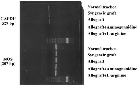
iNOS mRNA expression. Semiquantitative RT-PCR was
used to measure relative iNOS mRNA levels after normaliza-tion against levels of the reference gene GAPDH mRNA. In syngeneic grafts, induction of iNOS mRNA synthesis was seen at 3 d returning to the level of nontransplanted tracheas (Table III, Fig. 5). In nontreated allografts compared with syngeneic grafts, a significant induction of iNOS mRNA was observed at 10 d, coinciding with the peak inflammatory response. Ami-noguanidine treatment did not reduce iNOS mRNA levels but instead was associated with significant upregulation of iNOS mRNA at 30 d, indicating that the inhibition of NO production may inhibit negative feedback of NO to iNOS mRNA synthe-sis (6). Supplementation of iNOS pathway with L-arginine in-creased iNOS mRNA expression at all time points compared with nontreated allografts.
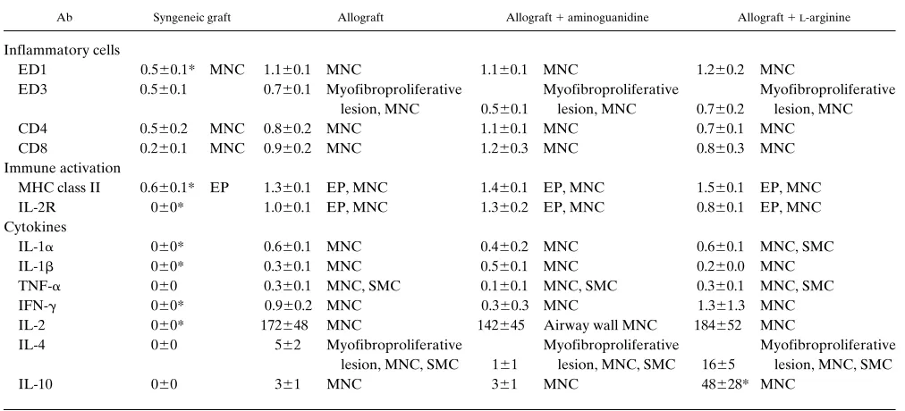
Airway wall inflammation and intragraft cytokine expres-sion of tracheal transplants. In nontreated allografts, a
promi-nent airway wall infiltration of ED11 and ED31 macrophages, CD41, and CD81 T cells was observed at 3 and 10 d after
transplantation, subsiding thereafter. Many of the infiltrating inflammatory cells showed immune activation with expression of IL-2R and MHC class II. Also, strong epithelial MHC class II expression was recorded. Aminoguanidine or L-arginine treatment did not alter the intensity of airway wall inflamma-tion or the immune activainflamma-tion of inflammatory cells. Allograft expression of proinflammatory cytokines, especially of TNF-a, IFN-g, IL-1, and IL-2 was upregulated compared to syngeneic grafts (Table IV, Fig. 6). Aminoguanidine treatment did not significantly alter allograft cytokine expression profiles. L -argi-nine treatment, however, was associated with upregulation of Th2 cytokines, IL-4, and IL-10 in the airway wall during peak inflammation at 10 d (Table IV, Fig. 6).
Discussion
Recent experimental observations in rodents demonstrate that heterotopic tracheal allografts develop myofibroproliferative lesions similar to those seen in small and large airways of hu-man lung allografts with OB (2–4, 25, 26). At the early phase of OB in heterotopic rat tracheal allografts, the respiratory epi-thelium strongly expresses MHC class II, enabling a direct pre-sentation of foreign antigens to alloreactive T cells (4). In addi-tion, graft-infiltrating recipient-derived professional APC such as dendritic cells and macrophages may process donor alloan-tigens for indirect T and B cell activation, leading to clonal cell proliferation and Ab production, respectively (4, 27). Recruit-ment of CD41 and CD81 T cells and macrophages into the airway wall induces local cytokine and growth factor produc-tion leading to epithelial damage, proliferaproduc-tion of a-smooth muscle actin immunoreactive cells, and gradual occlusion of the airway lumen. In syngeneic grafts and allografts immuno-suppressed with sufficient doses of cyclosporin A, obliterative changes do not develop, indicating the key role of alloimmune response in this disease process (2, 4, 28).
NO is reported to be an important mediator of inflamma-tory responses in lungs (11). Recently, induction of NO pro-ducing enzyme iNOS and production of NO during rejection were demonstrated. It has also been shown that treatment with aminoguanidine, a preferential inhibitor of iNOS, significantly attenuated the pathogenesis of acute cardiac and lung allograft rejection (15, 16, 18, 19). Supplementation of NO in cardiac al-lograft ischemia-reperfusion injury prolonged graft survival, which may be due to the protective role of NO to endothelial cells (29, 30).
Although induction of iNOS mRNA and protein expres-sion have been demonstrated in chronic rejection and allograft arteriosclerosis (31, 32), the biological role of NO in this
dis-Table III. Intragraft iNOS mRNA Expression
Days after
transplantation n
Normal DA trachea
Syngeneic
graft Allograft
Allograft 1 aminoguanidine
Allograft 1
L-arginine
0 6 0.0560.03
3 6 0.1660.03 0.2460.03 0.2760.03 0.4160.06*
10 6 0.0960.02* 0.3460.03 0.4160.02 0.5560.05*
30 6 0.0660.01 0.0560.02 0.2460.05* 0.2360.02*
[image:8.612.57.556.72.156.2]Relative iNOS gene expression levels were measured with semiquantitative RT-PCR after normalization against levels of reference gene GAPDH. Values are given as mean6SEM. *P , 0.05 when compared with nontreated allografts (Kruskall-Wallis and Dunn tests).
Figure 5. Example of RT-PCR of iNOS mRNA and GAPDH
[image:8.612.57.300.511.660.2]ease process remains unknown. In models of ordinary athero-sclerosis, inhibition of NOS has been shown to accelerate atherogenesis (33) and to increase cardiac fibrosis (34), whereas supplementation of NOS pathway with L-arginine reduced hy-percholesterolemia and endothelial denudation-induced ath-erosclerosis (20, 35). The same beneficial effect with L -argi-nine, however, was not achieved in a chronic rejection model of femoral artery allografts in the rat (36).
In this communication we demonstrate a protective regula-tory role of NO in OB development, i.e., chronic rejection. Supplementation of NO pathway by L-arginine resulted in sig-nificant reduction in the proliferation of a-smooth muscle actin immunoreactive cells, and thereby in downregulation of tra-cheal allograft occlusion. L-arginine treatment also reduced epithelial damage and necrosis. In contrast, inhibition of iNOS by aminoguanidine treatment significantly accelerated devel-opment of OB, particularly early after transplantation.
In normal tracheas and syngeneic grafts, iNOS was local-ized in the epithelium, suggesting that it may be the main source of NO. This result is consistent with an earlier report demon-strating continuous NO production by epithelial iNOS (10, 11). In nontreated allografts, an increase in the relative intra-graft iNOS mRNA and mononuclear inflammatory cell iNOS immunoreactivity was demonstrated during progressive loss of respiratory epithelium and airway occlusion. The intragraft NO production was, however, decreased, possibly due to al-loimmune damage to the epithelium, and thus decreased
epi-thelial iNOS. The aminoguanidine treatment–enhanced OB was not associated with reduction of relative iNOS mRNA synthe-sis, but was linked to upregulation of relative iNOS mRNA at the end-point, indicating that inhibition of NO production may inhibit negative feedback of NO to iNOS mRNA synthesis (6). Aminoguanidine did not affect total intragraft NO production, but decreased iNOS immunoreactivity in smooth muscle cells. In contrast, the antiproliferative effect of L-arginine supple-mentation was linked with enhanced relative intragraft iNOS mRNA synthesis, iNOS immunoreactivity in capillary endo-thelial and smooth muscle cells, and intragraft NO production.
L-arginine treatment, however, did not affect the number of macrophages, known to be an important source of iNOS (37).
Our findings suggest that one of the possible mechanisms of NO in OB prevention may be mediated by smooth muscle cell proliferation inhibition or enhanced revascularization of grafts by better-preserved capillary endothelium (30), indi-cated by increased iNOS expression in smooth muscle cells and capillary endothelium during L-arginine treatment. On the growth factor level of smooth muscle cell proliferation, NO in-hibits platelet-derived growth factor (PDGF) expression (38), which is a major mitogen for smooth muscle cells, and may have an essential role in OB development (3, 39, 40). NO also ap-pears to inhibit smooth muscle proliferation directly through elevation of cyclic-GMP levels (41, 42).
[image:9.612.55.561.73.303.2]Several mechanisms exist whereby NO may modulate im-mune responses in the graft. Supplementation of L-arginine has
Table IV. Inflammatory Cell Subsets, Immune Activation, and Cytokine Expression 10d after Transplantation
Ab Syngeneic graft Allograft Allograft 1 aminoguanidine Allograft 1L-arginine
Inflammatory cells
ED1 0.560.1* MNC 1.160.1 MNC 1.160.1 MNC 1.260.2 MNC
ED3 0.560.1 0.760.1 Myofibroproliferative Myofibroproliferative Myofibroproliferative lesion, MNC 0.560.1 lesion, MNC 0.760.2 lesion, MNC CD4 0.560.2 MNC 0.860.2 MNC 1.160.1 MNC 0.760.1 MNC CD8 0.260.1 MNC 0.960.2 MNC 1.260.3 MNC 0.860.3 MNC Immune activation
MHC class II 0.660.1* EP 1.360.1 EP, MNC 1.460.1 EP, MNC 1.560.1 EP, MNC IL-2R 060* 1.060.1 EP, MNC 1.360.2 EP, MNC 0.860.1 EP, MNC Cytokines
IL-1a 060* 0.660.1 MNC 0.460.2 MNC 0.660.1 MNC, SMC IL-1b 060* 0.360.1 MNC 0.560.1 MNC 0.260.0 MNC TNF-a 060 0.360.1 MNC, SMC 0.160.1 MNC, SMC 0.360.1 MNC, SMC IFN-g 060* 0.960.2 MNC 0.360.3 MNC 1.361.3 MNC IL-2 060* 172648 MNC 142645 Airway wall MNC 184652 MNC
IL-4 060 562 Myofibroproliferative Myofibroproliferative Myofibroproliferative lesion, MNC, SMC 161 lesion, MNC, SMC 1665 lesion, MNC, SMC
IL-10 060 361 MNC 361 MNC 48628* MNC
Graft-infiltrating inflammatory cell subsets, immune activation, and expression of inflammatory cytokines in the grafts. EC, capillary endothelial cells; EP, epithelial cells; MNC, mononuclear inflammatory cells; SMC, smooth muscle cells. For IL-2, IL-4 and IL-10 the number of positive cells were scored per cross-section. For others, the intensity of staining was scored from 0 to 3. Values are mean6SEM. *P , 0.05, when compared with non-treated allografts (Kruskal-Wallis and Dunn tests).
been shown to reduce platelet aggregation (43, 44) and leuko-cyte adhesion and emigration (45). In this study, supplementa-tion of NO pathway or inhibisupplementa-tion of iNOS pathway had no ef-fect on the number of graft-infiltrating CD41 or CD81 T cells or macrophages, or on the level of immune activation with ex-pression of IL-2R or MHC class II. Supplementation of NO pathway by L-arginine, however, modulated the immune re-sponse towards Th2 cytokine IL-4 and IL-10 production, which has been linked with prolonged graft survival and tolerance in-duction (46). NO has been shown to inhibit the expansion of cloned Th1, but not Th2 cells (47). Furthermore, observations in iNOS-deficient mice demonstrate that a low concentration of endogenously generated NO by cNOS may be required for Th1 cell proliferation, but a high concentration of NO pro-duced by iNOS is necessary for prevention of potential overex-pansion of Th1 cells, which is implicated in a variety of autoim-mune diseases (48).
In conclusion, we demonstrate that NO has a protective role in the OB development: while NO pathway supplementa-tion by L-arginine attenuated obliterative changes, preferential inhibitor of iNOS pathway enhances these changes. Our re-sults indicate furthermore that potential mechanisms of NO in the prevention of OB include either inhibition of smooth mus-cle cell proliferation or altered immune response towards Th2 cytokines.
Acknowledgments
The authors wish to acknowledge the excellent technical assistance of E. Aaltola, R.N., M. Anttila, R.N., and E. Wasenius, R.N.
This study was supported by the Jalmari and Rauha Ahokas Foundation, the Finnish Medical Society Duodecim, the Helsinki University Central Hospital Research Funds, Helsinki, the Leiras Re-search Foundation, and the Farmos Pharmaceuticals, Turku, Finland.
References
1. Lemström, K., P. Koskinen, and P. Häyry. 1995. Molecular mechanisms of chronic renal allograft rejection. Kidney. Int. 48:S2–S10.
2. Morris, R.E., X. Huang, C.R. Gregory, M.E. Billingham, R. Rowan, R. Shorthouse, and G.J. Berry. 1995. Studies in experimental models of chronic re-jection: use of rapamycin (sirolimus) and isoxazole derivates (Leflunomide and its analogue) for suppression of graft vascular disease and obliterative bronchi-olitis. Transplant. Proc. 27:2068–2069.
3. Koskinen, P.K., E.A. Kallio, C.A. Bruggeman, and K.B. Lemström. 1997. Cytomegalovirus infection enhances experimental obliterative bronchiolitis in rat tracheal allografts. Am. J. Respir. Crit. Care Med. 155:2078–2088.
4. Koskinen, P.K., E.A. Kallio, R. Krebs, and K.B. Lemström. 1997. A dose-dependent inhibitory effect of cyclosporine A on obliterative bronchiolitis of rat tracheal allografts. Am. J. Respir. Crit. Care Med. 155:303–312.
5. Yousem, S.A., J.B. Berry, P.T. Cagle, D. Chamberlain, A.N. Husain, H.R. Hruban, A. Marchevsky, N.P. Ohori, J. Ritter, S. Stewart, and H.D. Taze-laar. 1996. Revision of the 1990 working formulation for the classification of pulmonary allograft rejection: lung rejection study group. J. Heart Lung
Trans-plant. 15:1–15.
6. Moncada, S., and A. Higgs. 1993. The L-arginine-nitric oxide pathway. N.
Engl. J. Med. 329:2002–2012.
7. Sessa, W.C. 1994. The nitric oxide synthase family of proteins. J. Vasc.
Res. 31:131–143.
8. Nathan, C. 1992. Nitric oxide as a secretory product of mammalian cells.
FASEB (Fed. Am. Soc. Exp. Biol.) J. 6:3051–3064.
9. Moncada, S., and E.A. Higgs. 1995. Molecular mechanisms and therapeu-tic strategies related to nitric oxide. FASEB (Fed. Am. Soc. Exp. Biol.) J. 9: 1319–1330.
10. Guo, F.H., H.R. De Raeve, T.W. Rice, D.J. Stuehr, F.B.J.M. Thunnis-sen, and S.C. Erzurum. 1995. Continuous nitric oxide synthesis by inducible ni-tric oxide synthase in normal human airway epithelium in vivo. Proc. Natl.
Acad. Sci. USA. 92:7809–7813.
11. Barnes, P.J. 1995. Nitric oxide and airway disease. Ann. Med. 27:389–393. 12. Yan, Z.-Q., G.K. Hansson, B.-E. Skoogh, and J.O. Lötvall. 1995.
Induc-tion of nitric oxide synthase in a model of allergic occupaInduc-tional asthma. Allergy
(cph). 50:760–764.
13. Tsuji, M., V.B. Dimov, and T. Yoshida. 1995. In vivo expression of monokine and inducible nitric oxide synthase in experimentally induced pulmo-nary granulomatous inflammation. Am. J. Pathol. 147:1001–1015.
14. Hamid, Q., D.R. Springall, V. Riveros-Moreno, P. Chanez, P. Howarth, A. Redington, J. Bousquet, P. Godard, S. Holgate, and J.M. Polak. 1993. Induc-tion of nitric oxide synthase in asthma. Lancet. 342:1510–1513.
15. Yang, X., N. Chowdhury, B. Cai, J. Brett, C. Marboe, R.R. Sciacca, R.E. Michler, and P.J. Cannon. 1994. Induction of myocardial nitric oxide synthase by cardiac allograft rejection. J. Clin. Invest. 94:714–721.
16. Langrehr, J.M., N. Murase, P.M. Markus, X. Cai, P. Neuhaus, W. Schraut, R.L. Simmons, and R.A. Hoffman. 1992. Nitric oxide production in host-versus-graft and graft-versus-host reactions in the rat. J. Clin. Invest. 90: 679–683.
17. Misko, T.P., W.M. Moore, T.P. Kasten, G.A. Nickols, J.A. Corbett, R.G. Tilton, M.L. McDaniel, J.R. Williamson, and M.G. Currie. 1993. Selective inhibition of the inducible nitric oxide synthase by aminoguanidine. Eur. J.
Pharmacol. 233:119–125.
18. Worrall, N.K., W.D. Lazenby, T.P. Misko, T.-S. Lin, C.P. Rodi, P.T. Manning, R.G. Tilton, J.R. Williamson, and T.B. Ferguson, Jr. 1995. Modula-tion of in vivo alloreactivity by inhibiModula-tion of inducible nitric oxide synthase. J.
Exp. Med. 181:63–70.
19. Shiraisi, T., S.R. DeMeester, N.K. Worrall, J.H. Ritter, T.P. Misko, T.B. Ferguson, Jr., J.D. Cooper, and G.A. Patterson. 1995. Inhibition of inducible ni-tric oxide synthase ameliorates rat lung allograft rejection. J. Thorac.
Cardio-vasc. Surg. 110:1449–1460.
20. Cooke, J.P., A.H. Singer, P. Tsao, P. Zera, R.A. Rowan, and M.E. Bil-lingham. 1992. Antiatherogenic effect of L-arginine in the hypercholester-olemic rabbit. J. Clin. Invest. 90:1168–1172.
21. Lemström, K.B., P.T. Aho, C.A. Bruggeman, and P.J. Häyry. 1994. Cy-tomegalovirus infection enhances mRNA expression of platelet-derived growth factor-BB and transforming growth factor-b1 in rat aortic allografts. Possible mechanism for cytomegalovirus-enhanced graft arteriosclerosis. Arterioscler.
Thromb. 14:2043–2052.
22. Chomczynski, P., and N. Sacchi. 1987. Single-step method of RNA isola-tion by acid guanidinum thiocyanate-phenol-chloroform extracisola-tion. Anal.
Bio-chem. 162:156–159.
23. Moshage, H., B. Kok, J.R. Huizenga, and P.L.M. Jansen. 1995. Nitrite and nitrate determinations in plasma: a critical evaluation. Clin. Chem. 41:892–896. 24. Schmidt, H.H.H.W., T.D. Warner, M. Nakane, U. Förstermann, and F. Murad. 1992. Regulation and subcellular location of nitric oxide synthases in RAW264.7 macrophages. Mol. Pharmacol. 41:615–624.
25. Hertz, M.I., J. Jessurun, M.B. King, S.K. Savik, and J.J. Murray. 1993. Reproduction of the obliterative bronchiolitis after heterotopic transplantation of mouse airways. Am. J. Pathol. 142:1945–1951.
26. Reichenspurner, H., X. Huang, V. Soni, R. Shorthouse, G. Berry, and R.E. Morris. 1995. Pathogenesis and treatment of obliterative airway disease af-ter heaf-terotopic tracheal allograft and xenograft transplantation. Surg. Forum. 156:456–458.
27. Uyama, T., J.B. Winter, G. Groen, C.R.H. Wildevuur, Y. Monden, and J. Prop. 1992. Late airway changes caused by chronic rejection in rat lung allo-grafts. Transplantation (Baltimore). 54:809–812.
28. King, M.B., J. Jessurun, S.K. Savik, J.J. Murray, and M.I. Hertz. 1997. Cyclosporine reduces development of obliterative bronchiolitis in a murine het-erotopic airway model. Transplantation (Baltimore). 63:528–532.
29. Pinsky, D.J., M.C. Oz, S. Koga, Z. Taha, M.J. Broekman, A.J. Marcus, H. Liao, Y. Naka, J. Brett, P.J. Cannon, et al. 1994. Cardiac preservation is en-hanced in a heterotopic rat transplant model by supplementing the nitric oxide pathway. J. Clin. Invest. 93:2291–2297.
30. Cooke, J.P., and P.S. Tsao. 1993. Cytoprotective effects of nitric oxide.
Circulation. 88:2451–2454.
31. Russel, M.E., A.F. Wallace, L.R. Wyner, J.B. Newell, and M.J. Kar-novsky. 1995. Upregulation and modulation of inducible nitric oxide synthetase in rat cardiac allografts with chronic rejection and transplant arteriosclerosis.
Circulation. 92:457–464.
32. Akyürek, L.M., B.C. Fellström, Z.-q. Yan, G.K. Hansson, K. Funa, and E. Larsson. 1996. Inducible and endothelial nitric oxide synthase expression during development of transplant arteriosclerosis in rat aortic grafts. Am. J.
Pathol. 149:1981–1990.
33. Naruse, K., K. Shimizu, M. Marumatso, Y. Toki, Y. Miyazaki, K. Oku-mura, H. Hashimoto, and T. Ito. 1994. Long-term inhibition of NO synthesis promotes artherosclerosis in the hypercholesterolemic rabbit aorta.
Arterio-scler. Thromb. 14:746–752.
34. Hou, J.H., H. Kato, R.A. Cohen, A.V. Chobanian, and P. Brecher. 1995. Angitensin II-induced cardiac fibrosis in the rat increased by chronic inhibition of nitric oxide synthase. J. Clin. Invest. 96:2469–2477.
35. McNamara, D.B., B. Bedi, H. Aurora, L. Tena, L.J. Ignarro, P.J. Kad-owitz, and D.L. Akers. 1993. L-arginine inhibits balloon catheter-induced inti-mal hyperplasia. Biochem. Biophys. Res. Commun. 193:291–296.
admin-istration of L-arginine does not inhibit arterial neointimal formation after over-whelming alloimmune injury. J. Heart. Lung Transplant. 15:58–66.
37. Langrehr, J.M., D.A. White, R.A. Hoffman, and R.L. Simmons. 1993. Macrophages produce nitric oxide at allograft sites. Ann. Surg. 218:159–166.
38. Kourembanas, S., L.P. McQuillan, G.K. Leung, and D.V. Faller. 1993. Nitric oxide regulates expression of vasoconstrictors and growth factors by vas-cular endothelium under both normoxia and hypoxia. J. Clin. Invest. 92:99–104. 39. Hertz, M.I., C.A. Henke, R.E. Nakhleh, K.R. Harmon, W.A. Marinelli, J.M.K. Fox, S.H. Kubo, S.J. Shumway, R.M. Bolman, III, and P.B. Bitterman. 1992. Obliterative bronchiolitis after lung transplantation: a fibroproliferative disorder associated with platelet derived growth factor. Proc. Natl. Acad. Sci.
USA. 89:10385–10389.
40. Al-Dossari, G.A., J. Jessurun, R. Morton Bolman, III, R.K. Vibhu, M.B. King, J.J. Murray, and M.I. Hertz. 1995. Pathogenesis of obliterative bronchioli-tis: Possible role of platelet derived growth factor and basic fibroblast growth factor. Transplantation (Baltimore). 59:143–145.
41. Kariya, K.-i., Y. Kawahara, S.-i. Araki, H. Fukuzaki, and Y. Takai. 1989. Antiproliferative action of cyclic GMP-elevating vasodilators in cultured rabbit smooth muscle cells. Atherosclerosis. 80:143–147.
42. Garg, U.C., and A. Hassid. 1989. Nitric oxide-generating vasodilators and 8-bromo-cyclic guanosine monophosphate inhibit mitogenesis and
prolifer-ation of cultured rat vascular smooth muscle cells. J. Clin. Invest. 83:1774–1777. 43. Radomski, M.W., R.M.J. Palmer, and S. Moncada. 1990. An L-arginine/ nitric oxide pathway present in human platelets regulates aggregation. Proc.
Natl. Acad. Sci. USA. 87:5193–5197.
44. Murohara, T., S.J. Parkinson, S.A. Waldman, and A.M. Lefer. 1995. In-hibition of nitric oxide biosynthesis promotes P-selectin expression in platelets.
Arterioscler. Thromb. Vasc. Biol. 15:2068–2075.
45. Kubes, P., M. Suzuki, and D.N. Granger. 1991. Nitric oxide: An endoge-nous modulator of leukocyte adhesion. Proc. Natl. Acad. Sci. USA. 88:4651– 4655.
46. Sayegh, M.H., E. Akalin, W.W. Hancock, M.E. Russel, C.B. Carpenter, P.S. Linsley, and L.A. Turka. 1995. CD28-B7 Blockade after alloantigenic chal-lenge in vivo inhibits Th1 cytokines but spares Th2. J. Exp. Med. 181:1869–1874. 47. Taylor-Robinson, A.W., F.Y. Liew, A. Severn, D. Xu, S.J. McSorley, P. Garside, J. Padron, and R.S. Phillips. 1994. Regulation of the immune response by nitric oxide differentially produced by T helper type 1 and T helper type 2 cells. Eur. J. Immunol. 24:980–984.