Journal of Chemical and Pharmaceutical Research, 2014, 6(5):96-103
Research Article
CODEN(USA) : JCPRC5
ISSN : 0975-7384
Experimental study on promoting the regeneration of rat sciatic
nerve using slow-release FK506-eluting peripheral nerve stent
Tan Ding
1*, Hu-ping Hao
2*, Chao Zhu, Jun-Jie Du and Zhuo-Jing Luo
#Institute of Orthopedics, Xijing Hospital, The Fourth Military Medical University, Xi’an, PR China
_____________________________________________________________________________________________ABSTRACT
Design and construction of biocompatible tissue-engineered stents for long segment peripheral nerve regeneration has become a significant research topic. However, in the early of damaged, the immigration and aggregation of inflammatory cells and factors can induce a secondary nerve injury and peroxide damage to the injured nerves, which in turn can result in severe neuronal apoptosis. The use of immunosuppressant in the injury site can reduce the inflammatory reactions and secondary injury, and build a conducive microenvironment for nerve regeneration. In this study, we prepared tissue-engineering peripheral nerve stent using FK506, where the release of fk506 into the periphery after microsphere degradation still needs to pass through the stent wall. Such double effect of slow-release significantly reduced the burst release and prolonged the full release to 17 days. In our rat model of repairing a 20 mm sciatic nerve defect, 3 months after the surgery, the SFI and electrophysiological testing results of the ipsilateral side in group 1(FK506+PLGA+Stent) showed no significant difference from those in the autologous nerve grafting group. Morphological results also indicated that in terms of either the number of regenerated axons or the degree of myelin maturity, the regeneration outcome in group 1 was close to that in autologous nerve grafting group and significantly superior over that in the group 2(Only Stent). The slow-release FK506-eluting stent introduced in this study is easy for clinical application and provides a new experimental evidence of promoting peripheral nerve regeneration with tissue-engineering methods.
Keywords: Peripheral nerve; Tissue engineering; Slow-releasing; Stent; FK506
_____________________________________________________________________________________________
INTRODUCTION
The repair and replacement of injured peripheral nerve segments have been major issues in clinical practice. In recent years, the use of tissue engineering in constructing peripheral nerve stents has made great progress [1-3]. The tissue engineered artificial nerve graft is mostly made of an extracellular matrix that has good cell affinity and a basic structure of a hollow non-uniform axial multi-channel catheter. At present, collagen is the most ideal material for the extracellular matrix. A collagen made single-channel nerve conduit produced by Integra and Synovis is commercially available and has been used, clinically.
However, when peripheral nerve injury and fracture occur, the truncated nerve surface and damaged neurons are exposed to the environment of body fluids, facing a severe local inflammatory response and the resulted infiltration of inflammatory cells. These inflammatory cells may easily pass the porous structure of the wall of tissue-engineered stent and enter into the interior microtubules; during their cleaning of myelin debris and necrotic cells, these cells also cause secondary inflammatory injury and peroxide damage to the axons, hampering the nerve regeneration[4-6]. The subsequent proliferation of connective tissue and scar tissue formation may further cause traumatic neuroma and impede the nerve regeneration.
FK506 also chemically stable with high solubility in organic solvents, and its pharmacological effects as an immunosuppressant are not altered by encapsulating in a microsphere as slow-releasing agent. In this study, FK506-PLGA microspheres were prepared and loaded onto the stent, which was then implanted into the experimental animal at the early phase of the peripheral nerve injury. The result of local slow-release of FK506 in reducing the secondary nerve injury was investigated.
EXPERIMENTAL SECTION
Reagents, animals, and instruments
PLGA (RG503H, LA/GA=50:50, Mw:13000-23000) was from Boehringer Ingelheim (Ingelheim, Germany); type I collagen (C9879) and gelatin (G9382) were from Sigma-Aldrich (St. Louis, MO, USA); geniposide was the product of Challenge Bioproducts (Taiwan, China), Mek-5216k Hematology Analyzer was from Nihon Kohden (Tokyo, Japan); ACUSON Cypress digital echocardiography system was from Siemens (Harrisburg, PA, USA); overhead blender RW20 was the product of IKA Works (Wilmington, NC, USA); alpha2-4 freeze dryer was from Martin Chirst (Osterode, Germany); laser scanning confocal microscope was the product of Leica (Wetzlar, Germany); scanning electron microscope (SEM) was from Hitachi (Tokyo, Japan); Viking IV electrophysiological apparatus was the product of Nicdlet (Madison, WI, USA); chromatography column was from Waters Corporation (Milford, MA, USA); and laser particle size analyzer was the product of Beckman (Pasadena, CA, USA). A total of 36 male SD rats, weighing 220±20 g, were provided by the Experimental Animal Center of Fourth Military Medical University, Xi'an, China.
Preparation of slow-releasing FK506-PLGA microspheres
PLGA(Poly(lactic-co-glycolic acid),PLGA)(4 g) was dissolved in 60 mL of dichloromethane, added with 500 mg of FK506 at a ratio of 1:10 (w/w), and mixed thoroughly by shaking. The oil phase of above mixture was added dropwise into 0.5% polyvinyl alcohol (PVA) solution to obtain homogenized O/W emulsion under the action of a high pressure homogenizer (3,500 rpm, 8 min); the emulsion was stirred continuously at room temperature (300 rpm, 5 h) to remove the volatile organic solvent, and was further centrifuged at 3,000 rpm for 5 min to collect the microspheres, which were then washed 3 times and finally freeze-dried to obtain the FK506-PLGA microsphere powder.
Determination of the morphology and the encapsulation efficiency of FK506-PLGA microspheres
Appropriate amount of PLGA microspheres was fixed onto a clean dry silicon wafer and coated with gold under vacuum condition. The morphology and surface features of PLGA microspheres were observed under a current of 10 mA and an accelerating voltage of 5 kV. The size distribution of microspheres was measured using laser particle size analyzer. The encapsulation efficiency of FK506 in the microspheres was determined using high performance liquid chromatography (HPLC). Briefly, 10 mg microspheres were dissolved in 1 mL of acetonitrile, sonicated for 10 min to dissolve, and centrifuged at 3,000 rpm for 15 min; the supernatant was collected and detected for drug content on Waters 2695 HPLC system under the following conditions: Waters symmetry C18 column (150 × 4.6 mm, 5 µm), isocratic elution, mobile phase (10 mM ammonium acetate-0.1% formic acid aqueous solution : 0.1% formic acid methanol solution [10:90]), flow rate 1 mL/min, injection volume 20 µL, and detection wavelength 210 nm.
Encapsulation efficiency (%) = (La/Lt) × 100 (La: actual drug content in microspheres; Lt: input content of drug.
Drug loading rate % = (Pt/Mt) × 100 (Pt: drug weight in microspheres; Mt: weight of microspheres.
Preparation of FK506-PLGA stent
Separately, chitosan (130 mg) and collagen I (340 mg) were weighed on an electronic scale, placed into 20 mL beaker, added with 10 mL of 3-time-distilled water and 20 µL of 4 % acetic acid (pH 3.2), sealed, and placed in a 4 ℃ refrigerator to dissolve for 24 h. The above 2 solutions were then stir-mixed with homogenizer in ice-water bath
stirred at 5,000 rpm for 60 min. The thoroughly-mixed solution was then divided evenly into 3 parts. According to the predetermined microsphere drug loading rate, in group 1, 35 mg FK506-PLGA microspheres were added into the mixed solution, to obtain a final FK506 content of 4 mg; and in group 2, no ingredients were added. After slightly stirring, the mixture was quickly injected into a silicone tube (3 mm in diameter and 20 mm in length), both ends of which were occluded with a lead wire. The variously-treated silicone tubes were slowly placed into liquid nitrogen along the axial direction using a homemade micro-speed adjusting instrument at a speed of 2×10⁻⁵ m/s. After fully submerged, the mold was incubated in liquid nitrogen for 4 h, then removed, and placed onto a prechilled aluminum pan, which was then transferred to a vacuum dryer to freeze-dry the tubes under vacuum condition for 8 h to obtain the scaffold of nerve stent, which was then crosslinked in 1% genipin solution at 37 ℃ for 24 h.
[10,11] The obtained
The drug-release from RAPA-PLGA stent in vitro
The stents from groups 1 were dissolved in 5 mL of phosphate buffered saline (PBS, pH 7.4), respectively, at 37 °C under continuous shaking (50 rpm). At 1-10 d, 1 mL of medium was sampled daily, and the lost volume was supplemented with equal amount of PBS. The solutions sampled at different time points were added with 1 mL of dichloromethane respectively, mixed for 1 min by vortex, and centrifuged at 3,000 rpm for 10 min. The organic phase was collected and evaporated at 40 °C under nitrogen. The dried residues of samples collected at different time points were redissolved in methanol and analyzed for free FK506 concentration on HPLC. The curves of in vitro drug-release were plotted.
Nerve bridging surgery in experimental animals
Male Sprague–Dawley rats (n = 36, body weight 220 ± 20 g) were randomly divided into three groups: receiving stent with FK506 and PLGA coatings, receiving stent without coating and receiving autologous graft. Each rat was anesthetized with intraperitoneal injections of 3% sodium pentobarbital. With an incision of the skin and fascia in the left femoral region, the sciatic nerve was separated by blunt dissection, and a 20 mm portion was cut off. The stent was aligned with the near and distal stumps of the severed sciatic nerve and the surface of the scaffolds was sutured to the epineurium using microsurgery technology. In the autologous graft group, the cut piece of 20mm sci-atic nerve was reverse sutured. After surgery, the wound was closed and the animal was allowed to recover. All animal handling and procedures met Animal Care and Use Committee guidelines. The procedures were approved by the Experimental Animal Administration Committee of The Fourth Military Medical University, which determined that the experiment appropriately minimized the number of animals used and limited their suffering.
Leukocyte collecting and counting
At 1 day before surgery and 60 days post-surgery, under the guidance of ultrasound, a micro-injector was placed to draw 20 µL body fluid (without blood) from the area of damaged sciatic nerve every other day, and the number of blood leukocytes was counted using the Mek-5216k Hematology Analyzer, until the number was stabilized at the level of the operation day for 3 consecutive days.
Test of the sciatic nerve function
Footprint analysis was performed on all rats at days 15, 30 and 40 after surgery and the Sciatic Functional Index (SFI) was used to evaluate recovery of hind limb function. Preoperatively, the rats were trained to walk down a wooden track (50 × 7 cm) into a darkened goal box. Postoperatively, the rats’ hind paws were painted with nontoxic finger paint, and any changes in their paw prints that resulted from nerve injury and denervation were recorded. The recordings continued until five measurable footprints were collected. The SFI were input into the formula of Bain [12] as follows:
SFI = 109.5(ETS-NTS)/NTS-38.3(EPL-NPL)/NPL+13.3(EIT-NIT) /NIT-8.8. SFI = 0 was considered normal, and SFI = -100 was full damage.
Electrophysiological testing of sciatic nerve
Eleven weeks after the surgery, Sciatic nerves were exposed after the above-mentioned surgery from both legs and subjected to electrophysiological examination with an electric physiological instrument in vivo. The nerve potential amplitude (AMP) and nerve conduction velocity(NCV) were detected.
Nerve morphology
Twelve weeks after the surgery, animals from each group were fixed by formalin perfusion. The bridging body with 2 mm of nerve tissues in both ends was collected, fixed in 0.1 M cacodylate buffer (pH 7.3) containing 1% osmium tetroxide for 1 h at room temperature, dehydrated, and embedded in resin. Semi-thin (1.0 µm) and ultrathin (50 nm) sections of the mid bridging body were prepared. The semi-thin sections were stained with 1% toluidine blue/1% borax solution and observed under an optical microscope. The ultrathin sections were stained with uranyl acetate and lead citrate, and observed by transmission electron microscopy (TEM). The status of axon regeneration in each group was evaluated through the mean diameter of the nerve fibers and the mean thickness of the myelin sheath.
Histological examinations
The regenerated nerve tissues were dissected 90 days after the surgery and cut into ultra-thin sections for toluidine blue staining and observations under a transmission electron microscope (TEM) by the standard protocols. They were then observed under a light microscope or by SEM for the density of axons and the thickness of the myelin.
Statistical analysis
significance.
RESULTS
Characterization of the RAPA-PLGA microspheres
[image:4.595.79.538.186.344.2]The lyophilized powder of slow-releasing FK506-PLGA microspheres appeared as round and smooth spheres under light microscope (Fig.1A). The average particle size of drug-loaded microspheres was 111.6 ±6.1nm, and the sizes distributed normally (Fig.1B). The calculated drug-loading rate and the encapsulation efficiency of PLGA microspheres were 8.43% and 81.04% respectively.
Fig 1: The FK506-PLGA microspheres appeared as round and smooth spheres under microscope (A). The average particle size of microspheres was 111.6±6.1 nm, and the sizes distributed normally (B). Scale bars represent 200 nm
Highly-bionic peripheral nerve stent
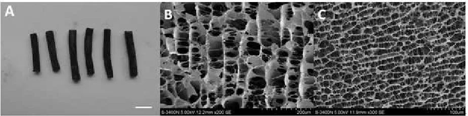
As described in the earlier section, collagen stents containing PLGA and FK506 were constructed by freeze-drying. Under SEM, a newly constructed scaffold was in a cylindrical shape (Fig. 2A). Its cross-section showed microtubules of uniform diameters (Fig.2B), and the longitudinal section displayed the parallel arrayed interstructure of microtubules (Fig. 2C). Since the diameters of these microtubules were about 20-80 mm, similar to that of the sciatic nerve, they may provide the passage for the nerve signal transduction that is essential for the function of nerve fibers.
Fig 2: The stent displayed a bluish-black cylindrical structure (A). Under SEM, the transversal section displayed a honeycomb-like structure with relatively-uniform diameter (B). Longitudinal sections displayed axially-oriented microtubules (C). Scale bars represent 1
cm
The in vitro release properties of the FK506-PLGA microsphere stent
As shown in Fig. 3, the burst release of FK506-PLGA microspheres was evident, i.e., approximately 40% FK506 were released within the initial 24 h, and the FK506 was completely released within 6 days. However, the double slow-release effects of PLGA microsphere and stent greatly attenuated the burst release of FK506 in group 1, in which less than 13 % of the free drug were released in the initial 24 h, displaying a gentle release curve, and the drug release lasted for 17 days, providing a sustained local drug release in the early phase of nerve injury.
Leukocyte counting
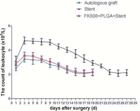
[image:4.595.76.539.473.588.2]increase. From 11th day after the surgery, the number of leukocytes started to decline, and after 23 days, the number returned to the preoperative level. For group 1 (FK506+PLGA+stent) and the control (autologous nerve grafting) group, the leukocyte number also peaked at day 3, which was 110% higher than that before the surgery; the number started to decline from the 7th day, and returned to the preoperative level at day 15.
Fig 3: The microspheres in group 1 (FK506-PLGA) was completely released till the 6th day. The double slow-release effects of PLGA microsphere and stent displayed a gentle release curve, and the drug release lasted 17 days
Fig 4: Postoperative peripheral leukocyte counts showed that there were various degrees of increase in peripheral leukocyte counts after the surgery. The group 1 (FK506+PLGA+stent) and autologous graft group showed the lowest peak leukocyte count and the fastest
recovery, followed by the group 2 (Stent)
The functional outcome of sciatic nerve
The sciatic nerve was damaged completely in each group immediately after the bridging surgery, and almost all the SFI values were nearly -100 and without significant differences among groups (Fig. 5). One month later, there were varying degrees of neural recovery in each group, and the recovery of SFI in autologous nerve grafting group and group 1 was relatively quicker, compared to the slow recovery in the group 2. Three months after the surgery, the group 1 and autologous nerve grafting group almost fully recovered, showing no significant difference, whereas the recovery of the group 2 was relatively slower.
The results of electrophysiological test
[image:5.595.190.426.365.552.2]Fig 5: Almost all the SFI values were nearly -100 and without significant differences among groups after the bridging surgery. From 30 and 90 d, the experimental group 1 showed no difference compared with autologous graft group, and both were significantly better than
the other 2 groups. All data were expressed as the mean±standard error of mean. *p<0.05, #p>0.05
[image:6.595.77.535.379.662.2]Electrophysiolgic examination (Values are expressed as mean±standard error,n=36)
Table 1: The NCV and AMP in the autologous graft group were similar to that of the normal nerve. The experimental group 1 was very closely related with autologous graft group, and there was no significant difference between both groups. Both NCV and AMP in the other 2 groups showed difference compared with autologous graft groups. All data were expressed as the mean±standard error of mean.
*p<0.05, #p>0.05
Group AMP(mv) NCV (m/s)
Autologous Graft 16.08±1.37 25.67±1.32 FK506+PLGA+Stent 14.21±2.01 * 15.32±0.92* Stent 10.56±1.89# 11.32±1.23#
Fig 6: In the autologous graft group, regenerative nerve fibers were dense with uniform morphology (A), and myelin sheaths were thick and nerve fibers were orderly aligned (D). The new fibers of experimental group 1 were moderately decreased (B), but the morphology was relatively irregular compared with the autologous graft group (E). And the nerve fibers of the group 2 were significantly less (C )
and thin (F) than the autologous graft group. Scale bars: A-C=10µm, D-F=2µm
The morphology of regenerated nerve
nerve grafting group had the thickest myelin and high degree of nerve maturity (Fig. 6D), followed by group 1 (Fig. 6E); although the myelin in groups 2 displayed an integrated structure and shape, it was thinner and with low maturity(Fig. 6F).
DISCUSSION
Peripheral nerve injury is often caused by open trauma and thus has severe wound contamination. Extensive and thorough wound cleaning should be carried out, followed by continuous drainage, and any medical equipment that can easily cause contamination spreading or bacterial parasitism should not be placed into the wound area. In this case, conventional tissue engineering peripheral nerve scaffolds cannot be used and subsequently early nerve regeneration cannot occur. Moreover, the contamination caused immigration and aggregation of inflammatory cells such as leukocytes can induce a secondary inflammation and peroxide damage to the injured nerves, which in turn can result in severe neuronal apoptosis. As a result, some axonal terminals can form neuromas in the early neuronal injury stage, which subsequently seriously affects nerve regeneration. In addition, the immigration of a large number of inflammatory cells into the internal microtubules of the stent can block the path of growth cones and subsequently baffle nerve regeneration.The severity of the immune response is associated with the degree of injury, the stronger the immune response, the severer the secondary injury, and the poorer the outcome of nerve regeneration.
FK506 is a hydrophobic macrocyclic-lactone isolated from a fermentation broth of Streptomyces tsukubaensis from Mount Tsukuba in northern Japan. Recently, the use of FK506 is of special interest in nerve regeneration, because it may be effective in the treatment of immune-mediated diseases[13,14]. Erlich[15] established a mouse model of acute closed head injury, their immunohistochemistry study indicated that the neuronal survival in treatment group was significantly increased compared with control group; they also indicated that the mechanism of immunosuppressant effect may be that it reduces local inflammation, enhances autophagy, and avoids the accumulation of harmful substances inside the cells, which promote neuronal survival and decrease neuronal apoptosis. Chen[16] reached the same conclusion with a rat model of spinal cord injury, immunosuppressant improved motor function through its autophagy-promoting, anti-inflammatory, and neuroprotective effects. Nitric oxide (NO) may exert antibacterial, anti-atherosclerosis, and other effects at µM level; however, under ischemic stroke, injury, or other abnormal conditions, glial cells and macrophages produce excessive NO, causing nerve damage.
PLGA is a lactic acid and glycolic acid polymer, with good biocompatibility and biodegradability in vivo; it's end degradation products are H2O and CO2. PLGA has been approved by FDA as biodegradable polymer for clinical
trials. Using PLGA microspheres to encapsulate a drug can protect the drug from degradation, postpone the drug release time, prolong the duration of drug action, and reduce the drug's toxicity and irritation.[17,18] In the present study, PLGA microspheres and stent ensured a double slow-release of FK506 that lasted 11 days after the surgery in the region of nerve damage (Fig. 3), mitigated the local aggregation of blood leucocytes (Fig. 4), and reduced the secondary injury to the truncated nerve ends. Our results showed that the SFI in group 1 (with slow-release of FK506) was recovered to a similar degree as that in the autograft group (Fig. 5B), a gold standard of peripheral nerve regeneration; 3 months after the surgery, group 1 showed no significant difference from the autograft group (Fig. 5D). Meanwhile, similar results were also observed in electrophysiological test, the NCV and potential amplitude showed no significant difference between group 1 and autologous nerve grafting group (Table 1). These results further confirmed that a local release of FK506 can reduce the secondary injury to nerves, providing a conducive microenvironment for the regenerating axons to cross the defective segment.
CONCLUSION
The FK506-PLGA microsphere-loaded bionic peripheral nerve stent employed in this study slowly released FK506 locally in the early phase of sciatic nerve regeneration, significantly reduced the secondary nerve injury, and evidently promoted the peripheral nerve regeneration.
Acknowledgements
This work was supported by grants from the National Natural Science Foundation of China (No. 81100900) and Shaanxi Provincial Program for Science and Technology Development (No. 2011K12-59). The authors have no other relevant affiliations or financial involvement with any organization or entity with a financial interest in or financial conflict with the subject matter or materials discussed in the manuscript apart from those disclosed. No writing assistance was utilized in the production of this manuscript.
REFERENCES
[2] Ding T, Luo ZJ, Zheng Y, et al. Injury.,2010,41:522-7.
[3] Ding T, Lu WW, Zheng Y, et al. Regen Med., 2011,6 (4), 437-447. [4] Bucky J, Ronald H, Philip G, et al. Neursci., 2005,25 (28), 6576-83. [5] Tonai T, Shiba K, Taketani Y, et al. J Neuroehem., 2001,78 (5),1064-72. [6] Tian DS, Xie MJ, Yu ZY, et al. Brain Res., 2007,1135 (1),177-85. [7] Ruben LV, Joaquim F, Xavier N, et al. Neurobio Dis.,2006,24 (3),443-54. [8] Kaori K, Kenji H, Goro F, et al. Neuroph armacology.,2005,48 (3),391-7. [9] Bruce GG, Heidi SG, Wang MS, et al. Neurosci Let.,1999,267 ( 1 ),33 -6 . [10] Sung HW, Huang RN, Huang LL. J. Biomater Sci Polym Ed.,1999,10, 63-78. [11] Huang RN, Sung HW, Tsai CC et al. J. Biomed Mater Res.,1998, 42, 568-576. [12] Bain JR, Mackinnon SE, Hunter DA et al. Plast Reconstr Surg.,1989, 83 (1), 129-38. [13] Esther U, Enrique V, Xavier N. Neuroscience Let., 2004,357 (2),99-102.
[14] Esther U, Dolores C, Bruce G, et al. Exp Neuro., 2003,183 (1),220-31.
[15] Erlich S, Alexandrovich A, Shohami E, et al. Neurobiol Dis., 2007,26 (1),86-93. [16] Chen HC, Fong TH, Hsu Pw, et al. J Surg Res., 2013,179 ( 1),e203-10.