Available online
www.jocpr.com
Journal of Chemical and Pharmaceutical Research, 2014, 6(5):1274-1280
Research Article
CODEN(USA) : JCPRC5
ISSN : 0975-7384
The application of hollow fiber-liquid phase microextraction based on green
chemistry to analyze carcinogenic nitrosamines in food samples
Yanuardi Raharjo*, Usreg Sri Handajani and Miratul Khasanah
Department of Chemistry, Faculty of Science and Technology, Universitas Airlangga,
Surabaya, Indonesia
_____________________________________________________________________________________________
ABSTRACT
A hollow fiber-liquid phase microextraction coupled with gas chromatography method has been developed for the determination of carcinogenic nitrosodiethylamine and nitrosodipropylamine in several food samples. In this method, a small amount of toluene as the acceptor phase was introduced to fill-up the channel of a polypropylene hollow fiber using a microsyringe while the hollow fiber was immersed in an aqueous donor solution. After extraction, the acceptor phase was withdrawn and injected for gas chromatographic analysis. Parameters that affect the extraction efficiency were studied including the organic solvent, length of fiber, volume of acceptor and
extraction time. The limits of detection for nitrosodiethylamine and nitrosodipropylamine were 330 µg L–1 and 20 µg
L–1 respectively. This method was applied successfully for the determination of nitrosodiethylamine and
nitrosodipropylamine in sausage, corned meat, fresh meat, and salted egg.
Keywords: Sample preparation, Liquid phase microextraction, Nitrosodiethylamine, Nitrosodipropylamine
_____________________________________________________________________________________________
INTRODUCTION
Membrane can be defined as a semi-permeable thin layer that serves as a specific filter to separate a mixture of components [1]. Application of membrane technology is growing very rapidly, i.e. applications in drinking water treatment and biomedical analysis. The use of membrane separation has many advantages such as less energy needs, does not alter the molecular structure of substances separated, can be operated at room temperature, and does not require additional chemicals during the separation process [2]. Using hollow fiber membrane is more desirable than a flat membrane; this is because the flat membrane has many drawbacks such as the frequent occurrence of membrane fouling causing reduction in its performance.
A hollow fiber membrane has a larger area per unit volume; therefore it is more effective to perform the separation. The surface area of hollow fiber membrane has a density of 3000 total M2/M3 compared to flat membranes which has a density of 400 totals M2/M3. Hollow fiber membranes also have a more robust mechanical structure than a flat membrane [3]. Hollow fiber membranes used for the extraction method is nonpolar fiber, so it is effective in extraction of target compounds that have similar polarity like nitrosamine group. Therefore, this membrane is widely used in the extraction process using a particular liquid phase microextraction (LPME) called Hollow Fiber-Liquid Phase Microextraction (HF-LPME). The sample preparation step is very important in the analysis of target compounds, especially for analyte in range parts per million (ppm) to parts per trillion (ppt) for example compounds such as nitrosamines e.g. nitrosodiethylamine (NDEA) and nitrosodipropylamine (NDPA).
effective way to prevent the growth of Clostridium botulinum bacteria, known as pathogens causing food poisoning. Nitrosamines compounds are nitrite reaction products which can damage to the liver and are carcinogens. Therefore, a reliable, validated and sensitive analytical method for the detection and determination of nitrosamines is essential to establish food safety.
Cancer is one of the biggest killers in the world are caused by environmental factors (90-95%) and genetic factors (5-10%). Knowledge of the cause of cancer is the most important thing that must be recognized in every society. Environmental factors that cause cancer due to the consumption of harmful substances such as tobacco and food additives and preservatives used in food and beverages (25-30%), obesity (30-35%), infections (15-20%), radiation, stress, lack of physical activity and the impact of environmental pollutants [6].
This research investigates application of HF-LPME as efficient samples preparation method which supports the principles of green chemistry for the determination of NDEA and NDPA in various diet samples obtained from the local Indonesian market such as sausage, corned meat, fresh meat and salted egg. Separation and quantification of analytes were performed by Gas Chromatography coupled with Flame Ionization Detector (GC-FID).
EXPERIMENTAL SECTION
2.1 Reagents, chemicals, and materials
Organic solvents (n-hexane, toluene, and carbon tetrachloride) analytical grade were supplied by Fluka (Buchs, Switzerland). Nitrosodiethyalmine and nitrosodipropylamine were purchased from Sigma Aldrich (Singapore). Accurel Q3/2 polypropylene hollow fiber membrane (600 µm I.D., 200 µm wall tickness, and 0.2 µm pore size) was purchased from Membrane (Wuppertal, Germany). Stock standard solutions (1000 mg L–1) of each analytes were prepared in methanol and water and were stored in the freezer at about -18 oC.
2.2 Instrumentation
The GC system used Agilent 6890 Series with flame ionization detector (FID). The column used HP-5 non polar (30m x 0,25 mm ,i.d, 0.25 µm film thickness) was purchased from Berca (Jakarta, Indonesia) which is composed of 5% diphenyl and 95% dimethylpolysiloxane was used in the analysis of the nitrosoamines under study. Nitrogen was used as the carrier gas at a flow rate 0.4 mL min–1 .
2.3 Liquid-phase microextraction procedure
The LPME device is shown in Fig. 1.
Fig.1. Device of HF-LPME
Yanuardi Raharjo et al
J. Chem. Pharm. Res., 2014, 6(5):1274-1280
______________________________________________________________________________
into the hollow fiber segment and the assembly was immersed in the 20 mL sample solution. The sample was stirred using a magnetic stirrer during the extraction process.
Liquid phase microextraction was developed as an alternative sample preparation method of the conventional methods of liquid - liquid extraction [7]. LPME technique is one of the sample preparation techniques, in which a single organic solvent microdrop can be used at the end of single microdrop microsyringe or placed in a hydrophobic porous membrane that separate single microdrop of donor phase through the membrane interface [8]. Pedersen and Rasmussen [9] used the perforated membrane to protect the organic solvent. Extraction method which stabilizes the organic solvent droplets with perforated membrane is called the HF - LPME technique. HF - LPME extraction method has the following advantages i) the use of the organic solvent in a very small amount which is placed in the hollow fiber enabling stable organic solvent as the extractor, and ii) the hollow fiber can also act as a filter. Accordingly, the HF - LPME technique is considered a simple, efficient and inexpensive technique for analytes extraction [5]. LPME method provides good recovery for the analytes present in a complex matrix selectively, which is ready for analysis without further treatment [10]. Liquid-phase microextraction represents a mean of green chemistry. It can reduce the consumption of hazardous organic solvents used. Furthemore, it is an efficient technique in sample preparation since it eliminates unwanted matrix components. Liquid-phase microextraction involves the distribution of an analyte between two immiscible liquid phases (donor and acceptor phase).
Polypropylene fiber has been widely used as mediator to transfer analyte from donor phase to acceptor phase [11]. Polypropylene has many advantages such as highly compatible with a broad range of organic solvents and with a pore size of approximately 0.2 µm, it strongly immobilizes in organic solvents [12].
RESULTS AND DISCUSSION
3.1 Gas Chromatography
Separations and quantification of analytes were achieved by GC coupled with flame ionization detector (FID). Helium and air as the carrier gas for FID and nitrogen as carrier gas through the column with total flow is 0.4 mL.min-1. An injection volume of 1µL with split less mode was used. The oven temperature program was as follows: 60 oC held for 2 min then raised to 120 oC at a rate 20 oC /min, then 200 oC was held for 2 min. The setting parameters were 250oC for the injector and 300 oC for the detector. The analytes were completely separated within 6 min (Fig. 2).
Fig.2. Chromatogram of 1) methanol; 2) NDEA; and 3) NDPA
Conditions: Concentration of NDEA and NDPA mixture 20 mg L-1; carrier gas: nitrogen; column flow: 40 mL min-1
3.2 Optimization of HF-LPME parameters
3.2.1 Extraction solvent
The choice of organic solvent as acceptor phase or extr
enrichment factor. A successful extraction is dependent on the ex
have good affinity for target compounds, have low solubility in water to prevent dissolution into the aqueous phase, and have low volatility which will restrict solvent evaporation during extraction [
considerations, n-hexane, carbon tetrachloride and physicochemical characteristics are shown in Table 1
The results of this optimization indicate that carbon tetrachloride as shown in Figure 3.
Fig.3. Acceptor solvent effect for the NDEA and NDPA
Conditions: Length of fiber: 2 cm; volume of extraction solvent: 3µL; sample volume: 20 mL; stirring rate: 300 rpm; extractio
a)
Fig.4. Volume of acceptor phase effect for the a) NDEA
Conditions: Length of fiber: 2 cm; acceptor phase: toluene; sample volume: 20 mL; stirring rate: 300 rpm; extraction time: 15
0 200 400 600 800 1000 A re a 0 20 40 60 80 100
0 1 2 3
A
re
a
Volume of Acceptor Phase (µL)
The choice of organic solvent as acceptor phase or extraction solvent is essential in LPME to achieve the highest enrichment factor. A successful extraction is dependent on the extraction solvent. Basically, extraction solvent must have good affinity for target compounds, have low solubility in water to prevent dissolution into the aqueous phase, and have low volatility which will restrict solvent evaporation during extraction [13,14
carbon tetrachloride and toluene were tested in preliminary experiments. physicochemical characteristics are shown in Table 1 and considered to be appropriate acceptor phases,
indicate that toluene can extract the analytes more efficiently than n in Figure 3. Therefore, toluene was used for subsequent extractions.
Fig.3. Acceptor solvent effect for the NDEA and NDPA used in this study
Conditions: Length of fiber: 2 cm; volume of extraction solvent: 3µL; sample volume: 20 mL; stirring rate: 300 rpm; extractio concentration of NDEA and NDPA: 30 ppm.
Table 1Characteristics of organic solvents [3]
Organic solvents Log Ko/w Boiling point (oC) n-hexane
toluene CCl4
3.90 – 4.11 2.69 2.04 69 110.6 76.72 b) .
Fig.4. Volume of acceptor phase effect for the a) NDEA and b) NDPA used in this study
Conditions: Length of fiber: 2 cm; acceptor phase: toluene; sample volume: 20 mL; stirring rate: 300 rpm; extraction time: 15 concentration of NDEA and NDPA: 30 ppm
0 200 400 600 800 1000
Toluene CCl4 n-hexane
Acceptor phase
NDEA NDPA
4 5
Volume of Acceptor Phase (µL)
0 200 400 600 800 1000 1200
0 1 2
A
re
a
Volume of Acceptor Phase (µL)
in LPME to achieve the highest traction solvent. Basically, extraction solvent must have good affinity for target compounds, have low solubility in water to prevent dissolution into the aqueous phase, 14]. On the basis of these toluene were tested in preliminary experiments. Their and considered to be appropriate acceptor phases,
analytes more efficiently than n-hexane and was used for subsequent extractions.
.
used in this study
Conditions: Length of fiber: 2 cm; volume of extraction solvent: 3µL; sample volume: 20 mL; stirring rate: 300 rpm; extraction time: 15 min;
and b) NDPA used in this study
Conditions: Length of fiber: 2 cm; acceptor phase: toluene; sample volume: 20 mL; stirring rate: 300 rpm; extraction time: 15 min;
2 3 4 5
Yanuardi Raharjo et al
J. Chem. Pharm. Res., 2014, 6(5):1274-1280
______________________________________________________________________________
3.2.2 Volume of extraction solvent
Enrichment factor value is dependent on the volume of acceptor and donor phase. Volumes of the donor and acceptor phases should be selected by taking into account several considerations. The combination of small acceptor phase volume and large donor phase volume will result in a high enrichment factor of the extraction. In this work, 1 to 4 µL of acceptor phase were tested. The peak areas of the analytes were found to vary with donor phase and acceptor phase volume (Fig. 4).
3.2.3 Length of fiber
The dispersion of acceptor phase is dependent on the length of fiber. Therefore, different lengths of fiber (1, 1.5, 2 and 2.5 cm) were tested. It was found that the best length of fiber was 1.5 cm (Fig. 5) because it gave the highest response factors. This length was used in subsequent extractions.
a) b)
Fig.5. Length of fiber effect for the a) NDEA and b) NDPA used in this study
Conditions: extracting solvent volume: 3 µL; acceptor phase: toluene; sample volume: 20 mL; stirring rate: 300 rpm; extraction time: 15 min; concentration of NDEA and NDPA: 30 ppm
3.2.4 Extraction time
A series of experiments were carried out to determine the effect of extraction time on extraction efficiency using the previously determined optimized conditions. The results show that the response for the analytes increased significantly with increase of extraction time of up to 10 minutes as shown in Fig.6. Extraction times of more than 10 min showed slight instability in analyte response probably because the organic phase as acceptor phase was saturated by the analyte and has reached equilibrium. Similar results have been reported by Shen and Lee [15] who noted that although longer exposure times to the acceptor solution generally result in increased extraction efficiency, it is not always practical to apply extended extraction times. As sampling times shorter than the total chromatographic time are often chosen in order to ensure high sample throughput, 10 min was chosen as optimum extraction time and used for subsequent extractions.
a) b)
.
Fig.6. Effect of extraction time for the a) NDEA and b) NDPA used in this study
Conditions: extracting solvent volume: 3 µL; acceptor phase: toluene; sample volume: 20 mL; stirring rate: 300 rpm; concentration of NDEA and NDPA: 30 ppm
0 10 20 30 40 50 60 70
0 1 2 3
A
re
a
Length of fiber (cm)
0 200 400 600 800
0 1 2 3
A
re
a
Length of fiber (cm)
0 20 40 60 80 100 120 140
0 20 40 60 80
A
re
a
Extraction time (min)
0 200 400 600 800 1000 1200 1400 1600
0 20 40 60 80
A
re
a
3.3 Validation method
[image:6.595.108.493.291.687.2]The optimal LPME parameters finally selected were as follows: toluene as extraction solvent, 1.5 cm for fiber length, 3 µL for volume of acceptor phase, and 10 min for extraction time. The recovery, detection limits, linear dynamic range and the calibration curves are listed in Table 2. Each measurement was performed in triplicate.
Table 2. Analytical performance of HF-LPME
Analyte Recovery (%)
Enrichment factor (EF)
Linearity range (mg L–1)
Correlation coefficient (r2)
LOD (µg L–1)
RSD (%) NDEA 101 20,000 5; 10; 15; 20 and 25 0.999 330 0.81
NDPA 100 20,000 5; 10; 15; 20 and 25 1.000 20 0.06
3.4 Real sample analysis
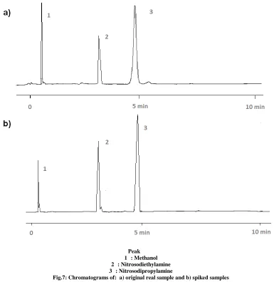
The developed extraction method was applied for the extraction of NDEA and NDPA from several food items e,g, sausage, corned meat, fresh meat, and salted egg. This method was successfully applied to the determination of NDEA and NDPA and the results indicated the presence of these hazardous NDEA and NDPA in sausage (3.25 and 0.06 ppm), corned meat (2.90 and 0.40 ppm), fresh meat (5.00 and 0.03 ppm) and salted egg (2.55 and 0.25 ppm) respectively. Spiked samples or standard addition method has been performed to verify the NDEA and NPDA peaks in the chromatograms.
Peak 1 : Methanol 2 : Nitrosodiethylamine 3 : Nitrosodipropylamine
Fig.7: Chromatograms of: a) original real sample and b) spiked samples
CONCLUSION
Yanuardi Raharjo et al
J. Chem. Pharm. Res., 2014, 6(5):1274-1280
______________________________________________________________________________
Acknowledgements
The authors are grateful to Indonesia Ministry of Education for financial support, Faculty of Science and Technology and Faculty of Pharmacy, Universitas Airlangga for the facilities.
REFERENCES
[1] J Mulder. Basic Principles of Membrane Technology, Kluwer, Academic Publishers ,USA, 1996
[2] ZF Cui, and HS Muralidhara. Membrane Technology First Edition. A Practical Guide to Membrane Technology and Applications in Food and Bioprocessing, Elsevier, The Netherlands, 2010
[3] N Peng; N Widjoyo; P Sukitpaneenit; MM Teoh; GG Lipscomb; T Chung; J Lai, Prog. Polym Sci. 2012 (37), 1401-1424.
[4] G Drabi-Markiewicz; B Dejaegher; ED Mey; S Impens; T Kowalska; H Paelinck; YV Heyden, Anal. Chim.
Acta, 2010 (657),123-130.
[5] S Yurchenko; U Molder, Food Chem., 2007 (100), 1713-1721.
[6] P Anand; AB Kunnumaraka; C Sandaram; KB Harikumar; ST Tharakan; OS Lai; B Sung; BB Aggarwal,
Pharm. Res., 2008, (25), 2097-2116.
[7] Y He; HK Lee, Anal. Chim. Acta, 1997, (69), 4634–4640. [8] AS Yazdi; A Amiri, Trends Anal. Chem., 2010, (29), 1–14.
[9] S Pedersen-Biergaard; KE Rasmussen, Anal. Chem. 1999, (71), 2650–2656 [10] JP Hsin; WH Ho, Anal. Chim. Acta, 2004, (527), 61–67
[11] Y Raharjo; MM Sanagi; WAW Ibrahim; AA Naim; HY Aboul-Enein, J. Sep. Sci. 2009, (32), 623-629 [12] KE Rasmussen; S Pedersen-Bjergaard, TrAC- Trends Anal. Chem. 2004, (23), 1-10.