Open Access
Technical innovations
A multimodality localization technique for radio-guided surgery
Seza A Gulec*, Erica Hoenie and Kristan Rheinheimer
Address: Goshen cancer Institute at Goshen Health System, Goshen, IN, USA
Email: Seza A Gulec* - [email protected]; Erica Hoenie - [email protected]; Kristan Rheinheimer - [email protected]
* Corresponding author
Abstract
Background: Intraoperative localization of image or endoscopy-detected lesions occasionally pose surgical challenges due to the small lesion size and/or difficult anatomic exposure. Identification of such lesions can be facilitated using a hand-held gamma probe with utilization of Tc-99m macroaggregate albumen (MAA) localization technique. The radiopharmaceutical injection can be performed using ultrasound (US) or endoscopy guidance.
Case presentations: The clinical use of the Tc-99m MAA protocol gamma probe-guided surgery was discussed in three representative cases. Surgical indication was diagnostic exploration in two patients with suspicious lymphadenopathy, and determination of extent of surgical resection in a patient with polyposis. Lesion localization with 100 microcurie (3.7 MBq) Tc-99m MAA prior to surgical exploration resulted in definitive localization of lesions intraoperatively.
Conclusion: The use Tc-99m MAA deposition technique at the site of surgical target is a highly efficient radio-guided surgery technique with definitive impact on the success of surgical exploration in selected indications.
Background
The role of gamma probes in surgical oncology practice has been well established [1-9]. Surgical performance with intraoperative gamma probe detection is critically dependent on target to surrounding background ratio (TBR). This ratio, for localization techniques that involve systemic administration of radiopharmaceuticals, is a function of radiopharmaceutical uptake and clearance kinetics. Probe's ability to discern the target signal also is a major technical factor in the clinical success. A mini-mum TBR of 1.5:1 is needed in the operative field for the operating surgeon to be comfortable that the differences between the target tissue and normal adjacent tissue are real [10]. Obtaining a satisfactory TBR is always a signifi-cant technical challenge with localization techniques
using systemic administration of radiopharmaceuticals. Administration of a locally-entrapped radiopharmaceuti-cal in or around the target tissue results in an ideal TBR.
Case presentations
Case 1
A 32 year-old woman with a history of T2-N1 left breast cancer, diagnosed 2 years ago and treated with mastec-tomy-immediate reconstruction, presented with a right axillary lymphadenopathy. Upon clinical exam, the lesion was non-palpable and measured approximately 1 cm by US. The lymphadenopathy persisted on a follow-up; FDG-PET imaging was negative. An US-guided FNA was non-definitive without evidence of malignancy. An increase in size of the node was noted at the subsequent follow-up. A
Published: 25 April 2007
World Journal of Surgical Oncology 2007, 5:43 doi:10.1186/1477-7819-5-43
Received: 1 February 2007 Accepted: 25 April 2007
This article is available from: http://www.wjso.com/content/5/1/43
© 2007 Gulec et al; licensee BioMed Central Ltd.
clinical decision was made for an excision biopsy. The node was injected with Tc-99m macroaggregate albumen (MAA) and lymphazurin blue 2 h prior to the planned operation. At surgery, the hot-spot was readily identified. Probe localization was distinctly focal over a level-I node, which was accessed with minimal dissection. Blue dye facilitated surgical exposure and dissection. Surgical pathology revealed chronic inflammatory changes (Figure 1).
Case 2
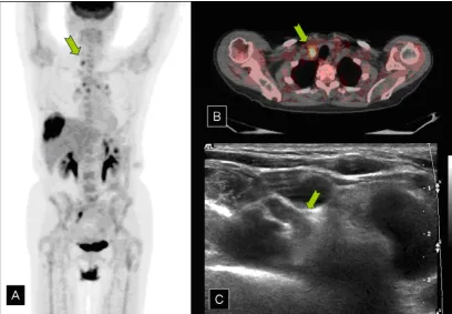
A 65 year-old woman with history of colorectal cancer (CRC) and non-Hodgkin's lymphoma of the scalp, pre-sented with mediastinal and retroclavicular lymphaden-opathy. CRC was diagnosed 2 years ago, and was treated with R-colon resection. Scalp lymphoma was diagnosed 6 months ago, when the patient presented with a 15 cm scalp lesion. Staging work-up at that time revealed posi-tive FDG uptake in the scalp lesion (SUV:18), multiple mediastinal and R-retroclavicular lymph node (SUV:3.2), and a 12 cm liver lesion (SUV: 8.5). Biopsy of the liver lesion was consistent with the colon primary. Nodal find-ings were concluded to indicate a stage III lymphoma, and a sequential intensity-modulated radiation therapy (IMRT) and chemotherapy using CHOP regimen was administered. There was a complete clinical and PET/CT response in the scalp lesion. Post-treatment FDG-PET/CT showed persistence of mediastinal and R-retrocla-vicular nodal uptake. The R-retroclaR-retrocla-vicular node was
injected with Tc-99m MAA and lymphazurin blue 2 h prior to the planned operation. At surgery, the hot-spot was readily identified. Probe localization was distinctly focal over an internal jugular-innominate vein confluence lymph node. The target was accessed through an incision made over the hot-spot. The line-of-sight provided a safe surgical dissection. The blue dye facilitated surgical expo-sure and dissection. Surgical pathology revealed a chronic granulomatous disease. The patient was restaged to have a stage I scalp lymphoma, and remained in complete remis-sion following treatment. Stage IV CRC was also con-cluded to be a liver-only disease, which allowed her to be considered for liver-directed therapy (Figure 2).
Case 3
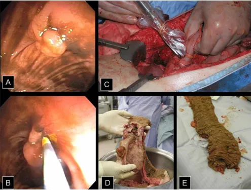
A 69 year-old man presented with anemia, subsequent colonoscopy revealed a right colon cancer and multiple polyps throughout the colon. Severe dysplastic changes were noted in a sessile polyp in the transverse colon and in a pedunculated polyp in the sigmoid colon of villous type. The descending colon polyp was completely excised colonoscopically and the site was submucosally injected with Tc-99m MAA and lymphazurin blue 18 h prior to a planned operation. At surgery, the ascending colon lesion was readily identified. None of the polyps were palpable. A slight, relatively diffuse discoloration of submucosal blue dye was noted at the site of injection. Probe localiza-tion was distinctly focal. A total abdominal colectomy was performed with 2-cm margins distal to the focal Tc-99m
a) Right axillary lymphadenopathy demonstrated on mammogram
Figure 1
MAA signal. Surgical pathology confirmed complete resection with free margins (Figure 3).
Gamma probe-guided surgery protocol
The patients receive an injection of 0.1 mCi (0.037 MBq) (in 0.1 ml solution) Tc-99m MAA the morning of planned surgery. Lymph node injections are given most conven-iently under US. For colon polyp localization, the
injec-tion using the same activity and volume is given endoscopically. Surgical exploration is scheduled within 6 hours post injection of the radiopharmaceutical. The injected activity might be doubled if surgery is planned 6– 12 hours after the injection (Table 1). Standard gamma probe settings (photopeak of 140 keV, Window of 20%, and a threshold of 136 keV) and operative technique are used in the operating room.
a) Retroclavicular lymph node demonstrated on composite PET image
Figure 2
a) Retroclavicular lymph node demonstrated on composite PET image. b) PET/CT demonstrating large liver lesion. c) Ultra-sound of lesion.
Table 1: Gamma probe-guided surgery protocol for Tc-99m MAA
Tc-99m MAA Lesion Localization with image/endoscopy guidance
Radiopharmaceutical Dose/Administration • Tc-99m MAA
• 0.1 mCi (.0037 MBq)/direct injection Standard Imaging Protocol • No nuclear medicine imaging needed Timing of Surgical Exploration • Within 6 h post-injection
Patient Preparation • Not required
Gamma Probe • Standard medium-energy gamma probe
System set-up • Analyzer Settings: Photopeak: 140 keV, Window: 20%, Threshold: 136 keV (In commercial systems this is a default setting)
• Verify calibration and settings of the system • Cover the probe with sterile plastic sleeve
Intra-operative Use • Probe survey at counts- per-second mode (Dynamic pitch range feed-back helpful)
Discussion
The use Tc-99m MAA deposition technique at the site of surgical target is a highly efficient radio-guided surgery technique with definitive impact on the success of surgical exploration in selected indications. Administration of the locally-entrapped Tc-99m MAA in or around the target tis-sue results in an ideal TBR. Tc-99m MAA, when injected into the tissues, remains almost stationary with a minimal local diffusion. Particle degradation, slow lymphatic absorption and phagocytosis constitute the principal mechanisms for MAA clearance. The biologic half life of MAA is approximately 6 hours. The effective half life of Tc-99m MAA is calculated at approximately 3 hours when the 6-hour physical half life of Tc-99m is factored in (1/2 TEff = 1/2 TBiol + 1/2 TPhys). A 0.1 mCi Tc-99m provides satis-factory signal intensity for gamma probe detection within a wide range of time frame. Surgical procedure can be scheduled any time after injection up to 6 hours.
(Sugges-tion: Surgical procedures can be scheduled any time dur-ing the 6 hours post injection.) Tc-99m MAA injection can be given under CT, US or endoscopic guidance. Lymphaz-urin blue (blue dye) can be added to the injectate to facil-itate dissection by providing a visual aid.
Conclusion
Major applications of the technique include localization of lymph nodes and colonic polyps. The technique may also be used in localization of non-palpable breast lesions as an adjunct to needle/wire localization techniques.
Competing interests
The author(s) declare that they have no competing inter-ests.
A and B) Sigmoid colon polyp and endoscopic injection of the base
Figure 3
Publish with BioMed Central and every scientist can read your work free of charge
"BioMed Central will be the most significant development for disseminating the results of biomedical researc h in our lifetime."
Sir Paul Nurse, Cancer Research UK
Your research papers will be:
available free of charge to the entire biomedical community
peer reviewed and published immediately upon acceptance
cited in PubMed and archived on PubMed Central
yours — you keep the copyright
Submit your manuscript here:
http://www.biomedcentral.com/info/publishing_adv.asp
BioMedcentral
Authors' contributions
SG – Design, Acquisition, analysis and interpretation of data, Drafting manuscript, Critical revision
EH – Analysis of data, Editing of the manuscript
KR – Acquisition of data
Acknowledgements
The information in this document was obtained in accordance with HIPAA regulations and with the approval of our Institutional Review Board. Patients consent was obtained for publication of their case records
References
1. Gulec SA, Moffat FL, Carroll RG: The expanding clinical role for intraoperative gamma probes. Nuclear Medicine Annual
1997:209-237.
2. Moffat FL, Vargas-Cuba RD, Serafini AN, Jabir AM, Sfakianakis GN, Sittler SY, Robinson DS, Crichton VZ, Subramanian R, Murray JH, et al.: Preoperative scintigraphy and operative probe scintime-try of colorectal carcinoma using technetium-99m-88BV59. J Nucl Med 1995, 36:738-745.
3. Krag DN, Meijer SJ, Weaver DL, Loggie BW, Harlow SP, Tanabe KK, Laughlin EH, Alex JC: Minimal-access surgery for staging of malignant melanoma. Arch Surg 1995, 130:654-660.
4. Gulec SA, Moffat FL, Carroll RG, Serafini AN, Sfakianakis GN, Allen L, Boggs J, Escobedo D, Livingstone AS, Krag DN: Sentinel node localization in early breast cancer. J Nucl Med 1998,
39:1388-1393.
5. Gulec SA, Moffat FL, Carroll RG, Krag DN: Gamma probe guided sentinel node biopsy in breast cancer. Quart J Nucl Med 1997,
41:251-261.
6. Bozkurt F, Ugur O, Hamaloglu E, Sayek I, Gulec SA: Optimization of gamma probe-guided parathyroidectomy. Am Surg 2003,
69:720-725.
7. Mariani G, Gulec SA, Rubello D, Boni G, Puccini M, Casara D, Manca G, Pelizzo MR, Sotti G, Erba P, Volterrani D, Giuliano AE: Preoper-ative localization and radioguided parathyroid surgery. J Nucl Med 2003, 44:1443-1458.
8. Schirmer WJ, O'Dorisio TM, Schirmer TP, Mojzisik CM, Hinkle GH, Martin EW: Intraoperative localization of neuroendocrine tumors with 125I-TYR(3)-octreotide and a hand held gamma-detecting probe. Surgery 1993, 114:745-752.
9. Ahlman H, Tisell L-E, Wangberg B, Nilsson O, Fjalling M, Forssell-Aronsson E: Somatostatin receptors on neuroendocrine tumors – a way to intraoperative diagnosis and localization. Yale J Biol Med 1994, 67:215-221.