GENETICS | REVIEW
Programmed Cell Death Initiation and Execution
in Budding Yeast
Randy Strich Department of Molecular Biology, Rowan University School of Osteopathic Medicine, Stratford, New Jersey 08055
ABSTRACTApoptosis or programmed cell death (PCD) was initially described in metazoans as a genetically controlled process leading to intracellular breakdown and engulfment by a neighboring cell . This process was distinguished from other forms of cell death like necrosis by maintenance of plasma membrane integrity prior to engulfment and the well-defined genetic system controlling this process. Apoptosis was originally described as a mechanism to reshape tissues during development. Given this context, the assumption was made that this process would not be found in simpler eukaryotes such as budding yeast. Although basic components of the apoptotic pathway were identified in yeast, initial observations suggested that it was devoid of prosurvival and prodeath regulatory proteins identified in mammalian cells. However, as apoptosis became extensively linked to the elimination of damaged cells, key PCD regulatory proteins were identified in yeast that play similar roles in mammals. This review highlights recent discoveries that have permitted information regarding PCD regulation in yeast to now inform experiments in animals.
KEYWORDScyclin C; apoptosis; oxidative stress; mitochondria; signal transduction
T
WO types of regulated cell death, necrosis and pro-grammed cell death, have been described in budding yeast (Lin and Austriaco 2014). Necrotic cell death was originally characterized as a simple collapse of the cell lead-ing to cell wall breakdown and ultimately lysis. However, more recent studies report the existence of a regulatory net-work governing necrotic cell death (Eisenberget al.2010). This review concentrates on programmed cell death (PCD) in yeast, which closely resembles the intrinsic or mitochondrial-derived apoptosis in multicellular organisms (Perrone et al. 2008). Mammalian apoptosis is initiated by accumulation of Bcl2homology 3 (BH3) containing proteins such as Bax on the mitochondrial outer membrane. Bax induces pore forma-tion leading to the release of cytochromec, which stimulates a cascade of proteases termedcysteine-dependentasp artate-specific proteasesor caspases (Danial and Korsmeyer 2004). Plants and fungi possess a related protease family called metacaspases (Uren et al. 2000). Metacaspases share se-quence and functional similarities but differ with respect to substrate recognition sites (asparagine/lysine rather than as-partic acid). Budding yeast possesses a single metacaspase(Yca1) and BH3 domain protein (Ybh3), which are both required for oxidative stress-induced PCD. Standard assays for PCD, such as double strand breaks or phophatidylserine externalization (Annexin V staining), routinely used to mon-itor apoptosis in metazoans, are also employed to assay PCD in yeast (Madeoet al.1997). However, following excessive damage, these PCD hallmarks may be joined by necrotic markers (e.g., propidium iodide permeability) (Yamaki et al. 2001). Therefore, it is important to note that these different cell-death modes can be observed simultaneously within a population and care should be used when judging the contribution that each death pathway has on overall cell viability.
Oxidative Stress, a Common Denominator for PCD Initiation
There are many stimuli, either externally or internally derived, able to induce PCD in yeast. For example, aging (Corte-Real and Madeo 2013), extreme pH environment (Ludovico et al. 2001), plant toxins (Narasimhan et al. 2001), defects in actin function (Gourlay and Ayscough 2006), osmotic stress (Silvaet al.2005), acetic acid (Ludovico et al.2002), the presence of lipid hydroperoxides (Alicet al. 2003), and prolonged mating-factor exposure (Severin and Hyman 2002) (although the exact nature of this cell death
Copyright © 2015 by the Genetics Society of America doi: 10.1534/genetics.115.179150
is in question) (Zhang et al.2006) all stimulate PCD. Al-though these stressors appear different, many have in com-mon the ability to generate internal reactive oxygen species (ROS). For example, a specific mutation inCdc48induces
PCD in yeast (Madeo et al. 1997) due to elevated ROS
(Madeoet al.1999) produced from defective mitochondria (Braun et al. 2006; Braun and Zischka 2008). Similarly, defects in endoplasmic reticulum (ER)-dependent protein folding also produces ROS (Tu and Weissman 2004) to levels sufficient to induce PCD (Haynes et al. 2004). In addition, defects in the electron transport chain (ETC) lead to ER-produced ROS through hyperactivation of the ER NADPH oxidase Yno1(Leadshamet al.2013). Thesefi nd-ings demonstrate the intricate relationships that have evolved between organelles that produce and respond to ROS-induced damage. The transcriptional response to, and the macromolecular damage caused by, oxidative stress in yeast are the subject of several excellent reviews (Avery
2011; Farrugia and Balzan 2012; Morano et al. 2012)
and will not be detailed here. Rather, given the universal nature of the oxidative stress response from yeast to humans, this review focuses on recent insights into the signaling systems that transduce the ROS signal and the effector proteins that coordinate the response between organelles in budding yeast.
External origins of ROS
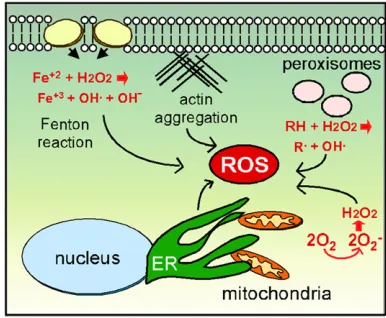
The cell maintains redox homeostasis by balancing low-level ROS produced by organelles or exogenous sources with an arsenal of antioxidant enzymes that neutralize reactive oxygen (e.g., superoxide dismutase, catalase) or repair oxi-dative damage (e.g., chaperones, DNA repair enzymes) once it occurs (Perroneet al.2008). However, increased internal ROS concentrations above a certain threshold lead to an accumulation of oxidized lipids, proteins, and DNA, collec-tively termed oxidative stress (Tsuzi et al. 2004; Drakulic et al. 2005; Temple et al. 2005). Exogenous sources of ROS occur in many forms including prooxidants such as H2O2(Vealet al.2007), exposure to heavy metals that stim-ulate superoxide production through the Fenton reaction (Liang and Zhou 2007; Nargund et al.2008), or treatment with certain anticancer drugs (Almeidaet al.2008) (Figure 1). Exogenous ROS can alter plasma membrane characteristics that trigger sensors able to induce signal transduction path-ways such as the cell-wall integrity pathway (Levin 2011) or the osmolarity-sensing pathway (Singh 2000; Bilsland et al. 2004) resulting in dramatic changes in the transcriptome. In addition, direct oxidation of transcription factors (e.g.,Yap1) promotes stress-responsive gene transcription (Delaunayet al. 2000; Kugeet al.2001).
Internal sources of ROS
Internal ROS is mostly derived from organelles performing their normal functions. The best studied and perhaps most important of these are the mitochondria (Figure 1). The mitochondrial function of ATP synthesis inherently produces
reactive oxygen through the leakage of electrons from the ETC. This amount of ROS is limited and thought to repre-sent a signaling molecule affecting many cellular processes (Guaragnellaet al.2012). However, mitochondrial dysfunc-tion via mutadysfunc-tions in ETC components, compounds that in-hibit ETC function, or loss of mitochondrial inner membrane integrity, can generate sufficient ROS concentrations to in-duce the oxidative stress response (Eisenberg et al.2007). For example, cytochromecmutants display ETC defects that generate H2O2(Barroset al.2003). In addition, stimulating Ras signaling induces high protein kinase A (PKA) activity, leading to loss of mitochondrial integrity and elevated in-ternal ROS (Hlavata et al. 2003; Hlavata et al. 2008; reviewed in Perroneet al.2008).
In addition to defects in internal processes, mitochondrial-derived ROS can be caused via indirect mechanisms as well. For example, mutations or drugs that reduce actin dynam-ics cause elevated mitochondrially derived ROS (Gourlay et al.2004). Interestingly, enhancing actin dynamics by de-leting a gene (SCP1) encoding a bundling protein reduces ROS (Gourlay et al. 2004). A second connection between actin and mitochondrial fitness is observed during parti-tioning of this organelle to daughter cells. Myosin motors direct mitochondria toward the bud along F-actin cables to facilitate organelle partitioning (Mishraet al.2014). In ad-dition, a retrograde actin cable force is present that directs cargo toward the mother. Healthy mitochondria can bind the motors with sufficient strength to navigate to the bud despite the retrograde force moving in the opposite direc-tion (McFaline-Figueroa et al. 2011). Pon and coworkers have likened this phenomenon to salmon swimming up-stream against the river current (Higuchiet al.2013). This
process assures healthy mitochondria migrate to the bud while defective and ROS leaking mitochondria remain in the mother. As described later, this phenomenon may have consequences in aging-induced PCD.
In addition to the mitochondria, the ER is also a source of reactive oxygen in the cell. The ER provides the critical function of folding newly synthesized proteins and then sorting them for various cellular addresses (Chen et al. 2013). ER protein folding utilizes specialized chaperones (protein disulfide isomerases and Ero1) and an oxidative environment (Pollard et al. 1998) resulting in conversion of oxygen to H2O2 (Zito 2015). Defects in protein folding trigger the well-studied unfolded protein response (UPR) that induces ERO1 transcription. Prolonged Ero1p expres-sion elevates ROS concentrations, ultimately leading to cell death (Hayneset al.2004). Interestingly, the UPR leads to ROS generation by both the ER and the mitochondria. For example, Yno1/Aim14, a NADPH-oxidase found in the ER,
generates ROS and promotes PCD (Rinnerthaler et al.
2012). Normally, Yno1-generated ROS concentrations are low and considered a signaling molecule in other fungi (Malagnac et al.2004). However, yeast strains overexpress-ing Yno1 produce sufficient ROS to induce PCD. Although Yno1is not part of the ER stress response, cytochrome oxidase c-defective mitochondria also raiseYno1activity by prevent-ing its normal turnover (Leadshamet al.2013). Similar to the engineered overexpression studies, elevatedYno1levels produce sufficient ROS to induce cell death. These studies highlight the intimate relationship between the ER and mi-tochondria with respect to ROS homeostasis.
Other organelles also contribute to oxidative stress. The peroxisome is important for b-oxidation of fatty acids that produce oxygen radicals and hydroperoxides (Manivannan et al. 2012). In addition, ROS are generated from peroxi-somes that are defective in either form or function. For ex-ample, loss ofPex6activity, a protein involved in peroxisome import, results in cells accumulating ROS to levels sufficient to induce cell death (Jungwirthet al.2008). However, these cells show hallmarks of necrosis rather than PCD, indicating that internally produced ROS can induce multiple types of cell death. As discussed below, signaling systems that trans-duce the ROS signal have been identified. It will be interest-ing to determine if ROS generated from the mitochondria, ER, or peroxisomes activate similar or different pathways to trigger the oxidative stress response.
Aging and PCD
Two types of aging, chronological and replicative, are studied in yeast. Chronological aging examines how long cells can remain alive in stationary phase and is thought to be analogous to quiescent, postdifferentiated mammalian cells (Braun and Westermann 2011; Corte-Real and Madeo 2013). Conversely, replicative aging determines the number of cell divisions an individual mother cell can undergo and has been proposed to serve as a model for stem cell-like divisions. Both aging types are controlled by genetic factors
as well as nutritional conditions, many of which impact mi-tochondrial function (Kaeberlein 2010; Corte-Real and Madeo 2013). Both replicative and chronological aging pro-cesses in budding yeast are driven by ROS accumulation that ultimately results in PCD (Laun et al.2001; Fabrizio et al. 2004; Herker et al.2004). For example, mother cells age through accumulation of oxidatively damaged proteins or mitochondria that are not passed on to their daughters (Aguilaniu et al. 2003; McFaline-Figueroa et al.2011). In addition, protein aggregates are retained in aging mothers (Rujano et al. 2006; Spokoini et al. 2012) thus allowing daughter cells to start with a clean aging slate. The cell has multiple avenues to counteract these aging hallmarks. For example, protein aggregates are recognized as cellular damage and are degraded through the activity of protein chaperones and the metacaspase Mca1/Yca1 (Hill et al. 2014). Similarly, chronologically aged cells induce the
NADP-dependent glutamate dehydrogenase Gdh3 that
detoxifies ROS and prevents PCD initiation (Lee et al. 2012). These studies, as well as many others, provide a di-rect link between ROS accumulation, PCD initiation, and longevity. In mammals, this question is more complex as oxidative stress-induced pathology is influenced by the pres-ence of cellular damage, and by other confounding factors including tissue type, stage in development, and the subcel-lular compartment in which the ROS originated (Cunningham et al. 2015). Therefore, the utility of yeast or mammalian tissue culture as a model to investigate some aspects of the free radical theory of aging may be limited.
Role of the mitochondrial dynamics in PCD execution
membrane (MOM) requires the receptorFis1(Mozdyet al. 2000; Tieu et al. 2002) and one of two adaptors, Mdv1 (Mozdy et al. 2000; Tieuet al. 2002; Cerveny and Jensen 2003) or Caf4 (Schauss et al. 2006; Motley et al. 2008). Interestingly, peroxisomefission also requires Fis1and one of two dynamin-like proteins, Vps1 or Dnm1; the latter seems only important in cultures grown on oleic acid (Hoepfneret al.2001; Kuraviet al.2006). For mitochondria, fission often occurs at sites of interaction with the ER (Friedman et al.2011). Many roles have been described for these junc-tions including sites of lipid transfer and Ca++ signaling (Michel and Kornmann 2012). Therefore, mitochondria–ER communication appears to be important for mitochondrial fission as well.
Of particular interest for this review, extensive mitochon-drial fragmentation is a common feature following exposure to many types of damage including oxidative stress. Stress-induced mitochondrial hyperfission is conserved from yeast to mammals and represents an early morphological adapta-tion of the stress response (Youle and van der Bliek 2012). Mitochondrial hyperfission has been associated with the re-lease of sequestered apoptotic factors (Frank et al. 2001; Breckenridgeet al. 2003) while preventingfission protects cells from PCD. For example, mutants lackingDnm1orFis1 are resistant to ROS-induced PCD (Fannjianget al.2004).
Although the basic fission machinery is required for stress-induced hyperfission, how their activity is enhanced occurs through an unlikely mechanism. In all eukaryotes examined, cyclin C (Ssn8) andCdk8(Ssn3) form a protein kinase that associates with the RNA polymerase II holoen-zyme to control transcription (Bourbon 2008) (Figure 2). In budding yeast, this kinase represses 100 genes that are induced in response to environmental stress (Cooper et al. 1997; Holstege et al.1998; van de Peppelet al.2005). To relieve cyclin C–Cdk8 repression, stressed cells translocate cyclin C from the nucleus to the cytoplasm where it is ulti-mately destroyed through activity of the Not4ubiquitin li-gase (Cooper et al.2012). However, cyclin C has a second function independent ofCdk8. Prior to its destruction in the cytoplasm, cyclin C associates with Mdv1 to induce exten-sive mitochondrial fragmentation (Cooper et al. 2014; reviewed in Strich and Cooper 2014). Deletion of its nuclear anchor, MED13, allows aberrant entry of cyclin C into the cytoplasm where it can inducefission in the absence of stress (Khakhinaet al.2014). These results indicate that cyclin C is both necessary and sufficient for hyperfission. Cyclin C-dependent hyperfission is directly related to the ability of the cell to induce PCD. Mutants lacking cyclin C are protected from ROS-induced PCD, whereasmed13Dmutants, in which the mitochondria are continuously fragmented, are hyper-sensitive to oxidative stress (Khakhina et al. 2014). It is important to note that continuous mitochondrial fission on its own is insufficient to induce cell death, although the health of this organelle suffers under these conditions through loss of mtDNA (Khakhinaet al.2014). These obser-vations indicate that mitochondrial fragmentation
potenti-ates the cell toward PCD initiation, but another stress signal is required to initiate this process. The role cyclin C plays in mitochondrialfission and PCD is remarkably well conserved. Mammalian cyclin C also translocates from the nucleus to the mitochondria in response to stress (Wanget al.2015). Knockout mouse embryonicfibroblast (MEF) cells revealed that cyclin C is required for stress-induced mitochondrial fission and apoptotic cell death. Finally, the yeast cyclin C is able to induce complete mitochondrial fragmentation when purified protein is added to permeabilized MEF cul-tures. In the other direction, the human cyclin C comple-ments the cell-death-resistance phenotype in cnc1D yeast mutants but not the transcriptional repression defect (Krasley et al.2006). This analysis represents an example in which the direction of information understanding apoptotic control flowed from yeast to mammalian studies.
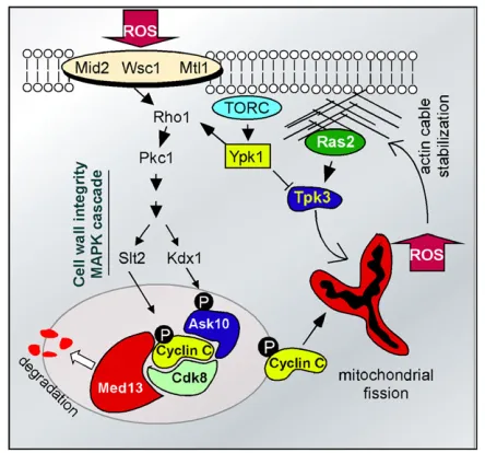
Signaling Pathways Directing ROS-Induced PCD: The Cell-Wall Integrity Pathway Controlling Cyclin C Nuclear Release
As indicated above, the failure to translocate cyclin C into the cytoplasm protects the cell from H2O2-induced PCD, while its aberrant release from the nucleus causes hypersen-sitivity to oxidative damage. Given the importance of this
decision, it is not surprising that the switch controlling cyclin C release is complex and appears to be composed of at least two arms. First, the nuclear anchor, Med13is destroyed in response to oxidative stress with kinetics similar to cyclin C release (Khakhina et al. 2014). This destruction is depen-dent on the 26S proteasome maturation factor Ump1, sug-gesting the involvement of ubiquitin-mediated proteolysis. Consistent with this model, the SCF ubiquitin ligase is re-quired for ROS-induced Med13destruction manner (K. F. Cooper, unpublished results). This result parallels a previous study in mammalian cells revealing a role for the SCF ligase in normalMed13turnover (Daviset al.2013). Currently, it is not known whether the yeast Med13degradation is es-sential for cyclin C nuclear release or whether its proteolysis serves to prevent retention of the cyclin if it reenters the nucleus.
The second arm of the cyclin C control pathway is me-diated by the cell-wall integrity MAP kinase pathway and includes a bifurcation at the MAP kinase step (Figure 2, see Table 1). The cell-wall integrity pathway transduces the oxidative stress signal from the cell wall to the nucleus to affect changes in transcription (Alicet al.2003; Stalevaet al. 2004; Vilellaet al.2005; Krasleyet al.2006; Petkovaet al. 2010) and actin remodeling (Pujol-Carrion et al. 2013). Treating cells with H2O2 activates two cell-wall receptor groups containing Wsc1 and either Mid2 or Mtl1 (Vilella et al.2005; Petkovaet al.2010; Jinet al.2013). The recep-tors in turn stimulate the Rho1p GTPase, which activates Pkc1and the cell-wall integrity MAP kinase pathway (Levin 2011). Recent studies revealed that theSlt2 MAPK directly phosphorylates cyclin C at Ser266 (Jin et al.2014). Elimi-nating this phosphorylation site prevents cyclin C cyto-plasmic translocation, while a phosphomimetic mutation enhances its translocation (Strich and Cooper 2014). The other branch contains the pseudokinaseKdx1that associates with Ask10(Jin et al.2014), a previously identified cyclin C-associating factor (Cohen et al.2003).Ask10is required for cyclin C cytoplasmic translocation (Jinet al.2014) and is phosphorylated in response to oxidative stress (Cohenet al. 2003). Surprisingly, Ask10p phosphorylation requires the two cell-wall-integrity pathway MAPKKs, Mkk1and Mkk2, and the pseudokinaseKdx1, but notSlt2(Cohenet al.2003;
Jinet al.2014), suggesting the presence of yet another sig-naling system controlling cyclin C release.
Signaling Systems Directing ROS-Induced PCD: The Ras Signaling Pathway
Ras2signaling also contributes to PCD initiation but in a man-ner different from the cell-wall integrity pathway. Rather than sensing and transducing the presence of ROS-induced dam-age, aberrant Ras signaling causes loss of mitochondrial in-tegrity and subsequent ROS release (Smethurstet al.2014). For example, aberrant actin aggregation, caused by specific actin monomer mutations or drugs that promote filament bundling, stimulatesRas2signaling leading to activation of protein kinase A subunitTpk3(Gourlayet al.2004; Gourlay
and Ayscough 2006; Leadsham and Gourlay 2010). Tpk3
activation leads to elevated ROS levels in the cell (Figure 2). This system sets up a potential feedback loop in which the mitochondrial-derived ROS drives more actin aggrega-tion through increased disulfide linkage of actin monomers (Haarer and Amberg 2004). Similarly, stationary-phase cells exposed to continuous Ras2 activation display ele-vated ROS levels and undergo PCD (Gourlay and Ayscough 2005). In both situations, constitutively active Ras results in a Tpk3-dependent loss of mitochondrial integrity and
elevated ROS. These findings are similar to results
obtained in mammalian cell culture in which prolonged RAS/RAF/ERK signaling also induces apoptosis (Cagnol and Chambard 2010).
The retrograde signaling pathway transduces information about mitochondrial activity and integrity to the nucleus to affect changes in gene expression (Jazwinski 2013). One component of this pathway is the target of rapamycin com-plex 2 (TORC2), which responds to cellular redox conditions through activation ofYpk1protein kinase (Nileset al.2014). Ypk1 stimulation activates the cell-wall integrity pathway through maintenance of sphingolipid levels required for proper localization of Rom2, the GAP activator of Rho1 (Niles et al.2014) (Figure 2). This pathway keeps internal ROS levels in check, thus preventing cyclin C cytoplasmic relocalization and destruction (Niles and Powers 2014). Ypk1also inhibitsTpk3activity thereby maintaining normal
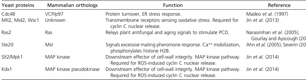
Table 1 Signaling molecules
Yeast proteins Mammalian orthologs Function Reference
Cdc48 VCP/p97 Protein turnover, ER stress response. Madeoet al.(1997)
Mtl2, Mid2, Wsc1 Unknown Transmembrane receptors sensing oxidative stress. Required for cyclin C nuclear release.
Jinet al.(2013)
Ras2 Ras Relays plant antifungal and aging signals to stimulate PCD. Narasimhanet al.(2005); Gourlay and Ayscough (2006) Ste20 Mst Signals excessive mating pheromone response. Ca++mobilization,
phosphorylates histone H2B.
Ahnet al.(2005); Severin (2002)
Slt2/Mpk1 MAP kinase Downstream effector of cell-wall integrity. MAP kinase pathway. Required for ROS-induced cyclin C nuclear release.
Jinet al.(2014)
Kdx1 MAP kinase pseudokinase Downstream effector of cell-wall integrity. MAP kinase pathway. Required for ROS-induced cyclin C nuclear release.
mitochondrial function and reducing excessive ROS produc-tion (Nileset al.2014). Thus, internal reactive oxygen levels are constantly monitored and adjusted to allow ROS to serve as a signaling molecule under certain situations.
Chromatin Modification and PCD Execution
Other, less-well-understood signaling pathways also play a conserved role in PCD execution. Ste20 is the founding member of the PAK (p21 activated protein kinase) protein family (Dan et al. 2001). A mammalian homolog of Ste20 (Mst1) (Creasyet al.1996) is activated by caspase cleavage and phosphorylateshistone H2Bon serine 14 (Cheunget al. 2003). This modification promotes chromatin condensation and apoptotic cell death. Similarly, in response to oxidative stress, yeast Ste20 also phosphorylates the analogous H2B residue (Ser10) (Ahnet al.2005) (Figure 3). Mutating H2B Ser10 to alanine protects the cell from H2O2-induced PCD, indicating an important role for this modification in the stress response. However, Ser10 phosphorylation occurs fol-lowing H2O2 exposure only when the adjacent lysine
(Lys11) is deacetylated by the Hos3 deacetylase (Ahn
et al.2006). Therefore, the stress signal must integrate both Hos3 andSte20activity. Conversely, monoubiquitylation of Lys123 on histone H2B by the Bre1ligase prevents Yca1-dependent H2O2-induced PCD induced by chronological ag-ing (Walter et al.2010). In addition to H2B,histone H3is also a target of posttranslational modifications that control
PCD execution. Methylation of H3 Lys4 (H3K4) by Set1
reduces aging-dependent PCD (Walter et al.2014). Consis-tent with thisfinding, deletingSET1renders cells more sensitive to oxidative stress, whereas mutating the demethylase,JHD2, makes cells more resistant. These results reveal a chromatbased tug-of-war between opposing signals that promote or in-hibit PCD execution.
Regulators of mitochondrial outer membrane permeability
Similarly to mammalian cells, mitochondrial outer mem-brane permeabilization represents the commitment step to PCD execution (Green and Kroemer 2004; Antignani and Youle 2006). The loss of inner and outer mitochondrial membrane integrity is required for release of proapoptotic factors such as cytochromec and two nucleases,Nuc1and Aif1(see Table 2). In mammalian cells, mitochondrial per-meability is regulated through the competing activities of prosurvival (e.g., Bcl-2) and proapoptotic (e.g., Bax, Bak) Bcl-2 homology (BH) family members (Green and Kroemer 2004). In response to proapoptotic stimuli, Bax is recruited to the mitochondrial outer membrane, where it, in conjunc-tion with Bak, forms pores that permeabilize the outer mem-brane. Therefore, the proper control of Bax and Bcl-2 activity is critical for the correct response to cellular damage. In yeast, loss of the BH-homology protein Ybh3 function reduces PCD efficiency in response to oxidative stress or aging, whereas its overexpression increases H2O2sensitivity
(Buttneret al.2011). In addition,Ybh3function requires the proposed BH3 domain and its activity is suppressed by ex-pression of the prosurvival human Bcl-XL (Buttner et al. 2011). Finally, similarly to mammalian Bax, which relocal-izes from the cytoplasm to the mitochondria (Lovell et al. 2008; reviewed in Renault et al. 2013), Ybh3translocates from the vacuole to the mitochondria in response to stress (Buttneret al.2011). Interestingly,Ybh3function requires two associated proteins, Cor1 and Mir1, a ubiquinol–cytochrome c oxidoreductase subunit, and a mitochondrial phosphate carrier, respectively (Buttneret al.2011). A similar function was confirmed for the mammalian orthologs of these pro-teins, QCR1 and PHC (Buttneret al. 2011). These results illustrate that, as with the cyclin C studies, the information obtained in analyzing yeast PCD is helping to instruct similar studies in mammalian cells.
Executioners of the programmed cell death pathway
The ultimate goal of mitochondrial outer membrane perme-ability is the release of proapoptotic proteins including cytochromec and two nucleases (Aif1andNuc1, see Table 2). Genetic studies have verified their role in PCD. Deleting these nucleases increases resistance to ROS-induced cell death, whereas their overexpression causes hypersensitivity (Wissing et al. 2004; Buttner et al. 2007). Similar to their mammalian counterparts Aif1 and EndoG, the yeast Aif1 and Nuc1 enter the nucleus and fragment chromatin. In mammalian cells, Aif1 activity requires association with the peptidylprolyl cis-transisomerase cyclophilin A (Cande et al. 2004). Likewise, yeastAif1 function is dependent on the yeast homolog of cyclophilin A (Cpr1) but not cyclophi-lin B (Cpr2). Taken together with the chromatin modifi ca-tion similarities, the nuclear changes in response to PCD execution are remarkably similar in yeast and mammals.
In mammalian cells, release of cytochrome c from mito-chondria activates the caspase 9 initiator protease, which resides in the Apaf-1 apoptosome complex (Riedl and Salvesen 2007). Yeast genetic evidence indicates that cyto-chrome c is partially required for efficient PCD (Ludovico et al. 2002; Giannattasio et al. 2008), although no Apaf-1 ortholog has been identified. Genetic studies have identified
several proteases that are required for PCD execution. Sim-ilarly to the caspase cascade in mammalian cells, Yca1 is activated by proteolysis and required for H2O2 and acetic acid-induced PCD (Madeo et al. 2002). Esp1 cleaves the cohesin Mcd1 in response to H2O2 treatment (Yang et al. 2008). Nma111, an ortholog of the human HtrA protease (Belangeret al.2009), cleavesBir1, the yeast ortholog of the mammalian inhibitor of apoptosis factor (Walter et al. 2006). Interestingly, these proteases exhibit full, partial, or no role in PCD execution depending on the stress (Liang et al.2008; Madeoet al.2009). These results suggest that different stimuli utilize specific caspases to execute the cell-death pathway. Alternatively, these proteases may perform overlapping activities masking their roles. The genetic anal-yses possible in yeast will be able to address whether func-tional overlap exists between proteases, or whether additional proteases exist that have not been ascribed a role in PCD control. In support of the latter possibility, protease activities that do not correspond to known caspase-like enzymes have been identified that are able to cleavefl uo-rescent substrates with specificities similar to mammalian caspases (Wilkinson and Ramsdale 2011).
Coordinating the oxidative stress response throughout the cell
The oxidative-stress response is a culmination of changes in gene expression, organelle structure/function, and the cytoskeleton (reviewed in Smethurst et al. 2014). The organellar communication between the nucleus and mito-chondria has been well studied (Hill and Van Remmen 2014; Shaughnessyet al. 2014). One example of this coor-dination, and insight into the complexity of the regulatory system governing this process, is demonstrated by examin-ing cyclin C–Cdk8activity in stressed and nonstressed cells. Several studies indicate both a prosurvival and prodeath role for cyclin C translocation from the nucleus to the cyto-plasm. As described above, transcriptome analysis and more directed studies indicate that cyclin C-Cdk8represses genes involved in the stress response (Cooperet al.1997; Holstege
et al.1998). Therefore, the stress-induced nuclear release of cyclin C inactivates Cdk8, which remains nuclear (Cooper et al. 2012). The inactivation ofCdk8allows complete and timely induction of meiotic (Cooper et al.1997) or stress-responsive (Cooperet al.2012) genes. In addition, cyclin C–
Cdk8 restricts H3K4 methylation (Law and Ciccaglione
2015), a chromatin mark that prevents PCD-induced chro-matin condensation (Walter et al.2014). As H3K4 methyl-ation is associated with transcriptional activmethyl-ation, these processes may well be related. Finally, cyclin C translocation to the cytoplasm induces extensive mitochondrial fragmen-tation, which may aid in the removal of damaged, ROS-leaking organelles (Youle and van der Bliek 2012). Therefore, derepressing stress response genes, enhancing H3K4 meth-ylation, and removing damaged mitochondria all protect cells from PCD-inducing insults. These findings would ex-plain why cnc1D mutants are more resistant to oxidative stress than either fis1D or dnm1D mutants (Cooper et al.
Figure 4 Stress-induced relocalization of PCD regulators. The direction of protein translocation is depicted in stressed cells. Ras2 and Ybh3 transit to the mitochondria from the plasma membrane and vacuole (Vac), respec-tively. Mdv1 and Dnm1 relocalize from the cytoplasm to the mitochondria to inducefission. Cyclin C and Mcd1 relocalization from the nucleus to the mitochondria inducesfission and loss of mitochondrial outer mem-brane integrity. Translocation of mitochondrial proteins Aif1, Nuc1, and Ndi1 stimulate chromatin breakdown in the nucleus.
Table 2 Executioner molecules
Yeast protein Mammalian orthologs Function Reference
Yca1/Mca1 Metacaspase Cleave proteins Madeoet al.(2002)
Nma111 HtrA2/Omi Nuclear serine protease required for ROS-induced PCD, cleaves Bir1. Fahrenkroget al.(2004)
Bir1 IAP Inhibitor of apoptosis. Substrate of Nma111. Walteret al.(2006)
Aif1, Ndi1 Aif/AMID Mitochondrial nuclease released following permeability. Required for chromatin destruction.
Wissinget al.(2004)
Esp1 Separin Caspase-like protease, cleaves the cohesion Mcd1. Yanget al.(2008)
Nuc1 EndoG Mitochondrial nuclease released following permeability. Required for chromatin destruction.
Buttneret al.(2007)
Kex1 Caspase-like Required for PCD in response to glycosylation defects, acetic acid, aging.
Hauptmann and Lehle (2008)
Cyclin C/Ssn3p Cyclin C Translocates to mitochondria following stress. Associates with mitochondrialfission machinery, required for mitochondrial fragmentation and permeability.
Cooperet al.(2012, 2014)
Ybh3 Bax Translocate to the mitochondria following stress. Induce
mitochondrial outer membrane permeability.
2014). However, the tipping point toward PCD is not mito-chondrial fission. Therefore, the cell requires an additional signal, perhaps mitochondrial recruitment ofYbh3, to initiate the cell death pathway. In this process, the cell has utilized cyclin C relocalization to affect changes in gene expression, chromatin remodeling, and mitochondrial dynamics.
Physiological role for PCD in a single-celled organism
Due to the lack of obvious counterparts (e.g., p53, Bcl-2), many early studies considered yeast an in vivotest tube in which to analyze mammalian apoptotic regulators free of complications from yeast-based PCD (Manon et al. 1997; Ligr et al. 1998; Lisa-Santamaria et al. 2009; Greenwood and Ludovico 2010; reviewed in Silva et al. 2011; Clapp et al. 2012). However, extensive studies in budding yeast, fission yeast, and other single-cell eukaryotes challenge this view (Shemarova 2010). The identification of conserved regulatory proteins such as cyclin C,Ybh3, andYca1in bud-ding yeast argues that PCD regulation is an ancient process. Several models have been put forth to explain the early evolutionary appearance of both and antiapoptotic pro-teins (Taylor-Brown and Hurd 2013). Given the prominent role of the mitochondria in PCD regulation, it is not surpris-ing that many models start at the conception of the eukary-otic cell with a bacterial parasite that eventually became endosymbiotic with its host. As cellular stress is a universal PCD initiator, one possibility is that ancient parasites recog-nized that their host was compromised and elicited cell death. This provided the bacteria a last gasp of nutrients as well as a free path to find another host (Nedelcu et al. 2011). As this relationship evolved to be less selfish and more mutually beneficial, the health of the newly identified mitochondria became coordinated with the rest of the cell.
As the eukaryotic cell and its symbiont became more intertwined, regulatory systems evolved to take advantage of this unique situation (Ameisen 2002). For example, pro-teins that regulate the newly evolving PCD would be pre-dicted to have “day jobs” required for normal cellular growth and development (Kroemer 1997). As described ear-lier, the yeast metacaspaseYca1helps resolve protein aggre-gates.Nuc1andAif1are important for mitochondrial RNA processing (Zassenhaus and Denniger 1994), while cyclin C regulates transcription in unstressed cells. However, it would be important for the cell to prevent the precocious activation of the PCD pathway until the proper stress signal occurs. To separate their cell death functions from their important day jobs, the cell utilizes regulated subcellular relocalization. For example, Ybh3 is found on the vacuole in unstressed cells but relocalizes to the mitochondria fol-lowing stress (Figure 4). Likewise, cyclin C translocates from the nucleus to the mitochondria upon stress. Conversely, Nuc1(Buttneret al. 2007),Aif1(Wissing et al.2004), and the AMID orthologNdi1(Liet al.2006) leave the mitochon-dria and are targeted to the nucleus where they fragment chromatin. In addition, Ras2translocates from the plasma membrane in response to loss of mitochondria activity
(Amigoniet al.2013) or defects in ETC function (Leadsham et al.2013). MitochondrialRas2accumulation increases ROS production and sensitizes cells to PCD (Amigoni et al.2013; Leadsham et al.2013). In addition, H2O2treatment induces the Esp1-dependent cleavage of the chromosomal cohesion Mcd1, resulting in a carboxyl terminal fragment that relocal-izes to the mitochondria to drive loss of mitochondrial integrity (Yanget al.2008). Therefore, the increased compartmentali-zation of the eukaryotic cell stages proteins at one address but allows their translocation to a different location in response to stress.
Why would yeast maintain a cell death pathway? Altru-ism has been argued to provide a selective pressure to maintain PCD based on the normal colony mode for yeast growth. For example, colonies contain regions of young and old cells (Vachova and Palkova 2005; reviewed in Gourlay et al.2006) with the death of older cells no longer capable of cell division providing metabolites for the younger cells. Sporulating colonies also provide evidence for more com-plex architecture in that zones of sporulating cells are sep-arated by vegetative layers (Piccirillo and Honigberg 2010). This patterning is consistent with cells possessing different “identities”based on their age, location within the colony, and environmental signals. Therefore, recycling the compo-nents of severely damaged or nonreplicative cells within a colony would maximize growth chances for younger, re-productive cells.
Future challenges for the singled-cell model community
As the single-celled eukaryotic community moves past the“if” and “why”questions concerning PCD, attention can now be focused on “how.”It seems clear that as eukaryotes became more complex, additional layers of regulation were required to fulfill the requirements for tissue sculpting and removal of un-wanted immune cells and damaged cells. Although some of these regulatory systems may be missing in single-celled organ-isms, the basic switches that recognize damage, transmit the signals, and coordinate the responses between the different organelles appear well conserved. Therefore, understanding how organelle-to-organelle communication coordinates both the stress response and ultimately PCD initiation represents a key challenge for the community in the near future.
Acknowledgments
I thank Scott Moye-Rowley, Campbell Gourlay, and Katrina Cooper for helpful comments and Katrina Cooper for permis-sion to include unpublished results. This work was supported by a grant from the National Institutes of Health (GM113052).
Literature Cited
Ahn, S. H., W. L. Cheung, J. Y. Hsu, R. L. Diaz, M. M. Smithet al., 2005 Sterile 20 kinase phosphorylates histone H2B at serine 10 during hydrogen peroxide-induced apoptosis in S. cerevisiae. Cell 120: 25–36.
Ahn, S. H., R. L. Diaz, M. Grunstein, and C. D. Allis, 2006 Histone H2B deacetylation at lysine 11 is required for yeast apoptosis induced by phosphorylation of H2B at serine 10. Mol. Cell 24: 211–220.
Alic, N., V. J. Higgins, A. Pichova, M. Breitenbach, and I. W. Dawes, 2003 Lipid hydroperoxides activate the mitogen-activated pro-tein kinase Mpk1p in Saccharomyces cerevisiae. J. Biol. Chem. 278: 41849–41855.
Almeida, B., A. Silva, A. Mesquita, B. Sampaio-Marques, F. Rodrigues
et al., 2008 Drug-induced apoptosis in yeast. Biochim. Biophys. Acta 1783: 1436–1448.
Ameisen, J. C., 2002 On the origin, evolution, and nature of pro-grammed cell death: a timeline of four billion years. Cell Death Differ. 9: 367–393.
Amigoni, L., E. Martegani, and S. Colombo, 2013 Lack of HXK2 induces localization of active Ras in mitochondria and triggers apoptosis in the yeast Saccharomyces cerevisiae. Oxid. Med. Cell. Longev. 2013: 678473.
Antignani, A., and R. J. Youle, 2006 How do Bax and Bak lead to permeabilization of the outer mitochondrial membrane? Curr. Opin. Cell Biol. 18: 685–689.
Avery, S. V., 2011 Molecular targets of oxidative stress. Biochem. J. 434: 201–210.
Barros, M. H., L. E. Netto, and A. J. Kowaltowski, 2003 H(2)O(2) generation in Saccharomyces cerevisiae respiratory pet mutants: effect of cytochrome c. Free Radic. Biol. Med. 35: 179–188. Belanger, K. D., D. Walter, T. A. Henderson, A. L. Yelton, T. G.
O’Brienet al., 2009 Nuclear localisation is crucial for the proa-poptotic activity of the HtrA-like serine protease Nma111p. J. Cell Sci. 122: 3931–3941.
Bilsland, E., C. Molin, S. Swaminathan, A. Ramne, and P. Sunnerhagen, 2004 Rck1 and Rck2 MAPKAP kinases and the HOG pathway are required for oxidative stress resistance. Mol. Microbiol. 53: 1743–1756.
Bleazard, W., J. M. McCaffery, E. J. King, S. Bale, A. Mozdyet al., 1999 The dynamin-related GTPase Dnm1 regulates mitochon-drialfission in yeast. Nat. Cell Biol. 1: 298–304.
Bourbon, H. M., 2008 Comparative genomics supports a deep evolutionary origin for the large, four-module transcriptional mediator complex. Nucleic Acids Res. 36: 3993–4008. Braun, R. J., and H. Zischka, 2008 Mechanisms of
Cdc48/VCP-mediated cell death: from yeast apoptosis to human disease. Biochim. Biophys. Acta 1783: 1418–1435.
Braun, R. J., and B. Westermann, 2011 Mitochondrial dynamics in yeast cell death and aging. Biochem. Soc. Trans. 39: 1520– 1526.
Braun, R. J., H. Zischka, F. Madeo, T. Eisenberg, S. Wissinget al., 2006 Crucial mitochondrial impairment upon CDC48 muta-tion in apoptotic yeast. J. Biol. Chem. 281: 25757–25767. Breckenridge, D. G., M. Stojanovic, R. C. Marcellus, and G. C.
Shore, 2003 Caspase cleavage product of BAP31 induces mi-tochondrialfission through endoplasmic reticulum calcium sig-nals, enhancing cytochrome c release to the cytosol. J. Cell Biol. 160: 1115–1127.
Buttner, S., T. Eisenberg, D. Carmona-Gutierrez, D. Ruli, H. Knauer
et al., 2007 Endonuclease G regulates budding yeast life and death. Mol. Cell 25: 233–246.
Buttner, S., D. Ruli, F. N. Vogtle, L. Galluzzi, B. Moitzi et al., 2011 A yeast BH3-only protein mediates the mitochondrial pathway of apoptosis. EMBO J. 30: 2779–2792.
Cagnol, S., and J. C. Chambard, 2010 ERK and cell death: mech-anisms of ERK-induced cell death–apoptosis, autophagy and se-nescence. FEBS J. 277: 2–21.
Cande, C., N. Vahsen, I. Kouranti, E. Schmitt, E. Daugas et al., 2004 AIF and cyclophilin A cooperate in apoptosis-associated chromatinolysis. Oncogene 23: 1514–1521.
Cerveny, K. L., and R. E. Jensen, 2003 The WD-repeats of Net2p interact with Dnm1p and Fis1p to regulate division of mitochon-dria. Mol. Biol. Cell 14: 4126–4139.
Chen, S., P. Novick, and S. Ferro-Novick, 2013 ER structure and function. Curr. Opin. Cell Biol. 25: 428–433.
Cheung, W. L., K. Ajiro, K. Samejima, M. Kloc, P. Cheunget al., 2003 Apoptotic phosphorylation of histone H2B is mediated by mammalian sterile twenty kinase. Cell 113: 507–517. Clapp, C., L. Portt, C. Khoury, S. Sheibani, R. Eid et al.,
2012 Untangling the roles of anti-apoptosis in regulating programmed cell death using humanized yeast cells. Front Oncol 2: 59.
Cohen, T. J., K. Lee, L. H. Rutkowski, and R. Strich, 2003 Ask10p mediates the oxidative stress-induced destruction of the Saccha-romyces cerevisiaeC-type cyclin Ume3p/Srb11p. Eukaryot. Cell 2: 962–970.
Cooper, K. F., M. J. Mallory, J. B. Smith, and R. Strich, 1997 Stress and developmental regulation of the yeast C-type cyclin Ume3p (Srb11p/Ssn8p). EMBO J. 16: 4665–4675.
Cooper, K. F., M. S. Scarnati, E. Krasley, M. J. Mallory, C. Jinet al., 2012 Oxidative-stress-induced nuclear to cytoplasmic relocal-ization is required for Not4-dependent cyclin C destruction. J. Cell Sci. 125: 1015–1026.
Cooper, K. F., S. Khakhina, S. K. Kim, and R. Strich, 2014 Stress-induced nuclear-to-cytoplasmic translocation of cyclin C pro-motes mitochondrialfission in yeast. Dev. Cell 28: 161–173. Corte-Real, M., and F. Madeo, 2013 Yeast programed cell death
and aging. Front Oncol 3: 283.
Creasy, C. L., D. M. Ambrose, and J. Chernoff, 1996 The Ste20-like protein kinase, Mst1, dimerizes and contains an inhibitory domain. J. Biol. Chem. 271: 21049–21053.
Cunningham, G. M., M. G. Roman, L. C. Flores, G. B. Hubbard, A. B. Salmon et al., 2015 The paradoxical role of thioredoxin on oxidative stress and aging. Arch. Biochem. Biophys. 576: 32–38. Dan, I., N. M. Watanabe, and A. Kusumi, 2001 The Ste20 group kinases as regulators of MAP kinase cascades. Trends Cell Biol. 11: 220–230.
Danial, N. N., and S. J. Korsmeyer, 2004 Cell death: critical con-trol points. Cell 116: 205–219.
Davis, M. A., E. A. Larimore, B. M. Fissel, J. Swanger, D. J. Taatjes
et al., 2013 The SCF-Fbw7 ubiquitin ligase degrades MED13 and MED13L and regulates CDK8 module association with Me-diator. Genes Dev. 27: 151–156.
Delaunay, A., A. D. Isnard, and M. B. Toledano, 2000 H2O2 sens-ing through oxidation of the Yap1 transcription factor. EMBO J. 19: 5157–5166.
Drakulic, T., M. D. Temple, R. Guido, S. Jarolim, M. Breitenbach
et al., 2005 Involvement of oxidative stress response genes in redox homeostasis, the level of reactive oxygen species, and ageing in Saccharomyces cerevisiae. FEMS Yeast Res. 5: 1215–1228.
Eisenberg, T., S. Buttner, G. Kroemer, and F. Madeo, 2007 The mi-tochondrial pathway in yeast apoptosis. Apoptosis 12: 1011–1023. Eisenberg, T., D. Carmona-Gutierrez, S. Buttner, N. Tavernarakis, and F. Madeo, 2010 Necrosis in yeast. Apoptosis 15: 257–268. Fabrizio, P., L. Battistella, R. Vardavas, C. Gattazzo, L. L. Liouet al., 2004 Superoxide is a mediator of an altruistic aging program in Saccharomyces cerevisiae. J. Cell Biol. 166: 1055–1067. Fahrenkrog, B., U. Sauder, and U. Aebi, 2004 The S. cerevisiae
HtrA-like protein Nma111p is a nuclear serine protease that mediates yeast apoptosis. J. Cell Sci. 117: 115–126.
Farrugia, G., and R. Balzan, 2012 Oxidative stress and programmed cell death in yeast. Front Oncol 2: 64.
Frank, S., B. Gaume, E. S. Bergmann-Leitner, W. W. Leitner, E. G. Robert et al., 2001 The role of dynamin-related protein 1, a mediator of mitochondrial fission, in apoptosis. Dev. Cell 1: 515–525.
Friedman, J. R., L. L. Lackner, M. West, J. R. DiBenedetto, J. Nunnari
et al., 2011 ER tubules mark sites of mitochondrial division. Science 334: 358–362.
Giannattasio, S., A. Atlante, L. Antonacci, N. Guaragnella, P. Lattanzio
et al., 2008 Cytochrome c is released from coupled mitochondria of yeast en route to acetic acid-induced programmed cell death and can work as an electron donor and a ROS scavenger. FEBS Lett. 582: 1519–1525.
Gourlay, C. W., and K. R. Ayscough, 2005 Identification of an upstream regulatory pathway controlling actin-mediated apo-ptosis in yeast. J. Cell Sci. 118: 2119–2132.
Gourlay, C. W., and K. R. Ayscough, 2006 Actin-induced hyper-activation of the Ras signaling pathway leads to apoptosis in Saccharomyces cerevisiae. Mol. Cell. Biol. 26: 6487–6501. Gourlay, C. W., L. N. Carpp, P. Timpson, S. J. Winder, and K. R.
Ayscough, 2004 A role for the actin cytoskeleton in cell death and aging in yeast. J. Cell Biol. 164: 803–809.
Gourlay, C. W., W. Du, and K. R. Ayscough, 2006 Apoptosis in yeast–mechanisms and benefits to a unicellular organism. Mol. Microbiol. 62: 1515–1521.
Green, D. R., and G. Kroemer, 2004 The pathophysiology of mi-tochondrial cell death. Science 305: 626–629.
Greenwood, M. T., and P. Ludovico, 2010 Expressing and func-tional analysis of mammalian apoptotic regulators in yeast. Cell Death Differ. 17: 737–745.
Guaragnella, N., M. Zdralevic, L. Antonacci, S. Passarella, E. Marra
et al., 2012 The role of mitochondria in yeast programmed cell death. Front Oncol 2: 70.
Haarer, B. K., and D. C. Amberg, 2004 Old yellow enzyme pro-tects the actin cytoskeleton from oxidative stress. Mol. Biol. Cell 15: 4522–4531.
Hauptmann, P., and L. Lehle, 2008 Kex1 protease is involved in yeast cell death induced by defective N-glycosylation, acetic acid, and chronological aging. J. Biol. Chem. 283: 19151– 19163.
Haynes, C. M., E. A. Titus, and A. A. Cooper, 2004 Degradation of misfolded proteins prevents ER-derived oxidative stress and cell death. Mol. Cell 15: 767–776.
Herker, E., H. Jungwirth, K. A. Lehmann, C. Maldener, K. U. Frohlich
et al., 2004 Chronological aging leads to apoptosis in yeast. J. Cell Biol. 164: 501–507.
Higuchi, R., J. D. Vevea, T. C. Swayne, R. Chojnowski, V. Hillet al., 2013 Actin dynamics affect mitochondrial quality control and aging in budding yeast. Curr. Biol. 23: 2417–2422.
Hill, S., and H. Van Remmen, 2014 Mitochondrial stress signaling in longevity: a new role for mitochondrial function in aging. Redox Biol 2: 936–944.
Hill, S. M., X. Hao, B. Liu, and T. Nystrom, 2014 Life-span exten-sion by a metacaspase in the yeast Saccharomyces cerevisiae. Science 344: 1389–1392.
Hlavata, L., H. Aguilaniu, A. Pichova, and T. Nystrom, 2003 The oncogenic RAS2(val19) mutation locks respiration, indepen-dently of PKA, in a mode prone to generate ROS. EMBO J. 22: 3337–3345.
Hlavata, L., L. Nachin, P. Jezek, and T. Nystrom, 2008 Elevated Ras/protein kinase A activity in Saccharomyces cerevisiae reduces proliferation rate and lifespan by two different re-active oxygen species-dependent routes. Aging Cell 7: 148– 157.
Hoepfner, D., M. van den Berg, P. Philippsen, H. F. Tabak, and E. H. Hettema, 2001 A role for Vps1p, actin, and the Myo2p motor
in peroxisome abundance and inheritance in Saccharomyces cerevisiae. J. Cell Biol. 155: 979–990.
Holstege, F. C., E. G. Jennings, J. J. Wyrick, T. I. Lee, C. J. Hengartner
et al., 1998 Dissecting the regulatory circuitry of a eukaryotic genome. Cell 95: 717–728.
Ishihara, N., M. Nomura, A. Jofuku, H. Kato, S. O. Suzuki et al., 2009 Mitochondrialfission factor Drp1 is essential for embry-onic development and synapse formation in mice. Nat. Cell Biol. 11: 958–966.
Jazwinski, S. M., 2013 The retrograde response: when mitochon-drial quality control is not enough. Biochim. Biophys. Acta 1833: 400–409.
Jin, C., A. V. Parshin, I. Daly, R. Strich, and K. F. Cooper, 2013 The cell wall sensors Mtl1, Wsc1, and Mid2 are required for stress-induced nuclear to cytoplasmic translocation of cyclin C and programmed cell death in yeast. Oxid. Med. Cell. Longev. 2013: 320823.
Jin, C., R. Strich, and K. F. Cooper, 2014 Slt2p phosphorylation induces cyclin C nuclear-to-cytoplasmic translocation in re-sponse to oxidative stress. Mol. Biol. Cell 25: 1396–1407. Jungwirth, H., J. Ring, T. Mayer, A. Schauer, S. Buttner et al.,
2008 Loss of peroxisome function triggers necrosis. FEBS Lett. 582: 2882–2886.
Kaeberlein, M., 2010 Lessons on longevity from budding yeast. Nature 464: 513–519.
Khakhina, S., K. F. Cooper, and R. Strich, 2014 Med13p prevents mitochondrial fission and programmed cell death in yeast through nuclear retention of cyclin C. Mol. Biol. Cell 25: 2807–2816.
Krasley, E., K. F. Cooper, M. J. Mallory, R. Dunbrack, and R. Strich, 2006 Regulation of the oxidative stress response through Slt2p-dependent destruction of cyclin C inSaccharomyces cere-visiae. Genetics 172: 1477–1486.
Kroemer, G., 1997 Mitochondrial implication in apoptosis. To-wards an endosymbiont hypothesis of apoptosis evolution. Cell Death Differ. 4: 443–456.
Kuge, S., M. Arita, A. Murayama, K. Maeta, S. Izawa et al., 2001 Regulation of the yeast Yap1p nuclear export signal is mediated by redox signal-induced reversible disulfide bond for-mation. Mol. Cell. Biol. 21: 6139–6150.
Kuravi, K., S. Nagotu, A. M. Krikken, K. Sjollema, M. Deckerset al., 2006 Dynamin-related proteins Vps1p and Dnm1p control peroxisome abundance in Saccharomyces cerevisiae. J. Cell Sci. 119: 3994–4001.
Kurihara, Y., T. Kanki, Y. Aoki, Y. Hirota, T. Saigusa et al., 2012 Mitophagy plays an essential role in reducing mitochon-drial production of reactive oxygen species and mutation of mitochondrial DNA by maintaining mitochondrial quantity and quality in yeast. J. Biol. Chem. 287: 3265–3272.
Laun, P., A. Pichova, F. Madeo, J. Fuchs, A. Ellinger et al., 2001 Aged mother cells of Saccharomyces cerevisiae show markers of oxidative stress and apoptosis. Mol. Microbiol. 39: 1166–1173.
Law, M. J., and K. Ciccaglione, 2015 Fine-tuning of histone H3 Lys4 methylation during pseudohyphal differentiation by the CDK submodule of RNA Polymerase II. Genetics 199: 435– 453.
Leadsham, J. E., and C. W. Gourlay, 2010 cAMP/PKA signaling balances respiratory activity with mitochondria dependent apo-ptosis via transcriptional regulation. BMC Cell Biol. 11: 92. Leadsham, J. E., G. Sanders, S. Giannaki, E. L. Bastow, R. Huttonet al.,
2013 Loss of cytochrome c oxidase promotes RAS-dependent ROS production from the ER resident NADPH oxidase, Yno1p, in yeast. Cell Metab. 18: 279–286.
apoptosis in stationary phase cells. J. Biol. Chem. 287: 44221– 44233.
Levin, D. E., 2011 Regulation of cell wall biogenesis in Saccharo-myces cerevisiae: the cell wall integrity signaling pathway. Ge-netics 189: 1145–1175.
Li, W., L. Sun, Q. Liang, J. Wang, W. Moet al., 2006 Yeast AMID homologue Ndi1p displays respiration-restricted apoptotic activ-ity and is involved in chronological aging. Mol. Biol. Cell 17: 1802–1811.
Liang, Q., and B. Zhou, 2007 Copper and manganese induce yeast apoptosis via different pathways. Mol. Biol. Cell 18: 4741–4749. Liang, Q., W. Li, and B. Zhou, 2008 Caspase-independent
apopto-sis in yeast. Biochim. Biophys. Acta 1783: 1311–1319. Ligr, M., F. Madeo, E. Frohlich, W. Hilt, K. U. Frohlich et al.,
1998 Mammalian Bax triggers apoptotic changes in yeast. FEBS Lett. 438: 61–65.
Lin, S. J., and N. Austriaco, 2014 Aging and cell death in the other yeasts, Schizosaccharomyces pombe and Candida albicans. FEMS Yeast Res. 14: 119–135.
Lisa-Santamaria, P., A. M. Neiman, A. Cuesta-Marban, F. Molli-nedo, J. L. Revueltaet al., 2009 Human initiator caspases trig-ger apoptotic and autophagic phenotypes in Saccharomyces cerevisiae. Biochim. Biophys. Acta 1793: 561–571.
Lovell, J. F., L. P. Billen, S. Bindner, A. Shamas-Din, C. Fradinet al., 2008 Membrane binding by tBid initiates an ordered series of events culminating in membrane permeabilization by Bax. Cell 135: 1074–1084.
Ludovico, P., M. J. Sousa, M. T. Silva, C. Leao, and M. Corte-Real, 2001 Saccharomyces cerevisiae commits to a programmed cell death process in response to acetic acid. Microbiology 147: 2409–2415.
Ludovico, P., F. Rodrigues, A. Almeida, M. T. Silva, A. Barrientos
et al., 2002 Cytochrome c release and mitochondria involve-ment in programmed cell death induced by acetic acid in Sac-charomyces cerevisiae. Mol. Biol. Cell 13: 2598–2606. Madeo, F., E. Frohlich, and K. U. Frohlich, 1997 A yeast mutant
showing diagnostic markers of early and late apoptosis. J. Cell Biol. 139: 729–734.
Madeo, F., E. Frohlich, M. Ligr, M. Grey, S. J. Sigrist et al., 1999 Oxygen stress: a regulator of apoptosis in yeast. J. Cell Biol. 145: 757–767.
Madeo, F., E. Herker, C. Maldener, S. Wissing, S. Lachelt et al., 2002 A caspase-related protease regulates apoptosis in yeast. Mol. Cell 9: 911–917.
Madeo, F., D. Carmona-Gutierrez, J. Ring, S. Buttner, T. Eisenberg
et al., 2009 Caspase-dependent and caspase-independent cell death pathways in yeast. Biochem. Biophys. Res. Commun. 382: 227–231.
Malagnac, F., H. Lalucque, G. Lepere, and P. Silar, 2004 Two NADPH oxidase isoforms are required for sexual reproduction and ascospore germination in thefilamentous fungus Podospora anserina. Fungal Genet. Biol. 41: 982–997.
Manivannan, S., C. Q. Scheckhuber, M. Veenhuis, and I. J. van der Klei, 2012 The impact of peroxisomes on cellular aging and death. Front Oncol 2: 50.
Manon, S., B. Chaudhuri, and M. Guerin, 1997 Release of cyto-chrome c and decrease of cytocyto-chrome c oxidase in Bax-expressing yeast cells, and prevention of these effects by coexpression of Bcl-xL. FEBS Lett. 415: 29–32.
McFaline-Figueroa, J. R., J. Vevea, T. C. Swayne, C. Zhou, C. Liu
et al., 2011 Mitochondrial quality control during inheritance is associated with lifespan and mother-daughter age asymmetry in budding yeast. Aging Cell 10: 885–895.
Meeusen, S., R. DeVay, J. Block, A. Cassidy-Stone, S. Waysonet al., 2006 Mitochondrial inner-membrane fusion and crista main-tenance requires the dynamin-related GTPase Mgm1. Cell 127: 383–395.
Michel, A. H., and B. Kornmann, 2012 The ERMES complex and ER-mitochondria connections. Biochem. Soc. Trans. 40: 445– 450.
Mishra, M., J. Huang, and M. K. Balasubramanian, 2014 The yeast actin cytoskeleton. FEMS Microbiol. Rev. 38: 213–227. Morano, K. A., C. M. Grant, and W. S. Moye-Rowley, 2012 The
response to heat shock and oxidative stress in Saccharomyces cerevisiae. Genetics 190: 1157–1195.
Motley, A. M., G. P. Ward, and E. H. Hettema, 2008 Dnm1p-dependent peroxisomefission requires Caf4p, Mdv1p and Fis1p. J. Cell Sci. 121: 1633–1640.
Mozdy, A. D., J. M. McCaffery, and J. M. Shaw, 2000 Dnm1p GTPase-mediated mitochondrial fission is a multi-step process requiring the novel integral membrane component Fis1p. J. Cell Biol. 151: 367–380.
Muller, M., and A. S. Reichert, 2011 Mitophagy, mitochondrial dynamics and the general stress response in yeast. Biochem. Soc. Trans. 39: 1514–1519.
Narasimhan, M. L., B. Damsz, M. A. Coca, J. I. Ibeas, D. J. Yunet al., 2001 A plant defense response effector induces microbial ap-optosis. Mol. Cell 8: 921–930.
Narasimhan, M. L., M. A. Coca, J. Jin, T. Yamauchi, Y. Ito et al., 2005 Osmotin is a homolog of mammalian adiponectin and controls apoptosis in yeast through a homolog of mammalian adiponectin receptor. Mol. Cell 17: 171–180.
Nargund, A. M., S. V. Avery, and J. E. Houghton, 2008 Cadmium induces a heterogeneous and caspase-dependent apoptotic re-sponse in Saccharomyces cerevisiae. Apoptosis 13: 811–821. Nedelcu, A. M., W. W. Driscoll, P. M. Durand, M. D. Herron, and A.
Rashidi, 2011 On the paradigm of altruistic suicide in the uni-cellular world. Evolution 65: 3–20.
Niles, B. J., and T. Powers, 2014 TOR complex 2-Ypk1 signaling regulates actin polarization via reactive oxygen species. Mol. Biol. Cell 25: 3962–3972.
Niles, B. J., A. C. Joslin, T. Fresques, and T. Powers, 2014 TOR complex 2-Ypk1 signaling maintains sphingolipid homeostasis by sensing and regulating ROS accumulation. Cell Reports 6: 541–552.
Otsuga, D., B. R. Keegan, E. Brisch, J. W. Thatcher, G. J. Hermann
et al., 1998 The dynamin-related GTPase, Dnm1p, controls mitochondrial morphology in yeast. J. Cell Biol. 143: 333–349. Perrone, G. G., S. X. Tan, and I. W. Dawes, 2008 Reactive oxygen species and yeast apoptosis. Biochim. Biophys. Acta 1783: 1354–1368.
Petkova, M. I., N. Pujol-Carrion, J. Arroyo, J. Garcia-Cantalejo, and M. Angeles de la Torre-Ruiz, 2010 Mtl1 is required to activate general stress response through Tor1 and Ras2 inhibition under conditions of glucose starvation and oxidative stress. J. Biol. Chem. 285: 19521–19531.
Piccirillo, S., and S. M. Honigberg, 2010 Sporulation patterning and invasive growth in wild and domesticated yeast colonies. Res. Microbiol. 161: 390–398.
Pollard, M. G., K. J. Travers, and J. S. Weissman, 1998 Ero1p: a novel and ubiquitous protein with an essential role in oxida-tive protein folding in the endoplasmic reticulum. Mol. Cell 1: 171–182.
Pujol-Carrion, N., M. I. Petkova, L. Serrano, and M. A. de la Torre-Ruiz, 2013 The MAP kinase Slt2 is involved in vacuolar function and actin remodeling in Saccharomyces cerevisiae mutants affected by endogenous oxidative stress. Appl. Environ. Microbiol. 79: 6459–6471.
Rapaport, D., M. Brunner, W. Neupert, and B. Westermann, 1998 Fzo1p is a mitochondrial outer membrane protein essen-tial for the biogenesis of functional mitochondria in Saccharo-myces cerevisiae. J. Biol. Chem. 273: 20150–20155.
by Bcl-2 and Bcl-x(L): keep your friends close but your enemies closer. Int. J. Biochem. Cell Biol. 45: 64–67.
Riedl, S. J., and G. S. Salvesen, 2007 The apoptosome: signalling platform of cell death. Nat. Rev. Mol. Cell Biol. 8: 405–413. Rinnerthaler, M., S. Buttner, P. Laun, G. Heeren, T. K. Felderet al.,
2012 Yno1p/Aim14p, a NADPH-oxidase ortholog, controls ex-tramitochondrial reactive oxygen species generation, apoptosis, and actin cable formation in yeast. Proc. Natl. Acad. Sci. USA 109: 8658–8663.
Rujano, M. A., F. Bosveld, F. A. Salomons, F. Dijk, M. A. van Waarde
et al., 2006 Polarised asymmetric inheritance of accumulated protein damage in higher eukaryotes. PLoS Biol. 4: e417. Schauss, A. C., J. Bewersdorf, and S. Jakobs, 2006 Fis1p and
Caf4p, but not Mdv1p, determine the polar localization of Dnm1p clusters on the mitochondrial surface. J. Cell Sci. 119: 3098–3106.
Sesaki, H., and R. E. Jensen, 1999 Divisionvs.fusion: Dnm1p and Fzo1p antagonistically regulate mitochondrial shape. J. Cell Biol. 147: 699–706.
Severin, F. F., and A. A. Hyman, 2002 Pheromone induces pro-grammed cell death in S. cerevisiae. Curr. Biol. 12: R233–R235. Shaughnessy, D. T., K. McAllister, L. Worth, A. C. Haugen, J. N. Meyeret al., 2014 Mitochondria, energetics, epigenetics, and cellular responses to stress. Environ. Health Perspect. 122: 1271–1278.
Shemarova, I. V., 2010 Signaling mechanisms of apoptosis-like programmed cell death in unicellular eukaryotes. Comp. Bio-chem. Physiol. B BioBio-chem. Mol. Biol. 155: 341–353.
Silva, R. D., R. Sotoca, B. Johansson, P. Ludovico, F. Sansonetty
et al., 2005 Hyperosmotic stress induces metacaspase- and mitochondria-dependent apoptosis in Saccharomyces cerevisiae. Mol. Microbiol. 58: 824–834.
Silva, R. D., S. Manon, J. Goncalves, L. Saraiva, and M. Corte-Real, 2011 The importance of humanized yeast to better understand the role of bcl-2 family in apoptosis:finding of novel therapeutic opportunities. Curr. Pharm. Des. 17: 246–255.
Singh, K. K., 2000 The Saccharomyces cerevisiae Sln1p-Ssk1p two-component system mediates response to oxidative stress and in an oxidant-specific fashion. Free Radic. Biol. Med. 29: 1043–1050.
Smethurst, D. G., I. W. Dawes, and C. W. Gourlay, 2014 Actin: a bio-sensor that determines cell fate in yeasts. FEMS Yeast Res. 14: 89–95. Spokoini, R., O. Moldavski, Y. Nahmias, J. L. England, M. Schuldiner
et al., 2012 Confinement to organelle-associated inclusion structures mediates asymmetric inheritance of aggregated pro-tein in budding yeast. Cell Reports 2: 738–747.
Staleva, L., A. Hall, and S. J. Orlow, 2004 Oxidative stress acti-vates FUS1 and RLM1 transcription in the yeast Saccharomyces cerevisiae in an oxidant-dependent Manner. Mol. Biol. Cell 15: 5574–5582.
Strich, R., and K. F. Cooper, 2014 The dual role of cyclin C con-nects stress regulated gene expression to mitochondrial dynam-ics. Microbial Cell 1: 318–324.
Taylor-Brown, E., and H. Hurd, 2013 Thefirst suicides: a legacy inherited by parasitic protozoans from prokaryote ancestors. Parasit. Vectors 6: 108.
Temple, M. D., G. G. Perrone, and I. W. Dawes, 2005 Complex cellular responses to reactive oxygen species. Trends Cell Biol. 15: 319–326.
Tieu, Q., V. Okreglak, K. Naylor, and J. Nunnari, 2002 The WD repeat protein, Mdv1p, functions as a molecular adaptor by interacting with Dnm1p and Fis1p during mitochondrialfission. J. Cell Biol. 158: 445–452.
Tsuzi, D., K. Maeta, Y. Takatsume, S. Izawa, and Y. Inoue, 2004 Regulation of the yeast phospholipid hydroperoxide glutathione peroxidase GPX2 by oxidative stress is mediated by Yap1 and Skn7. FEBS Lett. 565: 148–154.
Tu, B. P., and J. S. Weissman, 2004 Oxidative protein folding in eukaryotes: mechanisms and consequences. J. Cell Biol. 164: 341–346.
Uren, A. G., K. O’Rourke, L. A. Aravind, M. T. Pisabarro, S. Seshagiri
et al., 2000 Identification of paracaspases and metacaspases: two ancient families of caspase-like proteins, one of which plays a key role in MALT lymphoma. Mol. Cell 6: 961–967.
Vachova, L., and Z. Palkova, 2005 Physiological regulation of yeast cell death in multicellular colonies is triggered by ammo-nia. J. Cell Biol. 169: 711–717.
van de Peppel, J., N. Kettelarij, H. van Bakel, T. T. Kockelkorn, D. van Leenen et al., 2005 Mediator expression profiling epistasis reveals a signal transduction pathway with antagonistic submod-ules and highly specific downstream targets. Mol. Cell 19: 511– 522.
Veal, E. A., A. M. Day, and B. A. Morgan, 2007 Hydrogen peroxide sensing and signaling. Mol. Cell 26: 1–14.
Vilella, F., E. Herrero, J. Torres, and M. A. de la Torre-Ruiz, 2005 Pkc1 and the upstream elements of the cell integrity pathway in Saccharomyces cerevisiae, Rom2 and Mtl1, are re-quired for cellular responses to oxidative stress. J. Biol. Chem. 280: 9149–9159.
Wakabayashi, J., Z. Zhang, N. Wakabayashi, Y. Tamura, M. Fukaya
et al., 2009 The dynamin-related GTPase Drp1 is required for embryonic and brain development in mice. J. Cell Biol. 186: 805–816.
Walter, D., S. Wissing, F. Madeo, and B. Fahrenkrog, 2006 The inhibitor-of-apoptosis protein Bir1p protects against apoptosis in S. cerevisiae and is a substrate for the yeast homologue of Omi/HtrA2. J. Cell Sci. 119: 1843–1851.
Walter, D., A. Matter, and B. Fahrenkrog, 2010 Bre1p-mediated histone H2B ubiquitylation regulates apoptosis in Saccharomy-ces cerevisiae. J. Cell Sci. 123: 1931–1939.
Walter, D., A. Matter, and B. Fahrenkrog, 2014 Loss of histone H3 methylation at lysine 4 triggers apoptosis in Saccharomyces cer-evisiae. PLoS Genet. 10: e1004095.
Wang, K., R. Yan, K. F. Cooper, and R. Strich, 2015 Cyclin C mediates stress-induced mitochondrial fission and apoptosis. Mol. Biol. Cell 26: 1030–1043.
Westermann, B., 2010 Mitochondrial fusion andfission in cell life and death. Nat. Rev. Mol. Cell Biol. 11: 872–884.
Wilkinson, D., and M. Ramsdale, 2011 Proteases and caspase-like activity in the yeast Saccharomyces cerevisiae. Biochem. Soc. Trans. 39: 1502–1508.
Wissing, S., P. Ludovico, E. Herker, S. Buttner, S. M. Engelhardt
et al., 2004 An AIF orthologue regulates apoptosis in yeast. J. Cell Biol. 166: 969–974.
Yamaki, M., T. Umehara, T. Chimura, and M. Horikoshi, 2001 Cell death with predominant apoptotic features in Sac-charomyces cerevisiae mediated by deletion of the histone chap-erone ASF1/CIA1. Genes Cells 6: 1043–1054.
Yang, H., Q. Ren, and Z. Zhang, 2008 Cleavage of Mcd1 by caspase-like protease Esp1 promotes apoptosis in budding yeast. Mol. Biol. Cell 19: 2127–2134.
Youle, R. J., and A. M. van der Bliek, 2012 Mitochondrialfission, fusion, and stress. Science 337: 1062–1065.
Zassenhaus, H. P., and G. Denniger, 1994 Analysis of the role of the NUC1 endo/exonuclease in yeast mitochondrial DNA re-combination. Curr. Genet. 25: 142–149.
Zhang, N. N., D. D. Dudgeon, S. Paliwal, A. Levchenko, E. Grote
et al., 2006 Multiple signaling pathways regulate yeast cell death during the response to mating pheromones. Mol. Biol. Cell 17: 3409–3422.
Zito, E., 2015 ERO1: a protein disulfide oxidase and H2O2
pro-ducer. Free Radic. Biol. Med. 83: 299–304.