Lipid Membrane Based Biosensors
Georgia-Paraskevi Nikoleli1, Dimitrios P. Nikolelis2*, Christina G. Siontorou3, Marianna-Thalia Nikolelis2, Stephanos Karapetis1
1Laboratory of Inorganic & Analytical Chemistry, School of Chemical
Engineering, Dept 1, Chemical Sciences, National Technical University of
Athens, 9 Iroon Polytechniou St., Athens 157 80, Greece
2Laboratory of Environmental Chemistry, Department of Chemistry, University
of Athens, Panepistimiopolis-Kouponia, GR-15771 Athens, Greece
3Laboratory of Simulation of Industrial Processes, Department of Industrial
Management and Technology, School of Maritime and Industry, University of
Piraeus
*To whom correspondence should be addressed
Email: [email protected]
Abstract: The exploitation of lipid membranes in biosensors has provided the ability to reconstitute a considerable part of their functionality to detect trace of
food toxicants and environmental pollutants. Nanotechnology enabled sensor
miniaturization and extended the range of biological moieties that could be
immobilized within a lipid bilayer device. This chapter reviews recent progress
in biosensor technologies based on lipid membranes suitable for
environmental applications and food quality monitoring. Numerous biosensing
applications are presented, putting emphasis on novel systems, new sensing
techniques and nanotechnology-based transduction schemes. The range of
analytes that can be currently detected include, insecticides, pesticides,
herbicides, metals, toxins, antibiotics, microorganisms, hormones, dioxins,
etc. Technology limitations and future prospects are discussed, focused on
the evaluation/ validation and eventually commercialization of the proposed
sensors.
1. Introduction
Biosensors, in general, translate a chemical or biochemical interaction
into a signal, eg., voltage, current, absorbance, etc. The sector is very
dynamic, continuously evolving and well-established, almost in all continents,
with remarkable infrastructure and human potential. To no surprise, most
research teams were fast to adopt nano-tools, processes and concepts in
order to solve their technology problems and optimize their product. Yet, size
reduction to nano-dimensions is neither straightforward nor problem-free. The
fabrication of sensor sub-systems or materials with nanometers sizes may be
adequately addressed with nano-manufacturing strategies (e.g.,
self-assembly or 3D printing), but the efficient communication between these
sub-systems might prove to be tricky. Biosensors have a large number of
applications in food analysis and environmental monitoring and provide
distinct advantages as compared to liquid and gas chromatographic
techniques such as fast response times, portability, high sensitivity and
selectivity, very small preparation of sample, etc. There is a clear difference
between the multiple-use and single-shot because the latter characterizes the
devices that are used only for one test.
Nanosensing currently involves many research areas, of which the
most important are the field of nano-material-based bio- and chemical
sensors. The use of nanomaterials has harmonized the scale between
biological species and transduction platforms, thus resulting in the
development of devices with higher rates of information flow. The range of
strategies, architectures and materials for biomedical sensing, for example
wiring. Therefore, the number of affordable devices has increased
tremendously and have been integrated into systems for market applications.
These applications include a large number of food toxicants and
environmental pollutants, such as cholera toxin, aflatoxin M1 and B1,
saxitoxin, carbamates, arochlor 1242, hydrazines, naphthalene acetic acid
(NAA), doping materials (such as dopamine, adrenaline and ephedrine), urea,
uric acid, etc.
Since Mueller’s et al. work on bilayer lipid membranes (BLMs) [1], the
number of biosensor devices based on lipid films for applications in food
toxicants detection or environmental pollutants monitoring has tremendously
increased. However, the so called “black” lipid films produced were very
fragile and were prone to electrical and mechanical breakage and were not
stable outside an electrolyte solution. This has prohibited their practical
applications. Recent advances in the preparation of stabilized lipid bilayer
have resulted in lipid membrane based devices for the detection of a large
diversion of toxicants and pollutants in real samples. Lipid membranes based
biosensors represent an appropriate biocompatible structure with rapid
response times, high sensitivity and selectivity, small size, portability, and
offer many advantages compared with the bulky analytical instrumentation
such as liquid chromatographic units. The new generation of stabilized lipid
membrane nanosensors has the potential to develop site-specific monitors
with respect to analytical performance, operational stability and response .
This work reviews the devices based on lipid films that were explored
for applications in various fields of science such as for biomedical
provides novel reports on the design and microfabrication of prototype lipid
membrane nanosensing devices for the rapid in the field detection of food
toxicants, environmental pollutants, for biomedical applications and the
challenges that lie ahead.
2. Methods for preparation biosensors based on lipid films
Over the last two decades, a variety of techniques have been proposed
for the construction of stabilized lipid membranes that are not susceptible to
electrical or mechanical failure. Most of these techniques provide lipid
membranes that are stable enough for practical applications, whereas their
less than 1 µm size can describe the resultant devices as nanosensors.
These biosensors have been used for electrochemical experimentation and
belong therefore in electrochemical biosensors. An exception is the
development of stabilized polymerized lipid films on a filter paper that switch
on and off their fluorescence and therefore belong to optical biosensors.
Below we provide an overview of the most common techniques for the
preparation of mini- or nano-biosensors based on lipid membranes.
2.1. Metal supported lipid membranes
Tien and Salamon [2] proposed a simple and reliable technique for the
preparation of stabilized bilayer lipid membrane (sBLM) using the freshly cut
tip of a Teflon coated metallic wire and taking advantage of the interaction
between the amphiphatic lipid molecule with the nascent metallic surface. The
procedure required the cutting of a Teflon-coated stainless steel metal wire
using a miniature guillotine. The tip of the wire is coated with lipid solution that
turns into a lipid film; when transferred in electrolyte (0.1 M KCl), the lipid film
spontaneously thins into a self-assembled lipid bilayer membrane (sBLM). A
more recent and easier version of this approach is shown in Figure 1.
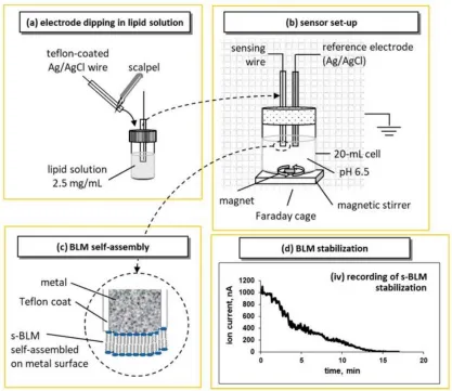
Figure 1
Representation of the device setup, and the lipid self-assembly process for
the preparation of metal supported sBLMs (not drawn to scale) based on the
original idea of [2]: (a) the tip of the sensing electrode is cut with a scalpel and immediately dipped in lipid solution before transferred in the electrolyte
(i.e., the sensing electrode and a Ag/AgCl reference electrode) in a
magnetically stirred 20-mL cell. The set-up is placed in a grounded Faraday
cage; 25 mV external DC potential is applied between the electrodes; the
ionic current through the BLM is measured with a digital electrometer. (c) Upon immersion, the lipid drop attached to the tip of the wire is
self-assembled into a bilayer; one layer is adsorbed on the metal surface and the
other faces the electrolyte. (d) Recording of the ion current decrease during the self-assembly process. The recording starts with the immersion of the
sensing electrode in the electrolyte solution [reprinted from ref. 3].
sBLMs have been fully characterized [2,4,5]. Device stabilization
depends upon the diameter of the wires and the organic solvent used [4,5].
Wires of 0.25 mm diameter should be avoided due to increased sensor noise;
the use of decane as a solvent should be also avoided as it enhances the
tendency for “black” lipid membranes that do not provide reproducible results.
Hexane solvent and silver wires with diameters of 0.5 and 1.0 mm provide
BLMs that are mechanically and electrically stable for over 48 hours.
Some attempts have been made to model the potential profile across
sBLMs and the structure of the lipid layer that faces the metal surface. A
plausible theory involves the interactions of oxygen atoms of the phosphate
groups of the lipid headgroups with the silver ions in the metal lattice [6,7].
Transmembrane ion mobility can be attributed to the presence of chloride ions
at the space between the metal and the inner lipid layer. There could be two
sources for chloride ions: through the lipid film during the initial BLM
stabilization process and through the partial wire insulation [4,5]. Chloride
experiments (against a Ag/AgCl reference electrode) [5] showed only small
voltages (relative to a silver wire against a Ag/AgCl reference electrode) when
the BLM had been removed using an organic solvent rinse. These results
suggest that (a) the metal surface is possibly coated with a thin layer of silver
chloride and (b) the lipid membrane actually consists of a network of nm-sized
BLMs [8].
2.2. Stabilized lipid films formed on a glass fiber filter
The preparation of stabilized lipid membranes supported on ultrafiltration
glass fiber filters has been reported in the literature [9] and allowed several
practical applications in real samples, such as the determination of aflatoxin M1
in milk and milk preparations [10]. The lipid membrane is formed on a
microporous filter glass fiber disk (namely GF/F glass microfiber, 0.9 cm in
diameter and 0.7 µm nominal pore size; Whatman Scientific Ltd., Kent, U.K.)
[9,10].
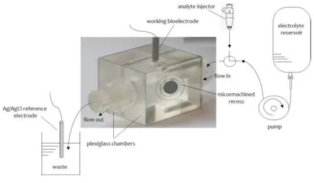
The experimental set up which was used for the formation of these
stabilized BLMs consisted of two plexiglas chambers separated by a thin
plastic partition (10 μm thick Saran-Wrap film). The plastic partition was folded
in half and a 0.32 mm hole was punched through the double layer of the
plastic film. A microporous glass GF/F microfiber disk was placed between the
two plastic layers, centered on the 0.32 mm hole. The partition containing the
filter was then clamped between the two plexiglas chambers. One of the
chambers had a circular shape (diameter 1.0 cm and depth 0.5 cm); this
chamber was connected with a carrier electrolyte flow system. An Ag/AgCl
reference electrode was immersed in the waste of the carrier electrolyte
perpendicular to the flow of the carrier solution. The upper hole of this cell was
circular (surface area of about 0.2 cm2) and the lower was elliptical (with
diameters 0.5 and 1.4 cm parallel and vertical to the flow of the carrier
electrolyte solution, respectively). The lower hole faced the opposing cell. An
Ag/AgCl reference electrode was positioned at the center of the cylindrical
cell. An external voltage of 25 or 50 mV d.c. was applied between the two
reference electrodes. A Keithley digital electrometer was used as a
current-to-voltage converter. A peristaltic pump was used for the flow of the carrier
electrolyte. Sample injections were made with a Hamilton repeating
dispenser. The electrochemical cell and electronic equipment were isolated in
a grounded Faraday cage. A simple scheme of the apparatus used is
presented in Fig. 2. Details for the procedure followed for the formation of the
stabilized BLMs can be found in [9,10], Briefly, a drop of the lipid solution (ca.
10 µL) was added to the electrolyte surface in the cylindrical cell near the
plastic partition. The level of the electrolyte solution was dropped below the
0.32 mm hole and then raised again within a few seconds. The formation of
the BLMs could be immediately verified by the ion current magnitude or/and
Figure 2
The experimental set-up used for the formation of stabilized lipid films on
glass fiber filters; the micromachined chambers are separated by a thin (12.5
μm thick) polyvinylidene chloride wrap and enclose the microfiber disk. For
more details, see text.[from ref. 3]
2.3. Polymer-supported bilayer lipid membranes
The preparation of polymer stabilized has been recently described in
literature. UV irradiation is much preferred [11,12], mainly because enzymes
retain their activity whereas heating the lipid mixture to 60 °C deactivates
them. Physicochemical methods, such as DSC, IR or Raman
spectrophotometry, indicated that the polymerization process requires 4
hours. This method facilitates a reliable and reproducible incorporation of
while the devices developed are stable outside the solution, i.e., in the air, for
more than 48 h.
The preparation of these stabilized lipid films was as follows [11,12]: 0.8
mL of a mixture containing 4% w/v egg phosphatidylcholine (PC) in n-hexane
were mixed with 0.07 mL of methacrylic acid, 0.8 mL of ethylene glycol
dimethacrylate, 8 mg of 2,2’-azobis-(2-methylpropionitrile) and 1.0 mL of
acetonitrile; n-hexane was used because it evaporates quickly leaving and the
films solvent-free. The mixture was sparged with nitrogen (1 min) and then
sonicated for 30 min. An aliquot of 0.15 mL of this mixture was spread on a
microfilter (microporous glass GF/F microfiber disk with a diameter of ca. 0.9
cm and nominal pore size of 0.7 µm) and irradiated using a UV deuterium
lamp. Raman spectrometry and differential scanning calorimetry (DSC) were
used to monitor the kinetics of the polymerization process. The measuring set
up was similar to that presented in Fig. 2. These membranes were stable in
storage in air for repetitive uses.
2.4. Polymer lipid films supported on graphene microelectrodes
Graphene nanomaterials have been extensively experimented upon
in an effort to seize their unique physicochemical properties: good sensing
ability, excellent mechanical and electrical properties, enhanced thermal
stability, large surface-to-volume ratio, improved biocompatibility, high
electron-transfer rates, limited toxicity and bio-safety. Their implementation in
electrochemical biosensing is quite beneficial as the large
response; the former might be proven critical for commercialization whereas
the latter allows for lower detectabilities while adequately handling biofouling
problems. Several nanobiosensors have been described using enzymes and
antibodies. A reliable system presented involves stabilized lipid films
wrapped around a copper wire containing graphene nanosheets [13,14].
These nanosensors have been implemented in the rapid detection of food
toxicants, environmental pollutants and toxins in real samples, such
insecticides [14], naphthalene acetic acid [15], cholera toxin [16], and
saxitoxin [17].
The preparation of graphene microelectrodes was as follows [13-17]:
using N-methyl-pyrrolidone (NMP) and mild sonication for 180 hours followed
by centrifugation at 700 rpm for 2 h yielded a homogeneous graphene
dispersion (∼0.4 mg/mL). The graphene suspension has been poured onto a
copper wire (0.25 mm in diameter) mounted on a glass fiber filter; the organic
solvent evaporated under a fan heater. The copper wire established the
connection for the extraction of voltage signals for the calibration curve.
Following a simple protocol, the drop wise dispersion of graphene suspended
in NMP solution has been utilized to scatter the graphene nanosheets on the
copper wire. The extended sonication time results in a good fraction of
monolayer sheets but with smaller lateral sizes.
The procedure of construction of these devices is in brief as follows
[13-17]: The stabilized lipid films were prepared by polymerization, as
described in [11,12]: 0.15 mL of a mixture containing 5 mg of a lipid powder
(w/w) of dipalmitoyl phosphatidyl choline (DPPC) (3.25 mg) were mixed with
0.070 mL of methacrylic acid, 0.8 mL of ethylene glycol dimethacrylate, 8 mg
of 2,2′-azobis-(2-methylpropionitrile) and 1.0 mL of acetonitrile. Phosphatidyl
choline (PC) is a more common lipid but because it can be oxidized by air and
does not provide reproducible results it has been replaced by DPPC. The
mixture was spumed with nitrogen for about 1 min and sonicated for 30 min.
This mixture could be stored in the refrigerator. For the preparation of the
stabilized lipid films, 0.15 mL of this mixture was spread on the glass filer
microfilter and irradiated. Raman spectrometry was used to monitor the
kinetics of the polymerization process [11,12].
The enzyme, antibody or receptor (“receptor”) was incorporated in
these BLMs prior to polymerization by spreading 15 µL of the “receptor”
suspension over the polymerization mixture. The filter-supported polymerized
lipid film was then mounted onto the copper wire containing graphene
nanosheets to produce the nano-device.
3. Applications of lipid film based biosensors in food analysis and environmental monitoring
The stabilized supported lipid membranes biosensors were used for the
detection of pesticides through flow injection analysis (FIA) [18]. The typical
pesticide studied was carbofuran. The analysis method was based on the
Carbofuran could be determined within the concentration range 10−7 - 10−9 M.
Interference studies used proteins and lipids that can be typically found in
foods. The results have shown no interferences from these compounds. The
sensor has been implemented in various real food samples, such as fruits,
vegetables and dairy products. The recovery ranged between 96% and 106%,
indicating no interferences from the sample matrix.
A paper was reported in the literature using a synthetic “receptor”
immobilized on supported lipid films on glass fiber filters. The supported lipid
films were modified by calixarenes and proved adequate for the sensitive and
rapid determination of various insecticides in fruit and vegetable samples [19].
Other devices similarly developed include a disposable chemosensor for the
selective and fast detection of food hormones such as naphthalene acetic acid
in fruits and vegetables [20] and a sensor for the detection of zinc in water [21].
A potentiometric urea lipid film based minisensor on graphene
nanosheets has been recently reported in the literature [22]. The structural
characteristics of graphene nanosheets have been extensively studied using
atomic force microscopy (AFM) and transmission electron microscopy (TEM).
The pre- and post-conjugated surfaces of graphene nanosheets has been
studied with UV-Vis and Fourrier transform IR (FTIR) spectroscopy. A
potentiometric urea biosensor has been developed (Figure 3) exhibiting good
reproducibility and reusability, high selectivity and fast response times (on the
order of 4 s), long shelf life under storage and a high sensitivity of ca. 70
mV/decade over the urea logarithmic concentration range from 1×10⁻⁶ M to
Figure 3. Photo of the lipid membrane based biosensor on graphene electrode developed for the potentiometric determination of urea (reprinted
from reference 23).
A nanosensor for naphthalene acetic acid (NAA) was based on stabilized
lipid films supported on a methacrylate polymer on a glass fiber filter with
incorporated auxin-binding protein 1 receptor [24]; the sensor has been tested
in reals samples of fuits and vegetables. Using a FIA system, NAA was injected
into the flowing carrier electrolyte solution and the flow stopped until an ion
current transient (peak) was obtained; the height of the peak could be correlated
to the concentration of the hormone in the sample. A micromolar detection limit
could be obtained. The analysis time was about 5 min. The effect of
interferences was studied using a wide range of compounds. The study
indicated no interferences from these compounds at concentration levels
usually found in real food samples. The sensor has been implemented for the
detection of NAA in fruits and vegetables and the reproducibility obtained was
A potentiometric carbofuran minisensor on graphene nanosheets with
incorporated lipid membranes has been reported [25]. The graphene
electrode was used to develop a carbofuran sensor using an artificial selective
receptor (resorcin[4]arene receptor) on stable lipid films. The detection range
was at the nanomolar levels, response times were ca. 20 s. The sensor was
easy to construct and exhibited good reproducibility, reusability, selectivity,
long shelf life and high electrode slope of ca. 59 mV/decade over the
carbofuran logarithmic concentration range from 10−6 to 10−3 M.
An atrazine lipid membrane sensor has been also described with
micromolar detection limit [26]. The interactions of atrazine with solventless
bilayer lipid membranes (BLMs) were found to be electrochemically
transduced by these films in the form of a transient current signal with
duration of seconds and reproducibly appearing within 1 min after the
membranes had been exposed to atrazine. The sensor could be optimized by
the introduction pf 35% (w./w.) DPPA in the lipid mixture and calcium ions in
the electrolyte solution; calcium induced alteration of the phase distribution of
DPPA doped membranes, thus increasing signals manifold. Alternatively,
doping PC membranes with platelet-activating factor (PAF; an ether analog of
PC) provided similar results.
The flow injection analysis of mixtures of the triazine herbicides
simazine, atrazine and propazine on PC/DPPA filter-supported BLMs has
been described in the literature [27]. When a sample containing a mixture of
these herbicides injected into the flowing electrolyte, a transient current signal
with a duration of seconds reproducibly appeared in less than two min after
concentration of the herbicides, which could be determined at µ range.
Repetitive cycles of injection of herbicides have shown no signal degradation
during each cycle. The time of appearance of the transient signal was
different for each triazine and increased to the order of simazine, atrazine and
propazine, thus permitting the simultaneous detection and analysis of these
triazines in mixtures.
A strategy was described in the literature that was based on monitoring
of changes of ion current through a lipid film with immobilized DNA probes
caused by interaction of these lipid membranes with hydrazine compounds
[28]. A s-BLM that was consisted of egg PC was deposited on a silver metal
electrode. The single stranded deoxyribonucleic acids used were thymidylic
acid icosanucleotide terminated with a C-16 alkyl chain to assist incorporation
into s-BLMs (dT20-C16), and deoxyadenylic acid icosanucleotide (dA20). These
s-BLMs with incorporated DNA interact with hydrazines, and it is possible to
monitor ppb levels of hydrazine, methylhydrazine, dimethylhydrazine and
phenylhydrazine. This BLM/DNA biosensor showed a highly sensitive,
selective, fast, and portable biosensor for monitoring these environmentally
and toxicologically significant compounds.
A paper appeared in the literature that describes the electrochemical
interactions of cholera toxin with polymerized lipid films incorporated with
ganglioside GM1 [29]. The analyte was injected into the flowing streams of a
carrier electrolyte solution, the flow of the solution stopped for 5 min and an
ion current transient was obtained. The magnitude of the signal could be
limits was 0.06 µM. Further work is directed to investigate the rapid detection
of other toxins used in bioterrorism and uses this novel ultrathin film
technology.
Switching to polymerized lipid membranes on graphene nanosheets,
Ganglioside GM1 provided better results, i.e., response time of ca. 5 min, and
detection limits of 1 nM [30]. The proposed sensor is easy to construct and
exhibits good reproducibility, reusability, selectivity, long shelf life and
sensitivity of 60 mV/decade of toxin concentration. The method was
evaluated, implemented and validated in lake water samples. The sensor is
currently adapted for the detection of other toxins.
A novel electrochemical biosensor based on a supported polymeric
lipid membranes with immobilized Sheep anti-PCB antibody for the rapid
determination of arochlor 1242 in flowing solution streams (FIA systems) has
been described [31]. The antibody was immobilized in the lipid membrane
during polymerization of the film; the injections of antigen were made into
flowing streams of a carrier electrolyte solution. The experimentation was
made in a stopped-flow mode; the lipid mixtures were composed of 15 %
(w/w) PA and 85% of DPPC to provide only one and only single transient
current signal with a peak height related to the concentration of the antigen.
Lipid films that were composed of 35 % DPPA were used to investigate the
regeneration of the active sites of antibody after complex formation; the
results showed adequate regeneration with intensive washing with the carrier
electrolyte solution. Repetitive cycles of injection of antigen have exhibited
A potentiometric saxitoxin minisensor based on graphene nanosheets
with incorporated lipid films and immobilized anti-STX (which is the natural
saxitoxin receptor) on stabilized lipid films was recently reported in the
literature [32]. A good selectivity and sensitivity for the detection of saxitoxin,
fast response times of ca. 5–20 min, and detection limits of 1 nM were
observed. The sensor is easy to construct, it has good reproducibility,
reusability, and selectivity, adequate storage stability and sensitivity (ca. 60
mV/decade over saxitoxin concentration). The method was evaluated and
validated in lake water and shellfish samples. This sensor can be easily
adapted for other toxins, as well.
An electrochemical biosensor that is suitable for the rapid and sensitive
screening of the sweetener sucralose based on surface-stabilized bilayer lipid
membranes (s-BLMs) composed of PC was recently described in the
literature [33]. The interactions of sucralose with s-BLMs provided an ion
current increase, that appeared within a few seconds after exposure of the
membranes to the sweetener. Differential scanning calorimetry was used to
investigate the mechanism of signal generation. The mechanism was found to
be related to changes of the electrostatic fields of the lipid membrane. These
studies have shown that there is an increase of the molecular area of the
lipids at the membranes and a stabilization of a gel phase structure; this was
due to adsorption of the sweetener in the membrane surface. The current
signal increases were correlated to the concentration of sucralose in bulk
solution in the µM concentration range. The present lipid film based biosensor
has provided a rapid response (order of seconds) to alterations of sucralose
of interactions of this sweetener with s-BLMs was evaluated by its
determination in granulated sugar substitute products.
A method that reports the FIA of mixtures of the artificial sweeteners
acesulfame-K, cyclamate, and saccharin using stabilized systems of
filter-supported BLMs has been proposed [34]. A transient current with duration of
seconds appeared in less than 1 min after exposure of the lipid membranes to
the artificial sweeteners. The peak height of the signal could be linearly
related to the concentration of artificial sweeteners, within a µM concentration
range; 30 analyses could be performed before the sensor showed signs of
signal degradation. The time of appearance of the signals was different for
each artificial sweetener and increased in the order of cyclamic acid,
acesulfame-K, and saccharin. This has allowed the simultaneous detection of
these artificial sweeteners in mixtures. Interference studies indicated no
interference from a large range of compounds commonly found in food
samples. The method has been implemented in real food samples (i.e.,
artificial sweetener tablets, diet soft drinks, wines, and yogurts) that contain
mixtures of artificial sweeteners. A comparison of results using this method
and that of an Official Method of Analysis showed good agreement between
the two methods.
Investigation of the transport phenomena through channels/pores it is
very important for various biological, medical, and technical applications. The
scope of a paper appeared in the literature is the development of nanofluidics
for the creation of biosensors capable of detecting single molecules and
manipulating them [35]. The detection of molecules was based on the
channel, which has a diameter comparable with the molecule size, the
current reduces. In order to improve transport properties of such channels,
their walls are often coated with a lipid bilayer, which behaves as
two-dimensional liquid and thus is capable of supporting transport phenomena.
Presently, this property of lipid membranes was utilized for the development
of a technique for detecting and controlling transport of single-stranded DNA
through channels formed by membrane cylinders with the luminal radii of 5–7
nm. It was demonstrated that in the conditions of small ion strength, the
appearance of a DNA molecule inside such channel is accompanied by an
increase of its ion conductivity and can be controlled by the polarity of the
applied voltage. The peak height of the current increase permits to evaluate
the number of DNA molecules inside the channels. It was also demonstrated
that upon adsorption of DNA molecules on the lipid bilayer surface, the
membrane cylinder behaves as a voltage-sensitive selective ion channel.
Biological membranes have been also studied. Eggshell, for example,
was used for the immobilization of urease in a potentiometric urea device [36].
Eggshell was treated with polyethyleneimine (PEI) to gain polycation
properties. Urease was adsorbed on the PEI treated eggshell membrane. A
SEM study was conducted in order to investigate the changes in surface
morphology and an FTIR study was carried out to observe the changes in IR
spectra after immobilization of the enzyme. The biosensor exhibited a
sigmoidal response for a urea concentration range from 0.5 to 10 mM. The
response time was 120 s. A single membrane could be used for 270 reactions
without loss of activity. The urease–eggshell membranes were stable for
Bilayer lipid membranes (BLMs) can be also produced by polymers
electrodeposited on a solid metallic support. Avidin–biotin interactions were
employed for enzyme immobilization on the BLM surface. A BLM glucose
biosensor based on glucose oxidase immobilized on a platinum support
modified with several polymers [37] showed improved storage stability and
better selectivity towards certain interfering electroactive species. Especially
good results were obtained for a mediated system in which the BLM was
formed on a Pt support covered with a layer of evaporated Nafion with
incorporated ferrocene. The stable and sensitive response with minimized
interference appears very promising for practical applications.
A chemiluminescence biosensor formed on a supported lipid layer
incorporated with ganglioside GM1 was reported for cholera toxin. The planar
supported lipid membrane was prepared as a biosensing interface via
spontaneous spread of ganglioside-incorporated phospholipid vesicles on the
octadecanethiol-coated gold surface [38]. The specific interaction of
multivalent toxin by ganglioside GM1 molecules enabled the implementation
of the sensor in a sandwiched format using a GM1 and horseradish
peroxidase (HRP) functionalized liposome probe, where the presence of the
toxin could be determined via the HRP-catalyzed enhanced
chemiluminescence reaction. This approach offers several advantages over
conventional strategies, especially as regards easy construction and renewal
of the sensing interface, small background noise (due to limited non-specific
adsorption of serum matrix constituents on the membrane), and effective
shared phospholipid reagents. The sensor could detect cholera toxin within
the range of 1 pg mL−1 to 1 ng mL−1 and a detection limit of 0.8 pg mL−1.
A work that reports a BLM-based nucleic acid biosensor supported by
modified patch-clamp pipette electrode was developed for staphylococcus
enterotoxins B (SEB) gene [39]. Hydrophobic dodecane tail (C12) modified
18 bp single-stranded DNA (ssDNA) probe was immobilized on the
membrane to yield linear correlations. The sensor was constructed by
selecting the ssDNA probe as the signal sensing element with the
concentration of 273.65 ng/mL. The electrochemical performance of the
biosensor for SEB detection was studied, showing a linear relationship
between the current and ln(concentration) from 20 to 5000 ng/mL, with a
detection limit of 20 ng/mL. In addition, the biosensor has shown a specific
response to SEB gene and no significant current alteration in the absence of
the SEB gene. AFM images were used to evaluate the microstructure of
BLMs, ssDNA immobilized on BLMs and BLMs after hybridization. The sensor
could be developed in a reliable tool for the detection of Staphylococcus
aureus, which produce SEB.
A nanostructured electrochemical biosensor was developed for
screening estrogenic substances using only the estrogen receptor (ER) [40].
ERs were immobilized in s-BLM modified with Au nanoparticles, and the
properties of the modified electrodes were characterized by cyclic
voltammetry and impedance spectroscopy. The results have shown that the
biosensor was able to detect 17β-estradiol (the natural estrogen) with a linear
correlation for the concentration range 5 - 150 ng/L and a detection limit of
bisphenol A and 4-nonylphenol with adequate sensitivity. The reliability of the
biosensor was good and the Au nanoparticles greatly enhanced the sensitivity
and stability of the sensor. The sensor was implemented for screening the
estrogenic activity of water samples and the results have been found to be in
good agreement with those determined by MCF-7 cell proliferation assay.
4. Conclusions and future prospects
The present paper describes a variety of approaches and strategies to
construct nanosensors based on lipid film technology and implement them for
food and environmental analyses. The recent technological advances include
the engineering of stabilized supported lipid film on graphene nanoelectrodes
with an incorporated “receptor” of any kind, natural or artificial. These films
remain stable in air and are suitable for the development of portable devices for
in the field applications. The sensors exhibit detection limits in the nM
concentration range. In effect, a portable unit that can be used for in-field and
market applications might be developed in the near future.
The results have shown that a variety of lipid film based detectors can be
reused after storage in air, even after few months, and can be reproducibly
fabricated with simplicity and low cost. These nanosensors have fast response
times and are easy to construct at quite lesser cost than chromatography-based
instrumentation; they can be also used as rapid hand-held detectors
complimentary to these methods for in-field and market measurements in foods
The present review describes biosensors based on lipid film technology
that can be used for the rapid detection of food toxicants and environmental
pollutants such as toxins, carbamates, hormones, polycyclic aromatic
hydrocarbons, etc and highlights their advantages which are high sensitivity
and selectivity, rapid response times, portability, etc. It is of common sense
that the use of nanotechnology to construct lipid membrane based biosensors
will provide devices with even improved characteristics.
Author Contributions
All authors contributed equally to this work.
Conflicts of Interest
The authors declare no conflict of interest.
References
1. Mueller, P., Rudin, D.O., Tien, H.T., Wescott, W.C. Reconstitution of cell
membrane structure in vitro and its transformation into an excitable system,
Nature 1962, 194, 979-980.
2. Tien, H.T.; Salamon, Z. Formation of self-assembled lipid bilayers on solid
substrates. J. Electroanal. Chem. Interfacial Electrochem. 1989, 22, 211–
3. Nikoleli, G.-P., Nikolelis, D., Siontorou, C.G., Karapetis, S. Lipid
membrane nanosensors for environmental monitoring: The art, the
opportunities, and the challenges. Sensors 2018, 18(1), 284;
4. Nikolelis, D.P.; Siontorou, C.G.; Krull, U.J.; Katrivanos, P.L. Ammonium
ion minisensors from self-assembled bilayer lipid membranes using
gramicidin as an ionophore. Modulation of ammonium selectivity by
platelet-activating factor. Anal. Chem. 1996, 15, 1735–1741.
5. Siontorou, C.G.; Nikolelis, D.P.; Krull, U.J.; Chiang, K.L. A triazine
herbicide minisensor based on surface-stabilized bilayer lipid
membranes. Anal. Chem.1997, 69, 3109–3114.
6. Hianik, T.; Dlugopolsky, J.; Gyepessova, M. Electrostriction of lipid
bilayers on a solid support. Influence of hydrocarbon solvent and d.c.
voltage. Bioelectrochem. Bioenerg. 1993, 31, 99–111.
7. Hianik, T.; Passechnik, V.I.; Sargent, D.F.; Dlugopolsky, J.; Sokolikova, L.
Surface potentials and solvent redistribution may explain the dependence
of electrical and mechanical properties of supported lipid bilayers on
applied potential and bilayer history. Bioelectrochem. Bioenerg. 1995, 37,
61–68.
8. Passechnik, V.I.; Hianik, T.; Ivanov, S.A.; Sivak, B. Specific capacitance
of metal supported lipid membranes. Electroanalysis 1998, 10, 295–302.
9. Nikolelis D.P., Siontorou C.G., Andreou V.G., Krull U.J. Stabilized
bilayer-lipid membranes for flow-through experiments. Electroanalysis. 1995, 7,
10. Andreou V.G., Nikolelis D.P. Flow injection monitoring of aflatoxin M1 in
milk and milk preparations using filter-supported bilayer lipid
membranes. Anal. Chem. 1998, 70, 2366–2371.
11. Nikolelis D.P., Raftopoulou G., Nikoleli G.-P., Simantiraki M. Stabilized
lipid membrane based biosensors with incorporated enzyme for repetitive
uses. Electroanalysis. 2006, 18, 2467–2474.
12. Nikolelis D.P., Raftopoulou G., Chatzigeorgiou P., Nikoleli G.-P., Viras K.
Optical portable biosensors based on stabilized lipid membrane for the
rapid detection of doping materials in human urine. Sens. Actuators B
Chem. 2008, 130, 577–582.
13. Nikoleli, G.-P.; Israr, M.Q.; Tzamtzis, N.; Nikolelis, D.P.; Willander, M.;
Psaroudakis, N. Structural characterization of graphene nanosheets for
miniaturization of potentiometric urea lipid film based
biosensors. Electroanalysis 2012, 24, 1285–1295.
14. Bratakou, S.; Nikoleli, G.-P.; Nikolelis, D.P.; Psaroudakis, N.
Development of a potentiometric chemical sensor for the rapid detection
of carbofuran based on air stable lipid films with incorporated
calix[4]arene phosphoryl receptor using graphene
electrodes. Electroanalysis 2015, 27, 2608–2613.
15. Bratakou, S.; Nikoleli, G.-P.; Siontorou, C.G.; Nikolelis, D.P.; Tzamtzis,
N. Electrochemical biosensor for naphthalene acetic acid in fruits and
vegetables based on lipid films with incorporated auxin-binding protein
receptor using graphene electrodes. Electroanalysis 2016, 28, 2171–
16. Karapetis, S.; Nikoleli, G.-P.; Siontorou, C.G.; Nikolelis, D.P.; Tzamtzis,
N.; Psaroudakis, N. Development of an electrochemical biosensor for
the rapid detection of cholera toxin based on air stable lipid films with
incorporated ganglioside GM1 using graphene
electrodes. Electroanalysis 2016, 28, 1584–1590.
17. Bratakou, S.; Nikoleli, G.-P.; Siontorou, G.C.; Nikolelis, D.P.; Karapetis,
S.; Tzamtzis, N. Development of an electrochemical biosensor for the
rapid detection of saxitoxin based on air stable lipid films with
incorporated Anti-STX using graphene
electrodes. Electroanalysis 2017, 29, 990–997.
18. Nikolelis, D. P., Simantiraki, M., Siontorou, G. C., Toth, K. Flow injection
analysis of carbofuran in foods using air stable lipid film based
acetylcholinesterase biosensor, Anal. Chim. Acta 2005, 537, 169-177 19. Nikolelis, D. P., Raftopoulou, G., Simantiraki, Μ., Psaroudakis, N.,
Nikoleli, G.-P., Hianik, T. Preparation of a selective receptor for
carbofuran for the development of a simple optical spot test for its rapid
detection using stabilized in air lipid films with incorporated receptor,
Anal. Chim. Acta 2008, 620, 134-141
20. Nikolelis, D. P., Ntanos, N., Nikoleli, G.-P., Tampouris, K. Development
of an electrochemical biosensor for the rapid detection of naphthalene
acetic acid in fruits by using air stable lipid films with incorporated
auxin-binding protein 1 receptor, Protein and Peptide Lett. 2008, 15, 789-794 21. Bratakou, S., Nikoleli, G-P. Siontorou, C.G., Karapetis, S., Nikolelis, D.P,
Tzamtzis, N. Electrochemical biosensor for naphthalene acetic acid in
protein receptor using graphene electrodes, Electroanalysis 2016, 28, 2171-2177
22. Nikoleli, G.-P.; Israr, M.Q.; Tzamtzis, N.; Nikolelis, D.P.; Willander, M.;
Psaroudakis, N. Structural Characterization of Graphene Nanosheets
for Miniaturization of Potentiometric Urea Lipid Film Based
Biosensors. Electroanalysis 2012, 24, 1285–1295.
23. Nikoleli, G.-P., Siontorou, C.G., Nikolelis, D.P., Bratakou, S., Karapetis,
S., Tzamtzis, N. Biosensors based on lipid modified graphene
microelectrodes, Carbon 2017, 3(1), 9; doi:10.3390/c3010009
24. Nikolelis, D. P., Raftopoulou, G., N. Psaroudakis, Nikoleli, G.-P.
Development of an electrochemical chemosensor for the rapid detection
of zinc based on air stable lipid films with incorporated calix4arene
phosphoryl receptor. Int. J. Environ. Anal. Chem. 2009, 89, 211-222 25. Bratakou, S.; Nikoleli, G.-P.; Nikolelis, D.P.; Psaroudakis, N.
Development of a potentiometric chemical sensor for the rapid
detection of carbofuran based on air stable lipid films with incorporated
calix[4]arene phosphoryl receptor using graphene
electrodes. Electroanalysis 2015, 27, 2608–2613.
26. Nikolelis, D.P.; Andreou, V.G. Electrochemical transduction of
interactions of atrazine with bilayer lipid
membranes. Electroanalysis 2005, 8, 643–647.
27. Nikolelis, D.P.; Siontorou, C.G. Flow injection monitoring and analysis
of mixtures of simazine, atrazine, and propazine using filter-supported
28. Siontorou, C.G.; Nikolelis, D.P.; Tarus, B.; Dumbrava, J.; Krull, U.J.
DNA biosensor based on self-assembled bilayer lipid membranes for
the detection of hydrazines. Electroanalysis 1998, 10, 691–694.
29. Nikoleli, G.-P., Nikolelis, D.P., Tzamtzis, N. Development of an
electrochemical biosensor for the rapid detection of cholera toxin using
air stable lipid films with incorporated ganglioside GM1. Electroanalysis
2011, 23(9), 2182-2189.
30. Karapetis, S.; Nikoleli, G.-P.; Siontorou, C.G.; Nikolelis, D.P.;
Tzamtzis, N.; Psaroudakis, N. Development of an electrochemical
biosensor for the rapid detection of cholera toxin based on air stable
lipid films with incorporated ganglioside GM1 using graphene
electrodes. Electroanalysis 2016, 28, 1584–1590.
31. Michaloliakos, A.I.; Nikoleli, G.-P.; Siontorou, C.G.; Nikolelis, D.P.
Rapid flow injection electrochemical detection of arochlor 1242 using
stabilized lipid membranes with incorporated sheep anti-PCB
antibody. Electroanalysis 2012, 24, 495–501.
32. Bratakou, S.; Nikoleli, G.-P.; Siontorou, G.C.; Nikolelis, D.P.; Karapetis,
S.; Tzamtzis, N. Development of an electrochemical biosensor for the
rapid detection of saxitoxin based on air stable lipid films with
incorporated Anti-STX using graphene
electrodes. Electroanalysis 2017, 29, 990–997.
33. Nikolelis D.P., Pantoulias S. A minisensor for the rapid screening of
sucralose based on surface-stabilized bilayer lipid membranes. Biosens
34. Nikolelis D.P., Pantoulias S. Selective continuous monitoring and
analysis of mixtures of acesulfame-K, cyclamate, and saccharin in
artificial sweetener tablets, diet soft drinks, yogurts, and wines using
filter-supported bilayer lipid membranes. Anal. Chem. 2001, 73(24), 5945-5952.
35. Chekashkina, K.V., Galimzyanov, T.R., Kuzmin, P.I. Akimov, S.A.,
Romanov, S.A., Pozmogova, G.E., Klinov, D.V., Bashkirov, P.V.
Detection of DNA molecules in a lipid nanotube channel in the low ion
strength conditions, Biochemistry (Moscow), Supplement Series A:
Membrane and Cell Biology, 2017, 11(3), 217–224.
36. D'Souza, S.F., Kumar, J., Jha, S.K., Kubal, B.S., Immobilization of the
urease on eggshell membrane and its application in biosensor, Mat. Sci.
& Engin. C 2013, 33, 850–854.
37. Trojanowicz, M., Miernik, A. Bilayer lipid membrane glucose biosensors
with improved stability and sensitivity, Electr. Acta, 2001, 46(7),
1053-1061.
38. .Chen, H., Zheng, Y., Jian, C.-Y., Wu, H.-L., Shen, G.-L., Yu, R.-Q. An
ultrasensitive chemiluminescence biosensor for cholera toxin based on
ganglioside-functionalized supported lipid membrane and liposome,
Biosens. & Bioelectr., 2008, 24(4), 684-689.
39. Liu, N., Gao, Z., Zhou, H.Y., You, M. Detection of SEB gene by bilayer
lipid membranes nucleic acid biosensor supported by modified
patch-clamp pipette electrode, Biosens. & Bioelectr., 2007, 22(9-10),
40. Xia, W., Li, Y., Wan, Y., Chen, T., Wei, J., Li, Y., Xu, S. ,Electrochemical
biosensor for estrogenic substance using lipid bilayers modified by Au