Resting-state functional connectivity in the brain and
its relation to language development in
preschool children
Der Fakultät für Biowissenschaften, Pharmazie und Psychologie
der Universität Leipzig
genehmigte
D I S S E R T A T I O N
zur Erlangung des akademischen Grades
doctor rerum naturalium
Dr. rer. nat.
Vorgelegt von
Yaqiong Xiao, M.Ed.
geboren am 07.04.1987 in Hunan/China
Dekan: Prof. Dr. Erich Schröger
Gutachter: Prof. Dr. Angela D. Friederici
Prof. Dr. Christian Fiebach
i
Acknowledgements
With the closure of the thesis, my heart is flooded with mixed feelings. I take this opportunity to express my sincere gratitude to some people without whom it would be impossible to have this thesis.
I am greatly thankful to Prof. Angela D. Friederici for giving me the wonderful opportunity to study in this well-known institute and work with excellent people. It is a great honor to be one of her doctoral students and actually, it is the greatest serendipity in my past life. It is just beyond my words to show my gratitude to her for having offered me stipend and supported me throughout my PhD studies over the past three years. I have been incredibly lucky to get her guidance on my research work. I appreciate her prompt response, and her invaluable comments and suggestion have really been beneficial to me throughout my research studies. Her insightful comments on the previous draft of the thesis have contributed significantly to the enrichment of the content. Moreover, her tireless efforts towards scientific work have inspired me all along. For me, she is not only a distinguished professor, supervisor, and leader but also a role model who is professional, gracious, and graceful.
I owe a lot to my advisor Dr. Jens Brauer who guided me in my PhD project and gave me direct help with my research work. I thank him very much for our countless discussions about data analyses during past years and his meticulous work on our previous publications. I appreciate it a lot that he has always been kind to me from the very beginning with his sincere approach and warm words. His patience and guidance were always a great support to me. I gradually learned from him about how to deal with tough questions from reviewers, how to work as hard as possible, and how to be an independent researcher. I am still on the road, but every piece of suggestion from him is precious for me and also valuable for my future career.
ii
My sincere thanks go to Dr. Daniel Margulies for providing me the great chance to attend the group meeting and journal club. I learned a lot from the presentations given in the group meetings and had fruitful discussions with his group members. In the past two years, attending the regular meetings enabled me to get access to the latest works in the field of resting-state fMRI and expanded my knowledge in this field. I also appreciate his contributions to the publications included in this thesis.
Over the past years, I have been very fortunate to get help and support from many colleagues. The first individual who comes to my mind is R. Muralikrishnan. We knew each other during my initial days when I came to the institute. During his stay, we had frequent conversations concerning daily life and research work. He always lent his help to me whenever I asked for it. His kind support was just like the light in the darkness, encouraging me to get through tough times. Even after he left, his extended help and support were still there for me. He might not be aware how important his unfailing support is for me and how much it has helped me over these years. My thanks should go to Xiangyu Long for his great help. His suggestions and comments on my data analyses were always useful. He is not only a colleague but also a tutor for me. He was generous enough to offer his help, and always tried to give answers to my endless questions. In some occasions when I felt puzzled and frustrated, he encouraged me as an experienced senior. I wholeheartedly appreciate him, and I still remember how much he has helped me and encouraged me in those struggling days. Dr. Thomas Gunter also deserves my heartfelt thanks. I appreciate his invitation to have Chinese lunch together and really enjoyed our conversation over lunch. He is very nice and always cares for the people around him. His willingness to offer help and support whenever they are needed is truly praiseworthy. I am the one who has benefited a lot from his kindness and favors.
My genuine thanks go to Dariya Goranskaya for providing me the warmth and kindness. We are of the same age, but her insightful thoughts always inspired me. I am fortunate to have such a
iii
smart, diligent, and self-confident officemate like her. Dariya was warmhearted to lend me a hand whenever I had questions or got into trouble. She was very generous and open-minded to share her knowledge, ideas, and even feelings. Her attention to my concerns was an important support to me, for which I cannot thank her more. Furthermore, her detailed comments on the previous draft of this thesis were very helpful. I also thank another officemate Benedict Vassileiou for his kind help with scripts in Matlab and some interesting discussion. My special thanks should go to Seung-Goo Kim for his kind help with some questions regarding data analysis. His extensive knowledge and research skills in the field of neuroscience and his thorough understanding of MRI methodologies inspired me a lot.
I am indebted to some more colleagues who have helped and supported me. They are Riccardo Metere, Mark Lanckner, Riccardo Cafiero, Marina Winkler, Emiliano Zaccarella, Kodjo Vissiennon, Chiao-Yi Wu, Caroline Beese, and Ulrike Kuhl. I am grateful to Peng Wang, Jing Jiang, Qianwen Miao, and Rui Zhang for their warm-heartedness and friendliness. I sincerely appreciate Lei Gao for being constantly in remote touch in the past years. Our discussion about data analyses and scientific research had ever excited me, and our online conversation had greatly supported me spiritually. During my studies, many people offered me extended support, amongst whom I especially thank Margund Greiner and Melanie Trümper (Secretary), Kerstin Flake and Andrea Gast-Sandmann (Graphics) as well as IT staff. In addition, I would like to thank the IMPRS NeuroCom for organizing various courses and Summer Schools, offering conference funding, and providing other support for my PhD studies. My grateful thanks go to our current coordinator Veronika Krieghoff and also former coordinator Katja Kirsche for lending me their help.
Last but not the least, I would like to thank my beloved parents from the bottom of my heart for their understanding and support all along in my life. I humbly believe that any progress in my life is beyond my persistence and hard work; instead, it comes from everyone who ever helped
iv
and inspired me mentally and supported me financially in my tough times. For those whose names are not mentioned here, I would say that you are in my mind and you have my gratitude all the time wherever I go.
In the end, I would like to encourage myself and any others who care for me with a famous old Chinese saying “Long as the way is I will keep on searching with my unbending will regardless of difficulties and hardships”.
v
Cogito ergo sum.
I think, therefore I am.
vii
ix
Bibliographishche Darstellung
Yaqiong Xiao
Resting-state functional connectivity in the brain and its relation to language development
in preschool children
Fakultät für Biowissenschaften, Pharmazie und Psychologie Universität Leipzig
Dissertation
114 pages, 199 references, 7 figures
Human infants have been shown to have an innate capacity to acquire their mother tongue. In recent decades, the advent of the functional magnetic resonance imaging (fMRI) technique has made it feasible to explore the neural basis underlying language acquisition and processing in children, even in newborn infants (for reviews, see Kuhl & Rivera-Gaxiola, 2008; Kuhl, 2010).
Spontaneous low-frequency (< 0.1 Hz) fluctuations (LFFs) in the resting brain have been shown to be physiologically meaningful in the seminal study (Biswal et al., 1995). Compared to task-based fMRI, resting-state fMRI (rs-fMRI) has some unique advantages in neuroimaging research, especially in obtaining data from pediatric and clinical populations. Moreover, it enables us to characterize the functional organization of the brain in a systematic manner in the
x
absence of explicit tasks. Among brain systems, the language network has been well investigated by analyzing LFFs in the resting brain.
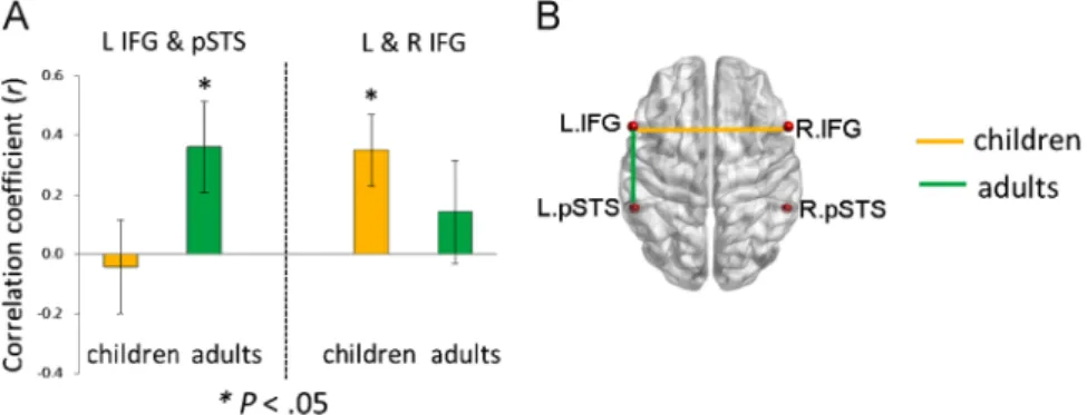
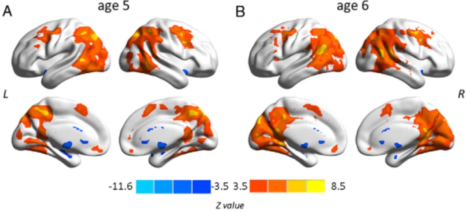
This thesis attempts to investigate the functional connectivity within the language network in typically developing preschool children and the covariation of this connectivity with children’s language development by using the rs-fMRI technique. The first study (see Chapter 2.1; Xiao et al., 2016a) revealed connectivity differences in language-related regions between 5-year-olds and adults, and demonstrated distinct correlation patterns between functional connections within the language network and sentence comprehension performance in children. The results showed a left fronto-temporal connection for processing syntactically more complex sentences, suggesting that this connection is already in place at age 5 when it is needed for complex sentence comprehension, even though the whole functional network is still immature. In the second study (see Chapter 2.2; Xiao et al., 2016b), sentence comprehension performance and rs-fMRI data were obtained from a cohort of children at age 5 and a one-year follow-up. This study examined the changes in functional connectivity in the developing brain and their relation to the development of language abilities. The findings showed that the development of intrinsic functional connectivity in preschool children over the course of one year is clearly observable and individual differences in this development are related to the advancement in sentence comprehension ability with age.
In summary, the present thesis provides new insights into the relationship between intrinsic functional connectivity in the brain and language processing, as well as between the changes in intrinsic functional connectivity and concurrent language development in preschool children. Moreover, it allows for a better understanding of the neural mechanisms underlying language processing and the advancement of language abilities in the developing brain.
xi
Contents
1 General introduction ... 1
1.1 A theoretical psycholinguistic framework of sentence processing ... 3
1.2 A neuroscientific model of language development ... 8
1.3 Neural basis underlying language processing ... 11
1.3.1 Ventral and dorsal pathway ... 11
1.3.2 Sentence processing in adults ... 14
1.3.3 Sentence processing in young children ... 18
1.4 Methodologies in brain research ... 21
1.4.1 BOLD functional magnetic resonance imaging ... 22
1.4.2 Resting-state fMRI ... 26
1.4.3 Approaches for rs-fMRI data analysis ... 30
1.4.3.1 RSFC and RSFC–behavior correlation analysis ... 30
1.4.3.2 Amplitude of low-frequency fluctuations ... 34
1.4.3.3 Degree centrality ... 36
1.5 The intrinsic language network ... 40
2 Publications ... 43
2.1 Development of a selective left-hemispheric fronto-temporal network for processing syntactic complexity in language ... 43
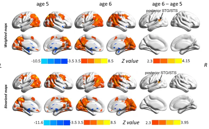
2.2 Longitudinal changes in resting-state fMRI from age 5 to age 6 covary with language development ... 55
xii
3 General discussion and outlook ... 69
3.1 General discussion of the findings ... 69
3.2 The outlook for future research ... 74
3.3 Conclusion ... 75 References ... 77 Summary ... 101 Zusammenfassung... 107 Appendices ... 113 Curriculum Vitae ... 113 Publications ... 114
1
1 General introduction
Language is one of the unique human capacities. Human infants learn their mother tongue in the first years of life easily and efficiently (for reviews, see Dehaene-Lambertz & Spelke, 2015; Friederici, 2005; Kuhl & Rivera-Gaxiola, 2008; Kuhl, 2010). A large body of research has shown the inherent competence of infants in language perception and acquisition. For example, early studies have observed that infants as young as 1 month of age are able to respond to speech sounds (Eimas et al., 1971) and they are also able to discriminate the acoustic cue underlying the phonemic distinction between voiced and voiceless stop consonants independent of relevant linguistic exposure at the age of 2 months (Streeter, 1976). Infants show a preference for phonemes in their native language by the age of 6 months, suggesting that the phonetic perception is shaped by the specific language that infants are exposed to, and it is detectable in the first half year of life (Kuhl et al., 1992). Language acquisition was found to be closely related to the statistical properties of the language input in 8-month-old infants (Saffran et al., 1996). Another study further investigated the statistical learning effect in language acquisition, and unveiled that infants aged 6 and 8 months are sensitive to the statistical distribution of speech sounds in the input language and this sensitivity influences speech perception (Maye et al., 2002). These findings demonstrate that infants are capable of distinguishing speech sounds and learning familiar speech patterns very quickly before the end of the first year.
The innate ability to acquire the mother tongue is regarded as a most fascinating puzzle in understanding the neural underpinnings of language, and a large number of theoretical and empirical studies have been presented to solve the mystery of language acquisition in young children. Over the past decades, with the advent of non-invasive neuroimaging techniques, there
2
have been numerous findings of the workings of the brain, which have allowed for a more comprehensive understanding of language acquisition and development in the early years of life.
The neural basis of language comprehension and production has been associated with superior temporal (Wernicke’s) and inferior frontal (Broca’s) cortical areas respectively. But, recent studies have reported a wide range of regions involved in language processing by employing blood oxygen level dependent (BOLD) functional magnetic resonance imaging (fMRI) (for a review, see Price, 2010). A recent study using the functional connectivity approach suggested a more extended network, including not only cortical regions in prefrontal, temporal, and parietal cortices but also subcortical regions in bilateral caudate, left putamen/globus pallidus, and subthalamic nucleus (Tomasi & Volkow, 2012). So far, most of the fMRI studies with regard to language processing have focused on adults, and fMRI data to characterize language processing in young children are still limited, not to mention data describing language-brain relationship in infants and young children (Skeide & Friederici, 2016).
In the past two decades, the resting-state fMRI (rs-fMRI) technique has been extensively used as a powerful tool to understand brain functions after the seminal study (Biswal et al., 1995). Different from traditional BOLD fMRI technique, no specific task is required for rs-fMRI, and data obtained in a relatively short scan length (as brief as 5 min) appear to be valid and reliable (Van Dijk et al., 2010). Thus, rs-fMRI has great advantages in acquiring data from pediatric and clinical populations who are not able to perform complex tasks or stay long in the scanner (for reviews, see Power et al., 2010; Uddin et al., 2010). Moreover, it provides an elegant way to characterize functional organizations of the brain in a systematic manner and to study various systems simultaneously (Cole et al., 2014; Smith et al., 2009). Among others, the language network has been well investigated by the analysis of low-frequency (< 0.1 Hz) fluctuations
3
(LFFs) in the resting brain (Tomasi & Volkow, 2012; Xiang et al., 2010). Xiang et al. (2010) first showed perisylvian language networks in a resting-state functional connectivity study, and Tomasi & Volkow (2012) demonstrated a widespread language network and its characterizations. These findings provide new insights into our understanding of the brain organizations involved in language functions.
So far, however, our knowledge of the functional brain connections within the language network with regard to language processing and their changes during the course of typical development in children is still very sparse, which does not allow us to relate the development of language abilities to brain maturation with age. Therefore, in the present thesis, using the rs-fMRI technique, I aimed to investigate the functional connectivity in the network of language-relevant brain regions and its relation to language processing in preschool children on the one hand, and to explore the developmental changes in this intrinsic connectivity and its response to language development in the typically developing brain on the other hand.
1.1 A theoretical psycholinguistic framework of sentence processing
Human language is not simply naturally acquired devoid of any context or pressure. Instead, language acquisition is a dynamic process interacting with multiple factors, including auditory patterns, articulatory patterns, social patterns, patterns implicit in the input, and pressures arising from general aspects of the cognitive system (for a review, see MacWhinney, 1998). Under this conception, the Competition Model was proposed based on cross-linguistic studies of sentence processing in young children, and it treats language learning as an emergent process with the goal of investigating the competitive relationships between lexical items, phonological forms, and syntactic patterns during language processing (Bates & MacWhinney, 1982, 1989; Bates et
4
al., 1984; MacWhinney, 1987). The term “competition” refers to the competition of assigning the actor’s role among several nouns in a sentence. For example, consider the sentence “The boy is petting the cat”: two nouns are involved in this sentence, and the competition occurs while deciding which one is the actor. In a sentence, the thematic roles could be basically characterized by proto-agent and proto-patient, and an agent carries more agentivity property than a patient based on the degree of agentivity (Dowty, 1991). Because of the contrary relationship between the agent and patient, there commonly exists a direct competition for the agent and patient identification.
The main focus of the Competition Model is the use of different cues for sentence comprehension. The cue is an information source that can be used by the language user to understand sentences in a language. When multiple cues are available for sentence interpretation, either competition or convergence between different cues will happen, which is dependent on the directions that different cues point toward. For instance, in the sentence “the carrot bites the cat”, word order and animacy cue point to different nouns as the agent of the action (word order implies “the carrot” while animacy cue suggests “the cat”) where competition occurs, but in the sentence “the cat bites the carrot”, both word order and animacy cue point to the same noun – “the cat” – as the agent where convergence happens.
An earlier study found that in case-inflected languages such as German, utterances of children rely on the dominant word order that serves as a cue in sentence comprehension before they have mastered the morphology of their language (Brown, 1973). However, findings from a cross-linguistic study concluded that it is not a universal predisposition for children to use word order as a cue to comprehend sentences. Rather, the use of a cue is dependent on the regularity and consistency of the language (Slobin & Bever, 1982), which is consistent with the claims of the
5
Competition Model. According to this model, learning of language forms is closely associated with the accurate recording of multiple exposures to words and patterns in different contexts, and sentence interpretation is supported by linguistic cues (Bates & MacWhinney, 1982; Bates et al., 1984; MacWhinney, 2012).
The model claims that the acquisition of a certain cue strategy during the course of language development is based on competition between the strength of cues, reflecting the extent of cues dominating or controlling language comprehension. The cue strength is primarily determined by cue validity, which is characterized by both availability and reliability. According to the model, the reliability of cues is defined as the proportion of times that the cue is correct over the total number of occurrences of the cue, and the availability of the cue is the proportion of times that the cue is available over the times the cue occurring; the product of cue reliability and cue availability is cue validity (MacWhinney, 2012). Cue reliability is the basic factor for cue validity, indicating that a presented cue is not misleading or ambiguous, and a cue with high reliability will lead to a correct functional choice. The availability of a cue represents to what extent it is available whenever needed, and a cue with high availability always exists for use. The most valid cues are those with both high reliability and availability, but cues with low reliability are not valid anymore even when they are highly available.
Previous behavioral results from cross-linguistic studies provide evidence for the different cue strategies in adults from different language backgrounds. In English, the dominant cue is the word order, i.e., subject-verb-object order; in German, case marking is the strongest cue, and the article serves as a cue to identify an agent; in Italian, agreement is the dominant cue; in Russian, case-marking, the case suffix in nouns, is the major cue; in Chinese, animacy cue is mostly used to point out the agent (Kempe & MacWhinney, 1998; Li et al., 1993; MacWhinney, 2001;
6
MacWhinney et al., 1984). Besides, the Competition Model has often been applied to the prediction of cue usage and its changes in young children. Given their immature language ability, cues are still inadequately processed in young children, but they can make full use of the cues to which they are frequently exposed, thereby showing different cue strategies across languages. For example, English-speaking children tend to use word order while Italian-speaking children tend to use animacy cue as young as 2 years of age (Bates et al., 1984), and Turkish-speaking children prefer case markers as cues with exposure to more reliable markers (Slobin & Bever, 1982). Moreover, ample behavioral evidence has shown that the factor affecting the cue strength varies with language development. At the very beginning, the acquisition of a cue is mainly dependent on cue availability because infants are only familiar with cues that are frequently presented in the language input, but with the increase of simultaneously presented multiple cues in the expanding language input, cue reliability becomes more important than cue availability and even dominant in the cue strength based on the proficiency level (MacWhinney, 2012).
Among languages, German is the focus of this thesis, which has been widely studied in order to confirm the validity of the Competition Model because of the language’s complexities of declension. The results successfully matched the model and showed the crucial role of cues in the acquisition of German declension (MacWhinney et al., 1989). Cues in German include case marking, subject–verb agreement, animacy, and word order, and among these cues case-marking is the strongest in adults (MacWhinney et al., 1984). In contrast to English, German has a relatively free word order and therefore cues other than word order are often needed to comprehend sentence information for early learners. The following example illustrates this:
7 (1) Canonical subject-initial sentence:
Der[NOM] Tiger zieht den[ACC] Fuchs.
“The tiger pulls the fox.”
(2) Non-canonical object-initial sentence:
Den[ACC] Fuchs zieht der[NOM] Tiger.
“The tiger pulls the fox.”
The functions of the noun phrases in the sentences above are marked by the nominative case (NOM) as a subject or the accusative case (ACC) as an object. The nominative case article “der” indicates “der Tiger” as the agent (actor) of the sentence, while the accusative case article “den” points to “den Fuchs” as the patient (receiver) of the action. As shown in the example sentences, the order of the noun phrases does not affect their functions, but the case marking (nominative or accusative) of the noun’s article conveys the information. In this example, flexible word order does not carry reliable cue for comprehending these sentences, but instead, case-marking cue has high availability. Based on the Competition Model, cue reliability is the main determinant of the cue strength, and low reliability of word order in German makes it hardly a dominant cue, whereas case-marking cue is a more practical candidate due to its high availability (MacWhinney et al., 1984). As mentioned earlier, however, a cue with high availability is not valid at all if its reliability is low.
Previous research has found that, compared to English and Italian, German has a strong preference for animate actors, indicating animacy serving as an important extra cue to rely on except for case marking (MacWhinney et al., 1984). A second-language learning study observed
8
that learners of German rely more on animacy information to supplement the case-marking cue when it is weak owing to the syncretism in the nominative and accusative cases of German feminine, neuter, and plural nouns (Kempe & MacWhinney, 1998). Research on German children 2-7 years showed that 2- to 3-year-olds rely mainly on the animacy cue and the case-marking cue is dominant from age 4 on, but agreement is the strongest cue afterward even in adults (Lindner, 2003). These empirical findings in the early stage of acquisition of German suggest that case-marking, despite its high availability, is not a valid cue under certain conditions because of its low reliability, and the animacy cue comes into play until the case-marking cue becomes more reliable.
1.2 A neuroscientific model of language development
During the past decades, the application of neuroimaging techniques, including electroencephalography (EEG)/event-related potentials (ERP), magnetoencephalography, fMRI, and near-infrared spectroscopy, has made it possible to explore the brain mechanisms underlying early language acquisition (for reviews, see Kuhl & Rivera-Gaxiola, 2008; Kuhl, 2010). Among these techniques, ERP has been the most widely used in investigating speech and language processing in infants and young children due to its operability and validity. Findings from ERP studies provide evidence for quantitative changes in language processing, demonstrating neurophysiological correlates of language acquisition in early childhood. In a review study, Friederici (2005) summarized the trajectory of language acquisition in infants from a number of previous ERP studies: infants are able to discriminate different phonemes in the first 2 months of life; they learn knowledge of stress patterns and phonotactic rules between 5–12 months; they develop phonotactic knowledge associated with lexical-semantic processes between 12–14
9
months and they are able to process semantics of words in picture contexts at 14 months; infants process words in sentential contexts around 30 months and they show electrophysiological response patterns to syntactic violations at 32 months. With respect to syntax acquisition, research has shown that 2-year-old toddlers are able to process syntactic information of sentences during listening to spoken sentences (Bernal et al., 2010), and they are also sensitive to syntactic errors in the form of phrase structure violations (Oberecker & Friederici, 2006).
Due to its relatively high spatiotemporal resolution, fMRI has been employed prevalently in adults and has even been applied to infants. However, given the difficulties of obtaining data from such young populations, only a few fMRI studies concerning language processing have been conducted in infants (Dehaene-Lambertz et al., 2002; Dehaene-Lambertz et al., 2006; Perani et al., 2011). Dehaene-Lambertz et al. (2002) performed an fMRI experiment in awake and sleeping 3-month-old infants to examine their brain responses while listening to sentences of their native language presented either forward or backward. The authors observed activations in adult-like left-lateralized regions, such as superior temporal and angular gyri. Another fMRI study investigated 3-month-old infants’ BOLD response to short sentences of their mother language presented in a slow event-related paradigm (Dehaene-Lambertz et al., 2006). This study observed slow BOLD responses in the bilateral superior temporal regions and Broca’s area, and found activations in Broca’s area engaged in processing repeated sentences. Perani et al. (2011) examined brain activations in 2-day-old newborns during listening to a story presented in three different conditions (i.e., normal speech, hummed speech, and flatted speech) and found strong activations in the bilateral superior temporal gyrus (STG) and inferior frontal gyrus (IFG) for both normal and hummed speech as reported in adults. In addition, this study confirmed activations in both hemispheric auditory cortices for normal speech, but with a right predominant
10
activation in the right hemisphere. These findings demonstrated primary evidence for the brain mechanisms of early language processing.
Recently, Skeide & Friederici (2016) proposed a model of the ontogeny of the cortical language network based on neuroimaging data from EEG and fMRI studies describing the acquisition of language in early years of life (Figure 1.1). According to this model, there are two main developmental stages. The first stage, referring to bottom-up processing, is primarily implemented in the bilateral temporal cortices and acquired in the early childhood. In this stage, with the exposure to language inputs and the pruning of neurons, infants are gradually able to deal with phonological, prosodic, and semantic information. The second stage is to develop the ability of sentence-level syntactic processing in a top-down manner, mainly involving the left inferior frontal and superior temporal cortices. By 2 years of age, children can already process semantic relations in a sentence (Friedrich & Friederici, 2010). At age 3, they are able to process syntactic information in sentences, but still inadequately. Thus, young children make use of semantic information in sentences as a cue to understand syntactically complex sentences until around 9 years of age (Skeide et al., 2014). This ability continues to increase into adulthood, as it is related to the cortical specialization of the left IFG and left posterior STG and it is also associated with the maturation of their underlying structural connection. More details on this point will be outlined in the following section.
11
Figure 1.1 The model of the ontogeny of the cortical language network (a), including two stages: bottom-up processes (colored in green) and top-down processes (colored in orange); the corresponding timeline of language acquisition (b; age in years). Arrows indicate the assumed flow of information along interconnecting white matter fiber tracts. aSTG: anterior superior temporal gyrus; aSTS: anterior superior temporal sulcus; pSTG: posterior superior temporal gyrus; pSTS: posterior superior temporal sulcus. (Figure adapted from Skeide & Friederici (2016)).
1.3 Neural basis underlying language processing
1.3.1 Ventral and dorsal pathway
The language processing framework was suggested to be characterized by two divergent processing streams (Hickok & Poeppel, 2000) and subsequently, it was extended to describe the
12
functional anatomy of language (Hickok & Poeppel, 2004). In this extended framework, a ventral pathway connecting the left superior temporal sulcus (STS) and posterior inferior temporal regions (i.e., parts of the middle temporal gyrus (MTG) and inferior temporal gyrus) is involved in sound-to-meaning mapping, and a dorsal pathway connecting the left inferior parietal and ventral frontal cortices is engaged in sound-to-motor mapping (Hickok & Poeppel, 2004). These two pathways have been widely acknowledged as the brain basis of language processing (e.g., Friederici, 2012a; Hickok & Poeppel, 2007; Rauschecker, 2012; Rauschecker & Scott, 2009; Saur et al., 2008). Combining fMRI and a diffusion tensor imaging approach, this model was further tested in terms of its neuroanatomical basis, which was identified to comprise a dorsal pathway connecting the superior temporal lobe and premotor cortices in the frontal lobe via the arcuate and superior longitudinal fasciculus (AF/SLF) and a ventral pathway connecting the middle temporal lobe and the ventrolateral prefrontal cortex via the extreme capsule (Saur et al., 2008).
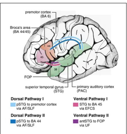
On the basis of numerous functional and anatomical neuroimaging studies, the dual-pathway model was refined and subdivided into double ventral and dorsal pathways with distinct functions in language processing (for reviews, see Friederici, 2011, 2012a) (Figure 1.2). One ventral pathway connecting BA 45 and the temporal cortex via the extreme capsule fiber system is related to sound-to-meaning mapping, while the other ventral pathway connecting the anterior STG and IFG is assumed to support processes of local syntactic structure building. One dorsal pathway connecting the posterior STG/STS to the premotor cortex via the AF/SLF is engaged in auditory-to-motor mapping, and the other dorsal pathway connecting the posterior STG/STS to BA 44 via the AF/SLF is involved in processing syntax, especially processing complex syntactic information.
13
Figure 1.2 Language processing pathways on the basis of structural connectivity, including two ventral and two dorsal pathways. BA: Brodmann area; p: posterior; ant: anterior; FOP: frontal operculum; AF: arcuate fasciculus, SLF: superior longitudinal fasciculus; EFCS: extreme fiber capsule system; UF: uncinate fasciculus. (Figure adapted from Friederici (2011)).
With regard to brain maturation, previous studies in newborn infants and young children have offered primary evidence for the different developmental rates of fiber tract connections underlying the ventral and dorsal pathways (Brauer et al., 2011; Brauer et al., 2013; Broce et al., 2015; Perani et al., 2011). It has been demonstrated that the ventral pathway is already present at birth and matures rapidly; however, one part of the dorsal pathway, connecting the temporal cortex to Broca’s area, is still underdeveloped at the age of seven years, while the other part of
14
the dorsal pathway connecting the temporal to the premotor cortices is already present at birth (for a review, see Friederici, 2012b). The immature fiber connection between the temporal cortex and Broca’s area is regarded to be associated with the inability of processing syntactically complex sentences in young children (for a review, see Friederici, 2012b). The close relationship between brain maturation and the concurrent development of sentence comprehension in children will be described in detail, and prior to this, the brain basis of sentence processing in adults will be introduced.
1.3.2 Sentence processing in adults
Over the last two decades, the neural mechanisms underlying sentence processing in adults have been well investigated using the fMRI technique. As the core of sentence comprehension, the process of syntactic information has been of a particular interest to researchers and has been studied thoroughly by different paradigms (e.g., word lists, sentence-level violations, and manipulation of word order).
In pioneering studies, it has been shown that sentence reading with varying syntactic complexity selectively activated Broca’s area in the left IFG by using positron emission tomography (PET) (Caplan et al., 1998; Stromswold et al., 1996). An early fMRI study, using three types of sentences that differ in structural complexity, revealed a modulation effect of sentence complexity in activations of Broca’s and Wernicke’s areas as well as their right homologs, but activations were much stronger in the left hemisphere (Just et al., 1996). These results showed primary brain networks involved in sentence-level syntactic processing; however, sentence-level semantic information, which generally interacts with sentence syntax, had not been taken into account yet. In order to differentiate the brain regions engaged in auditory language
15
comprehension, Friederici et al. (2000) varied the speech input in the presence or absence of semantic and syntactic information. The authors observed activations in the bilateral STG across different types of auditory language input, and additional activations in both left and right frontal cortices in violation conditions (i.e., syntactic speech and two word-list conditions) but not in the processing of normal speech. Moreover, the increase of activations in the bilateral anterior portion of the STG and deep portion of the left frontal operculum (FOP) was exclusively found in the focus of syntactic processes, suggesting an involvement of the left frontal and bilateral temporal cortices when processing syntactic information in sentences (Friederici et al., 2000). The differentiation of brain regions involved in processing sentence-level semantic and syntactic information was further investigated by using a violation paradigm, and it was demonstrated that semantic violations activated mainly the bilateral mid STG and the insular cortex, whereas the left anterior STG, the left posterior FOP, and the putamen were engaged in parsing syntactic violations (Friederici et al., 2003). Taken together, these findings suggest a temporo-frontal network related to both semantic and syntactic processes, but with distinct regions specialized for each process, such as the anterior STG specifically for syntactic processing (for a review, see Friederici, 2002).
ERP findings in adults suggest that the processing of syntactic violations can be divided into an early stage corresponding to processing phrase structure or agreement violations and a late stage corresponding to processing syntactic violations requiring syntactic repair or where temporarily ambiguous sentences require syntactic reanalysis (for reviews, see Friederici, 2002; Friederici & Kotz, 2003). Moreover, data from a number of experimental studies showed separable brain systems responsible for the early and late syntactic processes: the left anterior STG and left IFG are engaged in the early structure building processes, and the posterior STG and basal ganglia are
16
involved in late syntactic revision and integration processes (for a review, see Friederici & Kotz, 2003).
It has been demonstrated that the ability to deal with hierarchically structured sequences such as embedded structures like A[AB]B is unique in human, although non-human primates can also process and learn simple sequences like ABAB (Fitch & Hauser, 2004). The ability of comprehending the hierarchical structure is crucial for humans to understand more complex sentence information in language, but it requires additional computation compared to simple sequences and thus non-human primates are not able to process embedded structures (Fitch & Hauser, 2004). Following this study, Friederici et al. (2006a) investigated the functional differentiation of the brain regions responsible for comprehending these two grammars of different complexity and observed that the left FOP was involved in parsing both grammar types while BA 44 was additionally engaged in hierarchical information processing. This study also revealed differential structural connectivity that the left FOP is connected to the left anterior temporal lobe via the fasciculus uncinatus, whereas BA 44 is connected to the posterior STG in the left hemisphere via the fasciculus longitudinalis superior (Friederici et al., 2006a).
In order to further appreciate the brain basis of syntactically complex sentence processing, several studies have been performed by manipulating the argument hierarchies in sentences, such as the order of the arguments. In the examples shown in Chapter 1.1, reproduced here for convenience: “Der[NOM] Tiger zieht den[ACC] Fuchs” and “Den[ACC] Fuchs zieht der[NOM] Tiger”,
the first one with a sentence-initial nominative argument is considered a canonical subject-initial sentence, whereas the second one with a sentence-initial accusative argument is considered a non-canonical object-initial sentence. The object-initial sentence is syntactically more complex than the subject-initial sentence because of the non-canonical structure. Röder et al. (2002) used
17
syntactically easy and difficult sentences by varying the word order of nominal phrases in German, and reported the strongest effect of syntactic difficulty in the left IFG, i.e., BA 44/45. Friederici et al. (2006b) manipulated the complexity and grammaticality of sentences within the domain of word order variations in German, and observed that the pars opercularis of the left IFG (left IFGoper; BA 44) was especially involved in processing structural complexity of such sentences. Another study on sentence embedding (nested structures) in German also showed the activation in BA 44 that was independent of working memory load (Makuuchi et al., 2009). Furthermore, although the complexity of sentence structures is accompanied invariably by increasing working memory or task difficulty, it has been confirmed that the involvement of the left BA 44 in processing syntactically complex sentences is attributed to the hierarchical structure of sentences rather than the increasing working memory demand (Grewe et al., 2005; Makuuchi et al., 2009).
Notably, the activation in the left posterior STG/STS has been widely reported for complex syntactic processing (Bornkessel et al., 2005; Friederici et al., 2006a; Friederici et al., 2010; Friederici et al., 2009; Kinno et al., 2008; Newman et al., 2010; Santi & Grodzinsky, 2010). For example, it was found that the left posterior STG/MTG showed more enhanced responses to non-canonical object-initial sentences, the syntactically more complex sentence type, than non-canonical subject-initial sentences (Kinno et al., 2008). Friederici et al. (2009) observed activations in the bilateral posterior superior temporal cortex (i.e., left posterior STG/STS and right posterior STS) during complex sentences processing, implying the importance of the posterior superior temporal regions for processing sentential syntactic information. Comparing sentences containing a syntactic violation with syntactically correct sentences, it was observed that syntactic phrase structure violations engendered strong activation in the mid to posterior STG/STS (Friederici et
18
al., 2010). A meta-analysis based on a number of language-related neuroimaging studies revealed that higher syntactic processing demands primarily activate regions including BA 44/45 and the posterior STG/MTG in the left hemisphere (Hagoort & Indefrey, 2014).
Collectively, BA 44 together with the posterior STG/STS, constitutes a brain network in the left hemispheric inferior frontal and superior temporal areas for processing syntactic complexity of sentences (for a review, see Friederici, 2011). These findings have greatly enriched our understanding of the brain mechanisms underlying syntactic processing in adults, which could be regarded as a model of sentence processing in the human brain. Therefore, it provides a basis for studying typically developing children who are undergoing rapid development as their brains mature with age. Furthermore, it enables us to investigate the trajectory of language development from childhood to adulthood.
1.3.3 Sentence processing in young children
So far, mounting studies have investigated sentence processing in young children. However, research regarding different cues for sentence comprehension in early childhood is still sparse. As mentioned above, young children are still in the process of learning the valid cues for sentence comprehension, which has been examined by several behavioral studies (Chan et al., 2009; Dittmar et al., 2008; Gertner & Fisher, 2012; Lindner, 2003; Noble et al., 2011). For example, using a forced-choice pointing paradigm in English-learning children, Noble et al. (2011) found that, 2-year-olds can interpret agent and patient roles correctly in transitive sentences even with causal events, but cannot assign the conjoined agent in intransitive sentences with noncausal events until age 3. Dittmar et al. (2008) investigated cue usage in German children, and observed that 2-year-olds can understand sentences correctly when both word order
19
and case marking are valid cues and that 5-year-olds can use word order alone but not case marking alone; the latter became a dominant stand-alone cue only in 7-year-olds. An ERP study by Schipke et al. (2012) focused on the processing of case-marking and argument structures in German children aged 3, 4.5, and 6 years. The results showed that children at the age of 3 and 4.5 years use mainly a word-order strategy, although children at 4.5 years are already sensitive to case-marking cue; at the age of 6 years children can use case-marking cues, but still need extra effort for correct thematic role assignment. These studies indicate that the cue strategy in young children varies with the cue strength at different stages, which in turn reflects the development of sentence comprehension abilities with age.
In recent years, fMRI studies investigating the neural basis of sentence processing in young children have emerged. Brauer & Friederici (2007) studied sentence comprehension in terms of semantic and syntactic processes using violations in sentences, and observed function-specific activations in the left FOP and bilateral STG in adults for violations. Instead, children showed no specific activations in the bilateral STG for different language conditions, but compared to adults, they additionally engaged areas in the left IFG to support higher demanding processes. This study suggests that perisylvian regions, especially the left IFG and STG, play a crucial role in sentence comprehension, and these regions have not yet specialized in 5- to 6-year-old children whose brains are still immature. Further, Brauer et al. (2008) examined the time courses of the BOLD hemodynamic responses in fMRI during sentence processing, involving the bilateral inferior frontal cortex (IFC) and superior temporal cortex (STC). This study showed an overall later peak of BOLD responses and a temporal primacy of right over left hemispheric activations in children compared to adults. Moreover, it observed much later IFC responses than STC responses in children, but the difference is less pronounced in adults. These findings
20
indicate, due to slower and less automatic language processes, children need higher processing costs to comprehend sentences than adults, especially in the bilateral IFC. By using a multimodal approach and a syntactic processing task, Nuñez et al. (2011) showed a negative correlation between cortical thickness and the activation extent in the right IFG with age. The authors also reported a positive relationship between better syntactic performance and increased activation in the left IFG regardless of age, which suggests a decreased involvement of the right IFG for syntactic processing with age and an increased engagement in the left IFG with improved language skills.
The acquisition of case-marking cues for sentence interpretation was investigated by Knoll et al. (2012). This study tested typically developing German preschool children aged 6 years using auditorily presented short sentences which were either canonical subject-initial or non-canonical initial sentences. The results demonstrated that the left IFG was only activated for object-initial sentences but not for subject-object-initial sentences. In addition, individual differences showed a trend towards greater activation in the left IFG for object-initial sentences than for subject-initial sentences, but only in children with high grammatical performance. Yeatman et al. (2010) examined the brain activation network in response to increasing demands of complex sentences in children and adolescents aged 10-16 years using a cross-modal sentence-picture verification paradigm. This study reported greater activation of the IFG in response to increasing task demands in children with better language skills than children with average language abilities. In a recent study, sentence processing in children aged 3 to 4, 6 to 7, and 9 to 10 years was investigated using a paradigm that combined syntactic complexity and semantic plausibility (Skeide et al., 2014). Results found the syntax-semantics interaction in the left mid to posterior STG in 3- to 4- and 6- to 7-year-old children due to the reliance on semantic plausibility cues,
21
whereas children at 10 years of age were able to process syntax independent of semantics and showed adult-like activation in the left IFGoper. These findings suggest that the processing of syntax becomes gradually separated from the processing of semantics, and they also present a functional specification of cortical regions that support complex syntax processing with age. Subsequently, the relation between brain function and language performance as well as between brain structure and language performance was tested in the same cohort of children (Skeide et al., 2015). The authors observed that enhanced performance in processing syntactically complex sentences was not only positively correlated with activations of core language processing regions – left IFGoper and posterior STG, but also associated with the maturation of AF connecting these two regions (Skeide et al., 2015).
Taken together, these findings show that language-related regions, mainly the left hemispheric IFG and posterior STG, are related to the processing of sentential syntactic complexity in developing children and that, noteworthily, activations in these regions can predict children’s abilities to parse syntactically complex sentences (Skeide et al., 2015). It indicates that the investigation of individual differences might be useful for elucidating the neural mechanisms of language development in children.
1.4 Methodologies in brain research
In this section, I will firstly give an overview of the physical and physiological underpinnings of BOLD fMRI. On this basis, rs-fMRI will be introduced in detail as it is the main technique applied in the studies included in this thesis. Finally, I will outline several approaches used to analyze rs-fMRI data presented in Chapter 2.
22
1.4.1 BOLD functional magnetic resonance imaging
In the early 1990s, a seminal study by Ogawa et al. (1990) discovered that the BOLD contrast can provide maps of blood oxygenation in the brain by scanning anesthetized rodents at 7.0 Tesla MRI scanner. Shortly afterward, the BOLD contrast was firstly utilized in human neuroimaging and showed increases of the local signal during visual and motor stimulation, suggesting that the functional brain mapping is feasible by using intrinsic blood-tissue contrast (Kwong et al., 1992). At this time, other research independently published and reported similar findings in the human brain (Bandettini et al., 1992; Ogawa et al., 1992). These early studies consistently revealed that functional activation increases the regional cerebral blood flow (CBF), which results in an increased engagement of oxygenated blood.
Certain properties of blood hemoglobin in a magnetic field can explain the mechanisms behind the BOLD contrast. Continuing the pioneering study by Michael Faraday on the magnetic properties of hemoglobin (Faraday, 1846), Linus Pauling and Charles Coryell observed distinct magnetic susceptibilities of oxyhemoglobin and deoxyhemoglobin in a magnetic field (Pauling & Coryell, 1936). Specifically, they found that deoxygenated hemoglobin in veins disrupts the magnetic field like a little magnet because of the presence of iron in hemoglobin molecule, whereas oxygenated hemoglobin in arteries does not because oxygen neutralizes the effect of the iron (Pauling & Coryell, 1936). Several decades later, a breakthrough study by Thulborn et al. (1982) eventually bridged the gap between magnetic susceptibility of oxy- and deoxyhemoglobin and the fMRI signal reflected by brain oxygen consumption, paving the way for the successful application of fMRI in human neuroimaging.
23
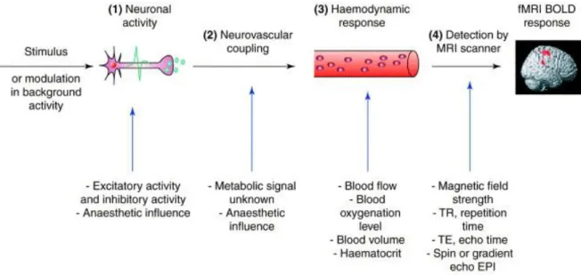
In general, the process of the fMRI BOLD signal production contains several key constituents (Figure 1.3). Neural activity responds to a stimulus presented externally, which triggers changes in the BOLD contrast, characterized by the hemodynamic response (HR). The link between neural activity and HR is known as neurovascular coupling, the nature of which remains largely unknown (for a review, see Villringer & Dirnagl, 1994). HR assumes the BOLD contrast depending on the dynamic deoxygenated hemoglobin amount of neuronal activity, but research has found that the BOLD signal could be better predicted by local field potentials (LFPs) rather than single- and multi-unit activity that occurs only transiently at the onset of the stimulus but do not persist over time, whereas LFPs not only occur transiently but also persist over time (Logothetis et al., 2001).
Figure 1.3 The process of the fMRI BOLD signal production. (1) neuronal activity induced by a stimulus
or modulation in background activity, with the engagement of both excitatory and inhibitory activity; (2) neurovascular coupling, characterized by the relationship between neuronal activity and hemodynamic response; (3) haemodynamic response with different representations, such as blood flow, blood oxygenation level, and blood volume; (4) fMRI signal detection by MRI scanner with multiple related
24
parameters, including the magnetic field strength, repetition time, echo time, spin or gradient echo echo-planar imaging. (Figure adapted from Arthurs & Boniface (2002)).
Despite the discovery of the linear correlation between neuronal activity and HR (Logothetis et al., 2001), in reality, it is still difficult to quantify HR because other physiological factors that are even harder to measure can also contribute to changes in deoxyhemoglobin concentration, such as vascular geometry, hematocrit, and basal oxygenation levels (Ogawa et al., 1993). Moreover, HR can vary widely across cortical areas (Soltysik et al., 2004). It has been recognized that the BOLD signal occurs at large draining veins as well as close by at the capillary level, and even possibly a few centimeters downstream from the neuronally active regions (Ogawa et al., 1993). In addition, the amount of the BOLD signal per se is potentially affected by many experimental parameters in fMRI scanning, including the magnetic field strength, echo time, repetition time, and imaging techniques such as spin- or gradient-echo echo-planar imaging. The quality of BOLD images is also susceptible to various artifacts, especially head motion and field non-homogeneities (Turner & Ordidge, 2000). These factors further affect the amount of the BOLD response that reflects a given HR.
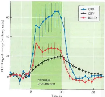
The nature of HR has been found to be related to changes in both CBF and cerebral blood volume (CBV) (Mandeville et al., 1999) (Figure 1.4). During HR, an initial dip occurs after the onset of neuronal activity due to the initial oxygen extraction out of nearby capillaries that results in a local increase in paramagnetic deoxyhemoglobin, hence a decrease in MR image intensity (Menon et al., 1995). After compensatory oxygen supply caused by increased neural activity, an increased inflow of oxygenated blood occurs. Continuous oxygen delivery results in a decrease of deoxygenated hemoglobin and a rise of the BOLD signal that reaches to a maximum value
25
after about five seconds, referred to as the peak of the HR function, and then extends to a plateau. The BOLD signal decreases quickly after neuronal activity stops, until below the baseline level where the signal lingers for an interval, known as post-stimulus undershoot. Based on a balloon model, the post-stimulus undershoot in the BOLD signal exists because CBF decreases more rapidly than CBV, and CBF returns to the baseline when the CBV remains elevated, leading to the presence of more deoxygenated hemoglobin (Buxton et al., 2004). When CBV slowly returns to the baseline level, the BOLD signal synchronously increases to the baseline, and the undershoot ends.
Figure 1.4 The hemodynamic response and its relationship with cerebral blood flow (CBF) and
cerebral blood volume (CBV). In the experiment, CBF and CBV were measured when stimulating the forepaw of a rat (Mandeville et al., 1999). Following the stimulus onset, the BOLD signal showed changes associated with both CBF and CBV. After the stimulus offset, a post-stimulus undershoot was observed, which might be due to the temporal mismatch between changes in CBF and CBV, leading to an increase of deoxyhemoglobin and thereby decreasing the BOLD signal. (Figure adapted fromHuettel et al. (2009)).
26
Regarding fMRI experiments, two other properties of the BOLD signal are relevant here: spatial resolution and temporal resolution. As the currently mainstay non-invasive neuroimaging technique, fMRI has great advantages over other functional imaging techniques, especially its excellent spatial resolution and reasonable temporal resolution. The spatial resolution of fMRI refers to its ability of distinguishing differences between nearby tissues, measured by the voxel size; a voxel is a three-dimensional cube. The spatial specificity of the fMRI signal could be improved by increasing the magnetic field strength (Logothetis, 2008). The temporal resolution is very important for many fMRI research questions, which is associated with the ability to accurately detect brain activity in response to a stimulus and depends on the applied repetition time. As mentioned above, the fMRI BOLD signal is detected from HR that is the result of neuronal activity, but hemodynamic changes do not occur following neuronal activity within tens of milliseconds; rather, they occur until after one to two seconds with a short delay because the BOLD signal estimates activity of slower changes in the vascular system. By resampling the BOLD signal into smaller repetition time, it allows for a better estimation of vascular changes and in turn, improves the interpretation of neural activity.
To date, as a valuable tool for the investigation of the human brain function, BOLD fMRI has been overwhelmingly employed in cognitive neuroimaging studies to broaden our understanding of the functional organization of the human brain.
1.4.2 Resting-state fMRI
In the early years of fMRI studies, the vast majority of task-based experiments only presented findings of activity increases and ignored activity decreases, although task-induced deactivations have been frequently observed (Gusnard & Raichle, 2001). Moreover, with regard to the brain’s
27
energy metabolism, early work found little impact of cognitive processing on CBF and oxygenation consumption in the brain (Sokoloff et al., 1955). For an adult, the brain represents about 2% of the total body weight while it consumes 20% of the whole energy (Clarke & Sokoloff, 1999), with just 1-10% of extra energy consumption for task performance (Shulman et al., 2014). These findings clearly show that the brain consumes most of its energy during rest rather than task state, suggesting a fundamental role of the intrinsic activity in the brain (Raichle & Mintun, 2006).
After the seminal study by Biswal et al. (1995), rs-fMRI quickly became prominent and shed new light on the brain mechanisms underlying human cognition. By using PET, decreased activations in a set of regions have been consistently observed during task performance in healthy human adults, independent of particular tasks (Shulman et al., 1997). These regions were further identified with a decrease of activation during externally oriented tasks while an increase of activation during the resting baseline, suggesting a baseline or default state of the brain, termed as “default mode” (Raichle et al., 2001). The identification of a default mode of the brain function demonstrated rs-fMRI as a powerful technique for exploring LFFs of the resting brain, and triggered broader and deeper investigations concerning the intrinsic functional system in the brain, which led to rapid development in the field of cognitive neuroimaging since the new millennium.
So far, there is no strict definition for “resting state”, but technically, it refers to the subject lying quietly but awake in the scanner, without performing an explicit task. In specific situations, subjects are asked to keep the eyes closed or open with or without visual fixation during scanning. Compared to traditional task-based fMRI research, in which external stimuli are presented to measure the neural activity and the subject’s response, rs-fMRI has some unique
28
advantages in terms of acquiring neuroimaging data from pediatric and clinical populations who are not able to perform complicated tasks in the scanner. Though still limited, novel findings have been reported in studies of infants and young children using the rs-fMRI technique (e.g., Cao et al., 2016; de Bie et al., 2012; Fair et al., 2008; Fransson et al., 2011; Fransson et al., 2007; Gao et al., 2009; W. Lee et al., 2013; Liu et al., 2008; Muetzel et al., 2016; Power et al., 2010; van den Heuvel et al., 2015; van den Heuvel et al., 2009), which expand our knowledge of functional changes during the early years of brain development. With the rs-fMRI technique, large samples of clinical populations with different diseases could be obtained for scientific research, although most of these patients may not be available or suitable for task-based fMRI studies (for a review, see Uddin et al., 2010). Moreover, rs-fMRI also allows for investigating the functional organization of the brain in a valid and relatively simple way and studying various systems in the brain, whereas only one specific system is usually examined in a task-based fMRI study. For instance, the intrinsic organization of the brain could be characterized by the default mode network (DMN) and its anti-correlated network in the absence of overt task performance (Fox et al., 2005). In independent component analysis (ICA)-based studies, 10 resting-state networks were found in adults, involving motor, visual, executive control, auditory, and memory regions besides the DMN, and these networks were reproducible across datasets (Damoiseaux et al., 2006); 5 unique networks were observed in infants, encompassing visual, sensorimotor, and auditory regions as well as an incomplete DMN (Fransson et al., 2007). Furthermore, first results have identified the functional architecture in very young infants (Fransson et al., 2011; Fransson et al., 2007) and even the topological development patterns prior to birth in the preterm infants brain (Cao et al., 2016). Importantly, rs-fMRI also provides the possibility of exploring
29
developmental changes in the brain’s functional architecture (Fair et al., 2008; Fair et al., 2009; Fair et al., 2007).
Despite the absence of explicit tasks, it has been widely demonstrated that brain networks that are engaged in cognitive tasks can also be reliably identified during resting state (Cole et al., 2014; Smith et al., 2009), suggesting a strong overlapping between the intrinsic functional connectivity and task-evoked activations in the brain. Notably, resting-state networks have been proved to be reliable and reproducible. For example, a persistent DMN was detected in different consciousness states (Greicius et al., 2008). The resting-state networks were still discernible during sleep and did not require active cognitive processes or conscious awareness (Fukunaga et al., 2006). Lately, a longitudinal study acquiring rs-fMRI data from a healthy subject weekly over 3.5 years reported high reproducibility of resting-state networks (Choe et al., 2015). Moreover, in a large sample (n = 536) of rs-fMRI data from children, resting-state networks were obtained by using ICA, and most of these networks were highly reproducible across different subsamples (Muetzel et al., 2016).
Intriguingly, recent research has revealed that an individual’s intrinsic connectivity can be used to distinguish that individual’s fundamental cognitive behavior (i.e., fluid intelligence) (Finn et al., 2015), and that the intrinsic activity in the resting brain can even predict individual differences during task performance (Tavor et al., 2016). These findings suggest the robust individual variability of the functional brain organization and therefore provide a critical foundation for correlating an individual’s intrinsic activity in the brain with that individual’s cognitive trait and the response to external task stimuli.
30
To date, most of the rs-fMRI studies have been performed in adults, but rs-fMRI studies in young children are still limited due to difficulties of data acquisition. In the present thesis, rs-fMRI data were acquired from typically developing children to investigate the intrinsic functional connectivity within the language network with respect to language processing as well as the development of the functional brain network over a one-year period and its behavioral relevance.
1.4.3 Approaches for rs-fMRI data analysis
As mentioned above, the rs-fMRI technique has some unique advantages in exploring the functional organization of the brain. During the past two decades, a growing body of neuroimaging research has focused on novel hypotheses by employing this powerful tool, and along this technique, increasing analytic methodologies for data analysis have also been developed in recent years (Margulies et al., 2010). Several approaches used for rs-fMRI data analysis in this thesis, including resting-state functional connectivity (RSFC), amplitude of low-frequency fluctuation (ALFF), and degree centrality, will be introduced in detail.
1.4.3.1 RSFC and RSFC–behavior correlation analysis
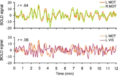
RSFC measures the level of synchronized activity of spontaneous fMRI time series recorded during rest, reflecting the level of functional connections between those spatially separated regions (Figure 1.5). In RSFC, high correlations indicate regions in the same system and selective correlations between regions are used to map the organization of brain systems. RSFC is particularly useful for featuring large-scale brain systems of widely distributed regions and connection strengths, offering new insight into the connectional architecture of the human brain (Van Dijk et al., 2010).