Comparison of Herpes Simplex Virus PCR with Culture for
Virus Detection in Multisource Surface Swab Specimens from
Neonates
Samuel R. Dominguez,a,bKristin Pretty,bRandy Hengartner,bChristine C. Robinsonb
aUniversity of Colorado, Department of Pediatrics, Section of Infectious Diseases, Aurora, Colorado, USA
bChildren's Hospital Colorado, Department of Pathology and Laboratory Medicine, Aurora, Colorado, USA
ABSTRACT The American Academy of Pediatrics currently recommends herpes sim-plex virus (HSV) culture or PCR for testing of swabs of the conjunctivae, mouth, na-sopharynx, and rectum (surface swabs) from neonates. The objectives of this study were to compare the performance and time to results of HSV PCR with those of HSV culture with surface swabs from neonates. Banked multisource surface swab samples that were collected from infants less than or equal to 30 days old from January 2017 to December 2017 and that had previously been cultured for HSV were identified and tested retrospectively by HSV PCR. Surface swab samples from 97 patients were included in the study. Of these 97 patients, 7 (7%) had clinical HSV disease. Of the 7 neonates with HSV disease, 3 (42.9%) had surface swabs positive by culture and 6 (85.7%) had swabs positive by PCR. Limiting the analysis to specimens that were positive only by culture or only by PCR, the specificity for both methods was 100%, but the sensitivity of PCR was 100%, whereas it was 50% for culture. During the study period, 341 HSV cultures and 426 HSV PCRs were performed. The median time from swab collection to reporting of results was 7.6 days (interquartile range [IQR], 7.1 to 7.9 days) for culture and 0.8 days (IQR, 0.6 to 1.0 days) for PCR. HSV PCR of surface swabs from neonates was considerably more rapid and sensitive than HSV culture without yielding false-positive results. Although larger studies are needed to support our findings, strong consideration should be given to utilize PCR instead of culture for the detection of HSV in surface swabs from neonates.
KEYWORDS HSV, neonatal, PCR, culture, surface swabs
N
eonatal herpes simplex virus (HSV) infections in the United States are relatively rare, with an estimated annual incidence of 9.6 per 100,000 births (1). Unfortu-nately, however, these infections are associated with significant morbidity and mortal-ity. In addition to obtaining a whole-blood and cerebrospinal fluid (CSF) sample for HSV PCR assay, the American Academy of Pediatrics (AAP) currently also recommends obtaining swab specimens from the conjunctivae, mouth, nasopharynx, and rectum (surface swabs) for HSV culture or PCR as part of the diagnostic workup for neonatal HSV disease (2, 3). Limited data exist regarding the sensitivity of PCR compared to culture for the diagnosis of HSV disease from neonatal surface swabs. The objective of this study was to compare the performance and time to results of PCR with those of conventional cell culture for the detection of HSV in surface swab samples obtained from children less than 30 days old.MATERIALS AND METHODS
Study design.The multisource surface swabs used in this study were single swabs of the conjunc-tivae, mouth, nasopharynx, and rectum and did not include samples from skin vesicles. Pooled multi-source surface swabs from patients less than or equal to 30 days old were collected in M4 viral transport medium (VTM) from January 2017 to December 2017 and submitted to the Children’s Hospital Colorado
Received16 April 2018 Returned for modification7 May 2018Accepted4 June 2018
Accepted manuscript posted online6 June 2018
CitationDominguez SR, Pretty K, Hengartner R, Robinson CC. 2018. Comparison of herpes simplex virus PCR with culture for virus detection in multisource surface swab specimens from neonates. J Clin Microbiol 56:e00632-18.https://doi.org/10.1128/JCM
.00632-18.
EditorMichael J. Loeffelholz, University of Texas Medical Branch
Copyright© 2018 American Society for Microbiology.All Rights Reserved. Address correspondence to Samuel R. Dominguez,
For a commentary on this article, seehttps://
doi.org/10.1128/JCM.00904-18.
crossm
on May 16, 2020 by guest
http://jcm.asm.org/
Clinical Microbiology Laboratory for prospective HSV culture. Each sample in VTM was vortexed, clarified by centrifugation, and inoculated within 24 h into culture, with the residual fluid frozen at⫺70°C. For culture, centrifugation-enhanced MRC5 shell vials were held for 18 to 24 h and MRC5 tubes were held for 7 days, with blind staining of shell vial coverslips and confirmation of a cytopathic effect in tubes, using fluorescein-labeled HSV-1- and HSV-2-specific monoclonal antibodies (Remel, PathoDx; Thermo Fisher Scientific, Waltham, MA). Banked samples from infants less than or equal to 30 days old collected from January 2017 to December 2017 were identified and tested retrospectively by HSV PCR. For PCR, each sample was thawed at room temperature, extracted on a Qiagen EZXL instrument (Germantown, MD) using the virus (version 2.0) minikit, and tested by the Luminex Multicode RTx HSV 1&2 PCR assay (Austin, TX) on an ABI 7500 instrument (Thermo Fisher Scientific, Waltham, MA). The Luminex assay is cleared by the U.S. Food and Drug Administration to test vaginal swabs and was validated by our laboratory (at Children’s Hospital Colorado) to test other specimen types. Our validation demonstrated a limit of detection of 1 to 5 copies of HSV-1 and 5 to 10 copies of HSV-2 per reaction in swabs, CSF, or blood.
Sensitivity and specificity were calculated using standard statistical methods and clinical truth as the gold standard. Clinical truth was defined as a positive HSV PCR result from a vesicle, blood, or CSF specimen and patient receipt of a full course (14 to 21 days) of intravenous (i.v.) acyclovir by the treating clinical team. Clinical and laboratory data were collected from the electronic medical record (EMR). The median time to results was calculated by subtracting the time from collection to the time to results reported in the EMR for all HSV PCRs and HSV cultures for all patients reported during 2017, and the times were compared using a Wilcoxon rank sum test. Statistical analyses were performed using SAS (version 9.4) software. The use of clinical specimens and data was approved by the Colorado Multiple Institutional Review Board.
RESULTS
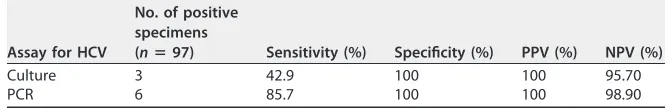
From 1 January to 31 December 2017, 102 multisource, surface swab samples from unique patients were submitted for HSV culture. Ninety-seven (95%) had sufficient sample remaining to perform PCR and were included in the study. Of the 97 patients, 7 (7%) had clinical HSV disease. The diagnostic workup and clinical presentations of these 7 neonates are shown in Table 1. All 7 neonates had a positive blood PCR result and were treated with a full course of i.v. acyclovir.
Of the 7 neonates who had clinical HSV disease, 3 (42.9%) had surface swabs that tested positive by HSV culture and 6 (85.7%) had swabs that tested positive by PCR. The 3 positive cultures were all positive by the shell vial assay at 24 h, and the relative PCR threshold cycle (CT) values ranged from 18.0 to 35.9 (Table 1). One patient with clinical HSV disease had
surface swabs that were negative by both PCR and culture. Compared to the clinical truth, the sensitivity, specificity, positive predictive value, and negative predictive value for culture and PCR are shown in Table 2. When limiting the analysis to specimens that were positive either only by culture or only by PCR, the specificity of both methods was 100%, but the sensitivity of PCR was 100%, whereas it was 50% for culture. During the study period, the laboratory performed 426 HSV PCRs and 341 HSV cultures for surface swabs and other specimens. HSV PCRs during this time were from patients of all ages and specimens, excluding neonatal multisource surface swabs and respiratory specimens. The median time to results was 0.8 days (interquartile range [IQR], 0.6 to 1.0 days) for PCR and 7.6 days (IQR, 7.1 to 7.9 days) for HSV cultures (P⬍0.001).
DISCUSSION
Although PCR is now the standard method to detect HSV in spinal fluid and blood, culture is still recommended by the AAP and, therefore, still used by many laboratories to detect the virus in surface swabs from neonates (3). This practice is primarily due to the lack of studies comparing the sensitivity of culture to that of PCR with surface specimens from newborns (2). Here we provide data that PCR has a higher sensitivity than culture, in agreement with previous studies of HSV detection in dermal, genital, ocular, mouth, and skin swabs from adults and older children (4–9). In addition, PCR did not exhibit any false-positive results. This high specificity is important, as concern exists that performing PCR shortly after birth could detect transient maternal contamination, as opposed to actively replicating virus (2, 10). However, this concern seems to be minimal, as most neonates evaluated for HSV are usually discharged from the hospital after birth but return days later for evaluation. Furthermore, false-positive PCR results, particularly those due to HSV-1, could occur due to contamination from other exogenous sources, resulting in unnecessary, prolonged acyclovir treatment. Concordance between HSV types by culture and PCR across specimen sites somewhat mitigates this concern.
on May 16, 2020 by guest
http://jcm.asm.org/
TABLE 1 Demographic, diagnostic, and clinical information for neonates with HSV disease a Age (days) Sex Result for HCV by:
PCR CT
Presence of: Other symptoms CSF WBC (no. of cells/mm 3) CSF WBC differential (%) Brain MRI finding c AST/ALT (U/liter) Duration of treatment with acyclovir (days) Culture of surface swabs PCR of surface swabs PCR of CSF b PCR of blood Vesicles Fever Apnea Seizures 8 M Pos HSV-1 Neg HSV-1 27.1 Yes (Pos by culture) No No No 7 27 L, 23 Mo, 50 Ma NT 32/24 14 15 F Pos HSV-2 Neg HSV-2 18.0 No Yes No No Liver failure 3 NT 1 10,003/2,573 Treated but deceased 16 M Pos HSV-2 HSV-2 HSV-2 22.5 No No Yes Yes 6 33 N, 8 B, 27 L, 29 Mo, 3 Ma 2 90/32 21 16 F Neg HSV-1 Neg HSV-1 35.9 No No Yes Yes 17 5 N, 39 L, 35 Mo, 18 Ma Normal 75/29 21 28 F Neg HSV-1 NT HSV-1 32.9 No Yes Yes No 2 NT Normal 1,032/365 21 19 F Neg HSV-2 Neg HSV-2 33.5 Yes (NT) No No Yes Retinitis 89 4 N, 60 L, 34 M o ,2M a 3 75/29 21 4 F Neg Neg Neg HSV-1 Neg No No No Yes Stroke 8 1 N, 34 L, 65 Mo 4 62/22 21 aF, female; M, male; Pos, positive; Neg, negative; NT, not tested; N, neutrophils; B, bands; L, lymphocytes; Mo, monocytes; Ma, macrophages; AST, aspa rtate aminotransferase; ALT, alanine aminotransferase; WBC, white blood cell count; MRI, magnetic resonance imaging. bAll patients with evidence of meningitis had repeat lumbar punctures prior to completion of acyclovir therapy, and all were HSV PCR negative. cBrain magnetic resonance imaging findings were classified as follows: 1, extensive diffuse restricted diffusion throughout the cerebral hemisphere s and, to a lesser degree, the brain stem and cerebellum, with associated T2 hyperintensities and a loss of gray-white matter differentiation; diffuse leptomeningeal enhancement; 2, multifocal areas of restricted diffu sion throughout both cerebral hemispheres; 3, extensive laminar necrosis and areas of prior cortical hemorrhage along the bilateral cerebral and cerebellar hemispheres, with scattered regions of cystic encephalomalacia, gr eatest in the left frontal and anterior temporal lobes; 4, deep right middle cerebral artery territory ischemia.
on May 16, 2020 by guest
http://jcm.asm.org/
PCR affords several distinct advantages over culture. PCR is now a widely available testing modality, and many laboratories no longer have the expertise or capacity to perform HSV culture. Importantly, PCR provides a greatly improved turnaround time to results compared to culture. At our institution (Children’s Hospital Colorado), which per-forms a commercially available HSV PCR assay once daily, PCR provides results a median of 6.8 days sooner than culture. Furthermore, turnaround times can be even longer at institutions that send swabs to a reference lab for culture because culture is not available in-house. This practice is very problematic for this patient population, as providers often wait for final results to rule out a diagnosis of HSV disease, during which time neonates are hospitalized and receive i.v. acyclovir therapy. Indeed, at our institution acyclovir treatment is continued in high-risk neonates until PCRs or cultures of samples from all sources are negative. Faster results could decrease hospital lengths of stay and minimize acyclovir exposure and the potential side effects associated with this medication. Indeed, recent studies have highlighted the increasing use of acyclovir over the past decade (11), the existing controversies regarding which neonates should be tested for HSV, and how these patients should be managed (12, 13). Replacing HSV culture with HSV PCR for surface swabs could simplify and streamline diagnostic algorithms by expediting results.
There are several limitations to our study. First, samples were frozen prior to performing PCR, which may have influenced its performance. Second, we utilized only one PCR assay, and as such, results may not be generalizable to other assays. Finally, although we collected samples for an entire year, our overall sample size was small and included only 7 neonates with clinical HSV disease.
In summary, our study is one of the first to compare and demonstrate the improved sensitivity of PCR over that of culture for the detection of HSV in multisource surface swabs from neonates. Furthermore, PCR demonstrated greatly improved turnaround times com-pared to culture, which affords important clinical advantages. Although larger studies are needed to support our findings, strong consideration should be given to ordering PCR instead of culture for the detection of HSV in surface swabs from neonates.
ACKNOWLEDGMENTS
This research did not receive any specific grant from funding agencies in the public, commercial, or not-for-profit sectors.
We have no competing interests to declare.
REFERENCES
1. Flagg EW, Weinstock H. 2011. Incidence of neonatal herpes simplex virus infections in the United States, 2006. Pediatrics 127:e1– e8.https://doi .org/10.1542/peds.2010-0134.
2. Kimberlin DW, Baley J. 2013. Committee on Infectious Diseases and Com-mittee on Fetus and Newborn. Guidance on management of asymptomatic neonates born to women with active genital herpes lesions. Pediatrics 131:e635– e646.https://doi.org/10.1542/peds.2012-3216.
3. American Academy of Pediatrics. 2018. Herpes simplex.InKimberlin DW, Brady MT, Jackson MA, Long SS (ed), Red book: 2018 report of the Committee on Infectious Diseases, 31st ed. American Academy of Pedi-atrics, Itasca, IL.
4. Espy MJ, Ross TK, Teo R, Svien KA, Wold AD, Uhl JR, Smith TF. 2000. Evaluation of LightCycler PCR for implementation of laboratory diagno-sis of herpes simplex virus infections. J Clin Microbiol 38:3116 –3118.
5. Filen F, Strand A, Allard A, Blomberg J, Herrmann B. 2004. Duplex real-time polymerase chain reaction assay for detection and quantifica-tion of herpes simplex virus type 1 and herpes simplex virus type 2 in genital and cutaneous lesions. Sex Transm Dis 31:331–336.https://doi .org/10.1097/00007435-200406000-00002.
6. Gitman MR, Ferguson D, Landry ML. 2013. Comparison of Simplexa HSV 1 & 2 PCR with culture, immunofluorescence, and laboratory-developed TaqMan PCR for detection of herpes simplex virus in swab specimens. J Clin Microbiol 51:3765–3769.https://doi.org/10.1128/ JCM.01413-13.
[image:4.585.38.372.94.149.2]7. Heaton PR, Espy MJ, Binnicker MJ. 2015. Evaluation of 2 multiplex real-time PCR assays for the detection of HSV-1/2 and varicella zoster virus directly from clinical samples. Diagn Microbiol Infect Dis 81: 169 –170.https://doi.org/10.1016/j.diagmicrobio.2014.11.012.
TABLE 2Performance of HSV culture and HSV PCR with neonatal multisource surface
swabs compared to clinical trutha
Assay for HCV
No. of positive specimens
(nⴝ97) Sensitivity (%) Specificity (%) PPV (%) NPV (%)
Culture 3 42.9 100 100 95.70
PCR 6 85.7 100 100 98.90
aPPV, positive predictive value; NPV, negative predictive value.
on May 16, 2020 by guest
http://jcm.asm.org/
8. Schmutzhard J, Merete Riedel H, Zweygberg Wirgart B, Grillner L. 2004. Detection of herpes simplex virus type 1, herpes simplex virus type 2 and varicella-zoster virus in skin lesions. Comparison of real-time PCR, nested PCR and virus isolation. J Clin Virol 29:120 –126.
9. Scoular A, Gillespie G, Carman WF. 2002. Polymerase chain reaction for diagnosis of genital herpes in a genitourinary medicine clinic. Sex Transm Infect 78:21–25.https://doi.org/10.1136/sti.78.1.21.
10. Turner R, Shehab Z, Osborne K, Hendley JO. 1982. Shedding and survival of herpes simplex virus from ‘fever blisters.’ Pediatrics 70:547–549. 11. Gaensbauer JT, Birkholz M, Pfannenstein K, Todd JK. 2014. Herpes PCR
testing and empiric acyclovir use beyond the neonatal period. Pediatrics 134:e651– e656.https://doi.org/10.1542/peds.2014-0294.
12. Brower L, Schondelmeyer A, Wilson P, Shah SS. 2016. Testing and empiric treatment for neonatal herpes simplex virus: challenges and opportunities for improving the value of care. Hosp Pediatr 6:108 –111. https://doi.org/10.1542/hpeds.2015-0166.
13. McGuire JL, Zorc J, Licht D, Hodinka RL, Shah SS. 2012. Herpes simplex testing in neonates in the emergency department. Pediatr Emerg Care 28:949 –955. https://doi.org/10.1097/PEC.0b013e3182 6c6daf.