Summary
Retinoic acid (RA) is a vitamin A-derived, non-peptidic, small lipophilic molecule that acts as ligand for nuclear RA receptors (RARs), converting them from transcriptional repressors to activators. The distribution and levels of RA in embryonic tissues are tightly controlled by regulated synthesis through the action of specific retinol and retinaldehyde
dehydrogenases and by degradation via specific cytochrome P450s (CYP26s). Recent studies indicate that RA action involves an interplay between diffusion (morphogen-like) gradients and the establishment of signalling boundaries due to RA
metabolism, thereby allowing RA to finely control the differentiation and patterning of various stem/progenitor cell populations. Here, we provide an overview of the RA
biosynthesis, degradation and signalling pathways and review the main functions of this molecule during embryogenesis.
Key words: Retinoids, Retinoic acid, Retinol dehydrogenase (RDH), Retinaldehyde dehydrogenase (RALDH), CYP26, Hindbrain, Forebrain
Introduction
Retinoic acid (RA) is derived from the liposoluble vitamin A (retinol). Vitamin A has long been known to be indispensable for vision, as its derivative retinaldehyde (Fig. 1) acts as a light-sensitive molecule, the isomerisation of which triggers the phototransduction process in photoreceptor cells of the retina (reviewed by Parker and Crouch, 2010). Many other functions have been assigned to this vitamin, and work in avian and rodent models has established that maternal vitamin A deficiency affects the embryo and foetus, leading to a complex spectrum of abnormalities (e.g. Gale et al., 1999; White et al., 2000; Wilson et al., 1953). About 25 years ago, the molecular basis of vitamin A action was elucidated when it was shown that its acidic metabolite, RA, acts as a ligand for transcription factors of the retinoic acid receptor (RAR) nuclear receptor superfamily, switching them from potential repressors to transcriptional activators.
Possible functions of RA during embryogenesis were first inferred by studying its teratogenic effects, i.e. how the administration of excess doses of RA, either globally or by local implantation using RA-impregnated beads, interferes with normal developmental processes. These studies have been performed in a wide range of species including amphibians, zebrafish, chick and rodents (e.g. Avantaggiato et al., 1996; Durston et al., 1989). Gene knockout studies then confirmed the crucial functions of RARs in mouse development (reviewed by Mark et al., 2009). Eventually, the enzymatic pathways that regulate embryonic RA synthesis from maternal retinol – or egg-stored retinoids – were characterised, and
it was found that another regulatory step involved the triggering of RA catabolism by a subfamily of cytochrome P450 enzymes. Altogether, the differential and often dynamic expression patterns of a small number of specific synthesizing and metabolising enzymes allow precise control of RA distribution within embryonic cell populations. A wide range of vertebrate models (from zebrafish to mouse) and experimental tools (including, for instance, reporter transgenes that reveal regions of active RA signalling, and selective antagonists for RARs or RA-synthesizing/metabolising enzymes) have been successfully used to decipher retinoid functions at the cellular and molecular levels.
Here, we first provide an overview of the pathways and proteins that regulate or mediate RA signalling during development, many of which appear to be highly conserved throughout vertebrate species. We then review the main functions of retinoid signalling during early embryonic development, first referring to the developing hindbrain as a system that has been most extensively studied with respect to RA functions and for which recent studies have refined our knowledge of the control of RA activity during rhombomeric segmentation. We further review extensive work that, over the last few years, has investigated how RA acts on progenitor cell populations in structures as diverse as the embryonic forebrain, the branchial apparatus and foregut derivatives, the neural plate and the posterior mesoderm during embryonic axial elongation. Understanding these functions and the underlying molecular events is of great importance, as retinoids are widely used in therapy and in many protocols for differentiating primary cultures or cell lines [including embryonic stem (ES) cells] into specific lineages (see Box 1). Retinoids thus hold promise for future use in stem cell-based therapy and regenerative medicine. Ongoing research will also guide more conventional clinical approaches, especially in the context of cancer chemoprevention or treatment (reviewed by Tang and Gudas, 2011).
The RA synthesis pathway
The only source of retinoids in most animals is diet derived, as these compounds cannot be synthesized de novo. In mammals, the main circulating retinoid is retinol bound to a carrier protein, retinol-binding protein 4 (RBP4) (see Box 2). Retinol homeostasis involves several proteins and enzymes that regulate its dietary uptake in intestinal cells and its storage mainly in liver hepatocytes and stellate cells (reviewed by D’Ambrosio et al., 2011). Maternal retinol transferred transplacentally is the major retinoid source for embryos of placental species. By contrast, oviparous species store vitamin A in the egg yolk and, according to the species, the main source can be retinol, retinaldehyde, or carotenoids such as beta-carotene (see Simoes-Costa et al., 2008). Work performed in zebrafish has demonstrated the importance of a beta-carotene cleavage enzyme (BCMO1) that acts tissue-specifically to generate embryonic retinaldehyde (Lampert et al., 2003).
Typically, in mammals, retinol-RBP4 is taken up by target tissues, and this uptake can be facilitated in some tissues by a transmembrane protein that is the product of the RA-inducible gene Development 139, 843-858 (2012) doi:10.1242/dev.065938
© 2012. Published by The Company of Biologists Ltd
Retinoic acid signalling during development
Muriel Rhinn* and Pascal Dollé*IGBMC (Institut de Génétique et de Biologie Moléculaire et Cellulaire), BP 10142, Illkirch, F-67404 France, and Inserm, U 964, CNRS, UMR 7104, Université de Strasbourg, France.
*Authors for correspondence ([email protected]; [email protected])
D
E
V
E
LO
P
M
E
N
STRA6(Kawaguchi et al., 2007). Interestingly, STRA6mutations constitute the only demonstrated cases of human mutations that affect a gene from the retinoid pathway and lead to a complex spectrum of developmental abnormalities (Pasutto et al., 2007). Other human mutations involving retinoid-binding proteins selectively affect visual function (e.g. Maw et al., 1997). Maternal RBP4 cannot cross the placenta; retinol thus diffuses across the yolk sac and placenta, where zygotic RBP4 synthesis occurs (Ward et al., 1997). Retinyl esters may also be significant retinoid sources for the embryo (Quadro et al., 2005).
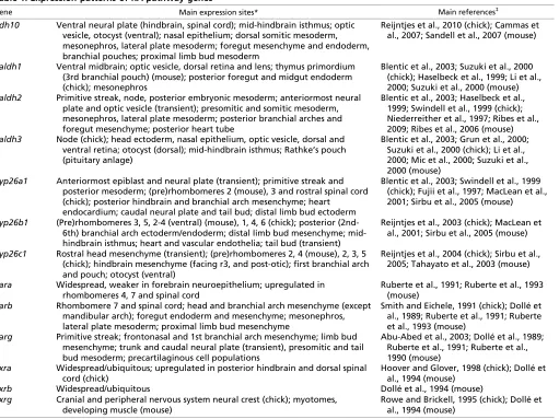
Enzymes that oxidize retinol to retinaldehyde (Fig. 1A) belong to two classes: the cytosolic alcohol dehydrogenases (ADHs) belonging to the medium-chain dehydrogenase/reductase family; and microsomal short-chain dehydrogenases/reductases (retinol dehydrogenases, RDHs) (reviewed by Pares et al., 2008). ADH5 (previously called ADH3) is ubiquitously expressed in the embryo and adult, whereas ADH1 and ADH7 (previously called ADH4) are tissue restricted (Ang et al., 1996). Null mouse mutants for Adh5display reduced viability and growth defects, which can be rescued by dietary supplementation with retinol (Molotkov et al., 2002a). No obvious phenotype is associated with the loss of ADH1 and ADH7 when mice are maintained on a vitamin A-sufficient diet (Deltour et al., 1999b). However, when large doses of retinol are administered to Adh1–/–mutants, the mice are more sensitive to vitamin A embryotoxicity (Molotkov et al., 2002b). This suggests that ADH enzymes might have a role in controlling the removal of excess retinol, rather than participating in RA synthesis. RDHs are well known to act during the visual cycle (Parker and Crouch, 2010). Rdh5–/–mice are viable but suffer from a delay in dark adaptation, consistent with a role in regenerating 11-cis-retinaldehyde after photobleaching. By contrast, Rdh10, which displays specific expression domains during development (Cammas et al., 2007; Romand et al., 2008) (Table 1), plays an important role in RA synthesis, as its loss-of-function is lethal between embryonic day (E) 10.5 and E14.5 (Sandell et al., 2007). Rdh10mutants (Fig. 1B,C) exhibit abnormalities characteristic of RA deficiency (Table 2), which can be partly rescued by maternal RA supplementation (Rhinn et al., 2011).
The next step in RA synthesis is the oxidation of retinaldehyde to RA (Fig. 1A), which is carried out by three retinaldehyde dehydrogenases (RALDHs): RALDH1, RALDH2 and RALDH3 (also known as ALDH1A1, ALDH1A2 and ALDH1A3). RALDHs display distinct and specific expression patterns that correlate with RA activity as detected by reporter transgenes (Table 1). Raldh2is the earliest RALDH to be expressed, and is found in the primitive streak and mesodermal cells, and later in somitic and lateral mesoderm, posterior heart tube and rostral forebrain (Niederreither Box 1. RA and stem cell differentiation
Unlike many other adult tissues, the nervous system of mammals has a limited ability to compensate for the loss of cells after lesions. Embryonic stem (ES) cells have long attracted attention as a potential source of cells that can be driven to differentiate into specific lineages for the development of cell therapy and pharmaceutical screens. The spontaneous development of neuronal cells from ES cells during in vitro culture is rather limited. Therefore, various protocols to increase the differentiation of neuronal cell types have been established. Recently, it was found that the addition of RA to rapidly proliferating mouse ES cells, cultured in suspension as embryoid bodies, leads to the selective generation of neural progenitors with characteristics of radial glial cells found in the developing central nervous system (Bibel et al., 2004; Plachta et al., 2004). These conditions led to a highly uniform population of Pax6-positive cells which, when further cultured in vitro, could differentiate into neurons that established synaptic contacts and exhibited electrophysiological properties similar to those of forebrain pyramidal neurons. When transplanted into the neural tube of chicken embryos, the RA-induced embryoid bodies contributed to spinal cord motoneurons and interneurons. In the future, these cellular systems could be used for studying commitment and neuronal specification in vitro, for pharmacological assays and drug screening, and for the selective isolation of differentiated neuronal cells or committed progenitors that may be used as a source for cell and tissue grafts.
All-trans-retinol
Retinaldehyde CH2
Retinoic acid All-trans-retinol
CH2
OH H
O O
C C
RDH10
ADH1,5,7 CYP1B1
CYP1B1
RALDH1, 2, 3
h
fl fl
ey
fn
fn
fl so
b1 b2
h h
b3
C D E F G
b1 A
[image:2.612.53.498.62.214.2]B
Fig. 1. Retinoic acid biosynthesis.(A)The oxidation of retinol into retinaldehyde and retinoic acid (RA). Enzymes responsible for each of the catalytic steps are shown, with enzymes that are crucial for normal development in bold. (B-G)The phenotypes of murine mutants lacking RDH10, RALDH2 or RALDH3 activity. Profile views of an E12.5 Rdh10–/–embryo (C), an E9.5 Raldh2–/–embryo (E, scanning electron micrograph), and a
histological section of the nasal cavities of an E18.5 Raldh3–/–mutant (G), are compared with their wild-type (WT) littermates (B,D,F). Rdh10–/– mutants exhibit abnormal facial development, lack of externally visible eyes (ey) and severe forelimb (fl) defects. Raldh2–/–mutants show an array of
abnormalities, with hypoplasia of the frontonasal region (fn) and telencephalic vesicles, lack of posterior branchial arches (b1-b3), defective heart (h) morphogenesis, shortened trunk region (bold arrow) with compacted somites (so), and absent forelimb buds. Raldh3–/–mutants fail to develop
choanae (CH; the ducts connecting the nasal and oral cavities), leading to lethal respiratory distress at birth. N, nasal cavity; NS, nasal septum; P, pharynx. Reproduced with permission: B,C (Rhinn et al., 2011); D,E (Niederreither et al., 1999); F,G (Dupé et al., 2003).
D
E
V
E
LO
P
M
E
N
et al., 1997). Raldh2–/–mouse mutants (Fig. 1D,E) die before
mid-gestation from defective heart morphogenesis and exhibit numerous abnormalities (Table 2) (Niederreither et al., 1999; Niederreither et al., 2001; Niederreither et al., 2000). Some of these abnormalities can be rescued by transient maternal RA supplementation from E7.5 to E8.5-9.5 (e.g. Mic et al., 2002; Niederreither et al., 2003). RALDH2 is solely responsible for embryonic RA synthesis until ~E8.5, and thereafter RALDH1 and RALDH3 contribute to RA synthesis in the eyes and olfactory system. Raldh3–/– mice have defects in nasal and ocular
development and die at birth due to respiratory distress (Fig. 1F,G) (Dupé et al., 2003). Raldh1–/–mutants, by contrast, are viable and
show minor defects in the dorsal retina (Matt et al., 2005; Molotkov et al., 2006).
The RA degradation pathway
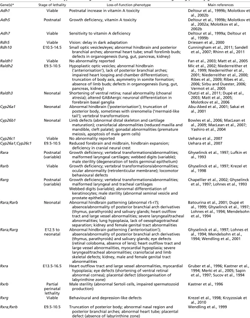
The distribution and levels of RA have to be tightly controlled during embryogenesis, and an important level of control is achieved through tissue-specific oxidative metabolism. Enzymes of the cytochrome P450 26 subfamily (CYP26A1, CYP26B1 and CYP26C1) catalyze reactions that convert RA into more polar metabolites, primarily 4-hydroxy-RA, which can be further oxidized to 4-oxo-RA (e.g. Chithalen et al., 2002) (Fig. 2A). Although 4-oxo-RA is able to bind RARs and interferes with embryonic patterning when administered exogenously (Pijnappel et al., 1993), both expression data and functional studies indicate that the role of CYP26-mediated RA metabolism is essentially to prevent inappropriate signalling in specific cell populations
(reviewed by Pennimpede et al., 2010). CYP26 enzymes display differential expression patterns (Table 1) that are often complementary to the RALDH expression domains. Cyp26a1and Cyp26c1 are the first to be expressed and are found in the rostralmost embryonic epiblast, and all three Cyp26 genes are expressed in a sequential manner during hindbrain development (MacLean et al., 2001; Sirbu et al., 2005) (see below). Cyp26a1is specifically expressed in tail bud tissues and Cyp26b1in distal limb bud mesenchyme (Yashiro et al., 2004). After mid-gestation, each Cyp26 gene displays complex patterns of expression in several developing organs, including the retina, inner ear and dental epithelium (Abu-Abed et al., 2002; Romand et al., 2006; Tahayato et al., 2003).
Genetic ablation of Cyp26a1and Cyp26b1results in phenotypes reminiscent of RA-induced teratogenesis (Table 2). Cyp26a1–/–
mice (Fig. 2B,C) display truncation of the posterior body region, abnormal hindbrain patterning and transformation of cervical vertebrae (Abu-Abed et al., 2001; Sakai et al., 2001). Cyp26b1–/–
mutants (Fig. 2D-G) exhibit severe limb malformations and facial abnormalities (MacLean et al., 2009; Yashiro et al., 2004). Cyp26c1loss-of-function embryos are viable, although compound inactivation of any other Cyp26 results in early embryonic patterning defects (Uehara et al., 2007; Uehara et al., 2009) (Fig. 2H,I). Disruption of the P450 oxidoreductase (POR), an enzyme required for P450 cytochrome activity, phenocopies many of these defects, underscoring the importance of CYP26 function during development (Ribes et al., 2007).
Collectively, these data show that CYP26 enzymes have major developmental functions, which are best described as preventing any detrimental (teratogenic) effect of endogenous RA in regions where it should not be allowed to signal. Interestingly, RA can control the expression of its own metabolising enzymes. RA treatments in vivo and in cultured cells lead to rapid upregulation of the Cyp26a1gene, which contains two functional RA-response elements (RAREs) (Loudig et al., 2005). These data have been integrated into mathematical models indicating how RA might participate in autoregulatory negative-feedback loops by inducing expression of catabolising enzyme(s) (White et al., 2007) (see below).
Gene regulation by RA
RA acts by binding to RARs, which are members of the nuclear receptor superfamily (reviewed by Rochette-Egly and Germain, 2009). There are three RARs (RAR, RARand RAR) that are conserved throughout vertebrates and that primarily bind all-trans-RA. RARs act in heterodimeric combinations with retinoid X receptors (RXR, RXRand RXR). RXRs bind a stereoisomer, 9-cis-RA, which, unlike all-trans-RA, is not detected endogenously in embryos or adult tissues (Mic et al., 2003); therefore, it was suggested that RXRs act mainly as scaffolding proteins to facilitate DNA binding of the RAR-RXR complex (see Chawla et al., 2001). Three receptors (RAR, RXR and RXR) have widespread expression patterns, whereas the others (RAR, RARand RXR) show more complex, tissue-specific expression (reviewed by Dollé, 2009) (Table 1). Thus, most tissues are potential targets of retinoid actions, although different heterodimeric complexes can transduce the RA signal. Gene knockout studies in mouse revealed a large degree of functional redundancy between RAR/RXR heterodimers, with developmental abnormalities usually occurring when two receptors are inactivated in combination, except in the case of RXRmutants, which die in utero due to heart defects (reviewed by Mark et al., 2009) (Table 2).
Box 2. Additional components of the retinoid pathway
Retinol-binding protein 4 (RBP4).Mainly synthesized in the yolk sac and postnatally in liver. It binds to retinol and delivers retinol from the liver to peripheral tissues.
Transthyretin (TTR).A serum protein that associates with RBP4-retinol to prevent RBP4-retinol degradation by the kidney. The TTR-RBP4-retinol complex transports TTR-RBP4-retinol in the circulation and delivers it to target tissues.
‘Ocular’ retinoid-binding proteins. These include retinaldehyde-binding proteins 1 and 3 (RLBP1 and RBP3), which are produced by retinal cells and are involved in the isomerisation and/or shuttling of retinol and retinaldehyde during the visual cycle.
Cytochrome P450 1B1 (CYP1B1). May catalyze the oxidation of retinol into retinaldehyde and RA. Human CYP1B1mutations are a major cause of congenital glaucoma, a severely blinding disease. Cellular retinol-binding proteins (CRBP-I and -II, also known as RBP1 and 2).These proteins belong to the fatty acid-binding protein (FABP) family. They bind both retinol and all-trans-retinaldehyde, and may function to control levels of intracellular retinol accumulation and esterification.
Cellular retinoic acid-binding proteins (CRABP-I and -II, also known as CRABP1 and 2). Bind all-trans-RA intracellularly. They solubilise and protect RA in the aqueous cytosol, although differential functions have been proposed, with CRABP-I presenting RA to metabolising (CYP26) enzymes, and CRABP-II favouring nuclear import and delivery of RA to RARs by direct protein-protein interactions.
Fatty acid-binding protein 5 (FABP5).Binds RA with a lower affinity than CRABPs, and may play a role in inducing a non-canonical RA signalling pathway. In cell lines, FABP5 favours RA binding to peroxisome proliferator-activated receptor /(PPAR/), which in turn can induce anti-apoptotic and proliferative responses when the FABP5 concentration exceeds that of CRABP-II.
D
E
V
E
LO
P
M
E
N
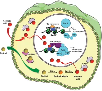
In the nucleus, RAR/RXR dimers bind to DNA motifs known as RAREs. RAREs consist of a direct repeat of a core hexameric sequence 5⬘-(A/G)G(G/T)TCA-3⬘ or of the more relaxed 5⬘ -(A/G)G(G/T)(G/T)(G/C)A-3⬘motif, separated by 1, 2 or 5 bp (see Balmer and Blomhoff, 2002). RAR/RXRs can bind RAREs even in the absence of ligand, thereby recruiting co-repressor complexes and maintaining target gene repression (Fig. 3). In the presence of ligand, a conformational change leads to the release of co-repressors and the recruitment of co-activator complexes. These induce chromatin remodelling, which decompacts the chromatin and facilitates the assembly of the transcription pre-initiation complex. A recent whole-genome chromatin immunoprecipitation-sequencing (ChIP-Seq) study performed in ES cells suggested that the presence of RA might also induce de novo RAR/RXR binding to numerous RAREs that are not bound by unliganded receptors (Mahony et al., 2011).
Numerous RAR target genes have been identified (see Balmer and Blomhoff, 2002), including genes from within the retinoid pathway, such as Rarb, Crbp1/2(Rbp1/2), Crabp1/2and Cyp26a1 (see Box 1 and Table 3). Also, several members of the Hox gene family, including Hoxa1, Hoxb1, Hoxb4 and Hoxd4, harbour RAREs, the function of which has been demonstrated in vivo (reviewed by Marshall et al., 1996). The number of putative target
genes is increasing rapidly through novel technologies. Recently, for example, Luijten et al. used rodent whole-embryo culture combined with RA treatments and performed microarray analysis to identify genes that were up- or downregulated by RA (Luijten et al., 2010). It should also be stressed that some of the effects of RA might involve binding to other nuclear receptors, such as PPAR/ (Schug et al., 2007).
RA functions during development
RA signalling during hindbrain development
Segmentation and patterning of the hindbrain are regulated by RA
[image:4.612.56.567.67.450.2]Numerous studies have focused on the embryonic hindbrain as an experimental paradigm for understanding RA regulatory effects. Hindbrain development involves the generation of seven to eight neuroepithelial compartments or rhombomeres (Fig. 4A), each with a distinct identity according to its anteroposterior (A-P) position (Kiecker and Lumsden, 2005). This segmentation underlies several events required for development of the brain stem, inner ear, branchial arches, and even the heart and large vessels, which are colonised by hindbrain-derived neural crest cells. Several Hox genes, according to their spatially restricted expression patterns, are required for the growth and/or positional identity of specific Table 1. Expression patterns of RA pathway genes
Gene Main expression sites* Main references‡
Rdh10 Ventral neural plate (hindbrain, spinal cord); mid-hindbrain isthmus; optic vesicle, otocyst (ventral); nasal epithelium; dorsal somitic mesoderm, mesonephros, lateral plate mesoderm; foregut mesenchyme and endoderm, branchial pouches; proximal limb bud mesoderm
Reijntjes et al., 2010 (chick); Cammas et al., 2007; Sandell et al., 2007 (mouse)
Raldh1 Ventral midbrain; optic vesicle, dorsal retina and lens; thymus primordium (3rd branchial pouch) (mouse); posterior foregut and midgut endoderm (chick); mesonephros
Blentic et al., 2003; Suzuki et al., 2000 (chick); Haselbeck et al., 1999; Li et al., 2000; Suzuki et al., 2000 (mouse) Raldh2 Primitive streak, node, posterior embryonic mesoderm; anteriormost neural
plate and optic vesicle (transient); presomitic and somitic mesoderm, mesonephros, lateral plate mesoderm; posterior branchial arches and foregut mesenchyme; posterior heart tube
Blentic et al., 2003; Haselbeck et al., 1999; Swindell et al., 1999 (chick); Niederreither et al., 1997; Ribes et al., 2009; Ribes et al., 2006 (mouse) Raldh3 Node (chick); head ectoderm, nasal epithelium, optic vesicle, dorsal and
ventral retina; otocyst (dorsal); mid-hindbrain isthmus; Rathke’s pouch (pituitary anlage)
Blentic et al., 2003; Grun et al., 2000; Suzuki et al., 2000 (chick); Li et al., 2000; Mic et al., 2000; Suzuki et al., 2000 (mouse)
Cyp26a1 Anteriormost epiblast and neural plate (transient); primitive streak and posterior mesoderm; (pre)rhombomeres 2 (mouse), 3 and rostral spinal cord (chick); posterior hindbrain and branchial arch mesenchyme; heart
endocardium; caudal neural plate and tail bud; distal limb bud ectoderm
Blentic et al., 2003; Swindell et al., 1999 (chick); Fujii et al., 1997; MacLean et al., 2001; Sirbu et al., 2005 (mouse)
Cyp26b1 (Pre)rhombomeres 3, 5, 2-4 (ventral) (mouse), 1, 4, 6 (chick); posterior (2nd-6th) branchial arch ectoderm/endoderm; distal limb bud mesenchyme; mid-hindbrain isthmus; heart and vascular endothelia; tail bud (transient)
Reijntjes et al., 2003 (chick); MacLean et al., 2001; Sirbu et al., 2005 (mouse)
Cyp26c1 Rostral head mesenchyme (transient); (pre)rhombomeres 2, 4 (mouse), 2, 3, 5 (chick); hindbrain mesenchyme (facing r3, and post-otic); first branchial arch and pouch; otocyst (ventral)
Reijntjes et al., 2004 (chick); Sirbu et al., 2005; Tahayato et al., 2003 (mouse)
Rara Widespread, weaker in forebrain neuroepithelium; upregulated in
rhombomeres 4, 7 and spinal cord
Ruberte et al., 1991; Ruberte et al., 1993 (mouse)
Rarb Rhombomere 7 and spinal cord; head and branchial arch mesenchyme (except
mandibular arch); foregut endoderm and mesenchyme; mesonephros, lateral plate mesoderm; proximal limb bud mesenchyme
Smith and Eichele, 1991 (chick); Dollé et al., 1989; Ruberte et al., 1991; Ruberte et al., 1993 (mouse)
Rarg Primitive streak; frontonasal and 1st branchial arch mesenchyme; limb bud
mesenchyme; trunk and caudal neural plate (transient), presomitic and tail bud mesoderm; precartilaginous cell populations
Abu-Abed et al., 2003; Dollé et al., 1989; Ruberte et al., 1991; Ruberte et al., 1990 (mouse)
Rxra Widespread/ubiquitous; upregulated in posterior hindbrain and dorsal spinal
cord (chick)
Hoover and Glover, 1998 (chick); Dollé et al., 1994 (mouse)
Rxrb Widespread/ubiquitous Dollé et al., 1994 (mouse)
Rxrg Cranial and peripheral nervous system neural crest (chick); myotomes,
developing muscle (mouse)
Rowe and Brickell, 1995 (chick); Dollé et al., 1994 (mouse)
*The stages covered correspond to embryogenesis, roughly from the onset of gastrulation to somitogenesis/early organogenesis. Expression at earlier stages (pre-implantation stages for mammals, blastula/morula) or during later organogenesis (foetal development in mammals) is not summarised.
‡Relevant references are quoted for two species (chick and mouse), with observations made only in one species indicated among the list of main expression sites.
D
E
V
E
LO
P
M
E
N
Table 2. Loss-of-function phenotypes resulting from targeted inactivation of retinoid signalling pathway genes in mice
Gene(s)* Stage of lethality Loss-of-function phenotype Main references
Adh1 Viable Postnatal increase in vitamin A toxicity Deltour et al., 1999b; Molotkov et
al., 2002b
Adh5 Postnatal Growth deficiency, vitamin A toxicity Deltour et al., 1999b; Molotkov et
al., 2002a; Molotkov et al., 2002b
Adh7 Viable Sensitivity to vitamin A deficiency Deltour et al., 1999a; Deltour et
al., 1999b
Rdh5 Viable Vision: delay in dark adaptation Driessen et al., 2000
Rdh10 E10.5-14.5 Small optic vesicles/eyes; abnormal hindbrain and posterior branchial arches; abnormal heart tube; small forelimb buds; defects in organogenesis (lung, gut, pancreas, kidney)
Cunningham et al., 2011; Sandell et al., 2007; Rhinn et al., 2011
Raldh1 Viable No abnormality reported Fan et al., 2003; Matt et al., 2005 Raldh2 E9.5-10.5 Hypoplastic optic vesicles; abnormal hindbrain
(‘anteriorisation’), lack of posterior branchial arches; impaired heart looping and chamber differentiation; truncation of body axis, asymmetry in somite formation; absence of limb buds; defects in organogenesis (lung, gut, pancreas, kidney)
Mic et al., 2002; Niederreither et al., 1999; Niederreither et al., 2001; Niederreither et al., 2000; Ribes et al., 2009; Ribes et al., 2006; Sirbu and Duester, 2006; Vermot et al., 2005
Raldh3 Neonatal Shortening of ventral retina; nasal abnormality (choanal atresia); altered GABAergic neuronal differentiation in forebrain basal ganglia
Chatzi et al., 2011; Dupé et al., 2003; Matt et al., 2005; Molotkov et al., 2006 Cyp26a1 Neonatal Abnormal hindbrain (‘posteriorisation’); truncation of
posterior body, sometimes with sirenomelia (‘mermaid-like tail’); vertebral transformations
Abu-Abed et al., 2001; Sakai et al., 2001
Cyp26b1 Neonatal Limb defects (abnormal distal skeleton and cartilage
maturation); craniofacial abnormalities (reduced maxilla and mandible, cleft palate); gonadal abnormalities (premature meiosis, apoptosis of male germ cells)
Bowles et al., 2006; MacLean et al., 2009; MacLean et al., 2007; Yashiro et al., 2004
Cyp26c1 Viable No abnormality reported Uehara et al., 2007
Cyp26a1;Cyp26c1 E9.5-10.5 Reduced forebrain and midbrain, hindbrain expansion; deficiency in cranial neural crest
Uehara et al., 2007
Rara Postnatal
(variable)
Growth deficiency; vertebral transformations/abnormalities; malformed laryngeal cartilages; webbed digits (variable); male sterility (degeneration of testis germinal epithelium)
Ghyselinck et al., 1997; Lufkin et al., 1993
Rarb Viable Growth deficiency; vertebral transformations/abnormalities;
ocular abnormality (retrolenticular membrane); locomotor behavioural defects
Ghyselinck et al., 1997; Krezel et al., 1998
Rarg Postnatal
(variable)
Growth deficiency; vertebral transformations/abnormalities; malformed laryngeal and tracheal cartilages
Webbed digits (variable); abnormal differentiation of keratinocytes; male sterility (abnormal seminal vesicle and prostate epithelia)
Chapellier et al., 2002; Ghyselinck et al., 1997; Lohnes et al., 1993
Rara;Rarb Neonatal Abnormal hindbrain patterning (abnormal r5-r7);
absence/abnormality of posterior branchial arch derivatives (thymus, parathyroids) and salivary glands; heart outflow tract and large vessel abnormalities; severe laryngeal/tracheal abnormalities; lung hypoplasia, lack of oesophagotracheal separation; kidney and female genital tract abnormalities
Batourina et al., 2001; Dupé et al., 1999; Ghyselinck et al., 1997; Lohnes et al., 1994; Mendelsohn et al., 1994
Rara;Rarg E12.5 to neonatal
Abnormal hindbrain patterning (‘anteriorisation’);
absence/abnormality of posterior branchial arch derivatives (thymus, parathyroids) and salivary glands; eye defects (retinal coloboma, absence of lens); heart outflow tract and large vessel abnormalities, myocardial hypoplasia; severe laryngeal/tracheal abnormalities; craniofacial and limb skeletal defects; kidney, male and female genital tract abnormalities
Ghyselinck et al., 1997; Lohnes et al., 1994; Mendelsohn et al., 1994; Wendling et al., 2001
Rxra E13.5-16.5 Heart outflow tract and large vessel abnormalities, myocardial
hypoplasia; eye defects (shortening of ventral retinal abnormal cornea); placental defect (disorganisation of labyrinthine zone)
Gruber et al., 1996; Kastner et al., 1994; Merki et al., 2005; Sapin et al., 1997; Sucov et al., 1994
Rxrb Partial
perinatal lethality
Male sterility (abnormal Sertoli cells, impaired spermatozoid production)
Kastner et al., 1996
Rxrg Viable Behavioural and depression-like defects Krezel et al., 1998; Krzyzosiak et
al., 2010 Rxra;Rxrb E9.5-10.5 Truncation of posterior body; abnormal nasal region and
posterior branchial arches; abnormal heart tube; placental defect (absence of labyrinthine zone)
Wendling et al., 1999
*All phenotypes refer to homozygous germline mutants with gene disruptions generated in embryonic stem (ES) cells. Some examples of compound (double homozygous null) mutations leading to severe embryonic abnormalities are also given. The double mutations were generated through mouse intercrosses, except for
Cyp26a1;Cyp26c1, for which the two neighbouring genes were deleted in ES cells.
D
E
V
E
LO
P
M
E
N
rhombomeres (reviewed by Marshall et al., 1996; Rijli et al., 1998). Treatment of pregnant mice or rats with excess RA leads to teratogenic changes in the hindbrain (Morriss, 1972). Interestingly, RA exposure at late gastrula/early neurula stages increases hindbrain size at the expense of other brain regions (Avantaggiato et al., 1996), whereas RA treatment at later stages specifically leads to a ‘posteriorisation’ of rhombomeres (r) 2-3 to an r4-r5 identity (see Marshall et al., 1996).
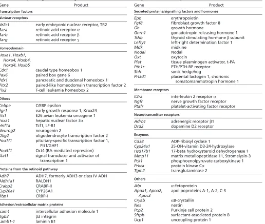
Evidence that endogenous retinoids are required for hindbrain patterning was found in vitamin A-deficient (VAD) quail embryos, in which the caudal hindbrain region (r4-r8) was misspecified into an enlarged r3, and more anterior rhombomeres expanded posteriorly (Fig. 4B) (Gale et al., 1999). Region-specific effects of RA deficiency have also been documented in Raldh2–/–mouse mutants, which lack rhombomeric segmentation and exhibit a severe reduction of the posterior hindbrain. Strikingly, the expression of genes normally restricted to r3-r4 [such as Hoxb1, Krox20 (Egr2)] spreads posteriorly in these mutants, whereas expression of r5-r7 determinants (Mafb, Hoxd4) is reduced or abolished (Fig. 4C) (Niederreither et al., 2000). These patterning defects have dramatic consequences for related developmental events, such as inner ear patterning, neural crest migration (leading to a lack of development of all branchial arches except for the first one), or neurite/cranial nerve differentiation. In zebrafish raldh2 mutants, a similar hindbrain anteriorisation is described (Begemann et al., 2001; Grandel et al., 2002). These results led to the conclusion that RA produced by RALDH2 in somitic mesoderm diffuses towards the hindbrain, acting as a classical ‘vertical’ signal to control patterning and regulate the expression of posterior rhombomeric determinants.
As RARs have partly redundant functions, hindbrain abnormalities are found only when at least two receptors are inactivated in combination. Interestingly, such compound inactivations have different outcomes on hindbrain patterning. Rara–/–;Rarg–/–mutants display severe malformations similar to
those of Raldh2–/– mutants (Wendling et al., 2001) (Fig. 4C). Rara–/–;Rarb–/–mutants show a different phenotype, in which only r5-r7 have abnormal boundaries, and genes normally restricted to r5-r6 spread posteriorly at the expense of r7 markers (Fig. 4D) (Dupé et al., 1999). Exposure of cultured wild-type embryos to BMS493, a pan-RAR antagonist, at early somite stages phenocopied the Rara–/–;Rarg–/– hindbrain defects, whereas an earlier treatment at the beginning of gastrulation yielded an Rara–/–;Rarb–/–-like phenotype (Wendling et al., 2001). This indicates that RARand/or RARmediates the early effects of RA on hindbrain patterning, whereas RAR functions later in development in setting up the size and caudal boundary of the r5/r6 territory.
Importantly, Cyp26 genes display differential, rhombomere-specific expression patterns. Cyp26a1 loss-of-function leads to subtle patterning defects, with an enlarged r4 and partial transformation of r3 to an r4-like identity (Abu-Abed et al., 2001; Sakai et al., 2001) (Fig. 4E). Hindbrain abnormalities were not observed in Cyp26b1–/–or Cyp26c1–/–mutants, although compound Cyp26a1;Cyp26c1inactivation leads to severe defects (lack of segmentation and posteriorisation of the prospective r1-r4 region) (MacLean et al., 2009; Uehara et al., 2007) (Fig. 4F). As described below, CYP26 enzymes are likely to act in a concerted manner to control RA diffusion and maintain specific pre-rhombomeric territories in an RA-free state.
The hindbrain: an experimental paradigm to study RA morphogenetic gradients
The striking effects of both retinoid excess and retinoid deficiency on hindbrain patterning have stimulated research into the underlying mechanisms and modes of RA action. Almost 25 years ago, RA was identified as the first candidate vertebrate morphogen, following the discovery of uneven concentrations of RA across the developing chick limb bud (Thaller and Eichele, 1987). Morphogens are signalling molecules that act non-cell-All-trans-retinoic acid
4-oxo-RA All-trans-retinoic acid
O
OH
OH
O C
CYP26A1, B1,C1
hl nt*
fl
fl h hl
hd
hd
tl B
A
C D E
F G
b1 b1
b2
b2 h
h
H I
4-hydroxy-RA
(Conjugation, elimination)
la
la md
p p
md
mx mx
fp
[image:6.612.53.511.61.228.2]fp
Fig. 2. RA metabolism and degradation. (A)All-trans-retinoic acid is converted by CYP26 enzymes (CYP26A1, CYP26B1 and CYP26C1) into the more polar metabolites 4-hydroxy-RA and 4-oxo-RA, which eventually become conjugated (mainly as glucuronates) and are eliminated (by excretion). (B-I)Phenotypes of null mutants for Cyp26a1(C) and Cyp26b1(E,G) and of the compound Cyp26a1;Cyp26c1mutant (I), compared with their wild-type (WT) littermates (B,D,F,H). At prenatal stages (E18.5), Cyp26a1–/–mutants have an open neural tube (nt*) and severe posterior body
truncation with abnormal positioning of the hindlimbs (hl). Cyp26b1–/–mutants exhibit a spectrum of facial, laryngeal and vertebral abnormalities.
Compound inactivation of Cyp26a1and Cyp26c1is early embryonic lethal and leads to dramatic head truncation, with associated hindbrain and neural crest alterations. F,G show skeletal preparations visualised by Alizarin Red/Alcian Blue staining; H,I are scanning electron micrographs. b1, b2, branchial arches; ey, eye (abnormally open in the Cyp26b1–/–mutant); fl, forelimb; fp, frontal plate; h, heart; hd, head; la, larynx; md, mandible; mx,
maxillary; np, nasal process; p, parietal bone; tl, tail. The asterisk in G indicates abnormally fused cervical vertebrae. Reproduced with permission: B,C (Abu-Abed et al., 2001); D-G (MacLean et al., 2009); H,I (Uehara et al., 2007).
D
E
V
E
LO
P
M
E
N
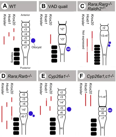
autonomously in a concentration-dependent fashion to assign positional identities to fields of cells (Wolpert, 2011). Over the last 10 years, several groups have tried to gain insights into how the spatial distribution of RA is regulated. Below, we discuss the ‘classical’ and newer aspects of this regulation and establishment of possible RA gradient(s), the main emerging concept being that several enzymatic activities (e.g. of RDHs and CYP26s) are required in addition to RALDHs to dynamically control and shape RA distributions within the embryo.
Does RA match the definition of a morphogen? As a small lipophilic molecule (Fig. 1) it is able to diffuse across cell membranes. Furthermore, it is soluble in water up to ~200 nM (Szuts and Harosi, 1991), i.e. within its physiological concentration range, so diffusion gradients might also occur in the extracellular space. Within the hindbrain, RA can control gene expression in a concentration-dependent manner, as demonstrated by concentration-dependent effects of RA treatments (e.g. Durston et al., 1989). Treatment of chick or zebrafish embryos with the RA antagonist BMS493 showed that more posterior rhombomere boundaries require progressively higher concentrations of RA for their correct positioning (Dupé and Lumsden, 2001; Maves and Kimmel, 2005). The RA source for hindbrain patterning is local and corresponds to presomitic mesoderm (PSM) and somites expressing Raldh2 near the caudal hindbrain (Fig. 5). These observations might support a role for RA acting as a diffusible morphogen in the pre-segmented hindbrain, perhaps diffusing from its mesodermal source via cell-cell contacts. Unlike other established morphogens [e.g. FGF8 or sonic hedgehog (SHH)], a gradient for RA has never been demonstrated, mainly owing to technical limitations. Recently, White et al. (White et al., 2007) visualised RA indirectly in zebrafish using a fluorescent marker under the control of RAREs (rare:yfp). In transgenic embryos, YFP expression was clearly graded from posterior to anterior regions of the hindbrain. This expression is lost in RA-deficient mutant
embryos, but can be restored by the prior transplantation of cells overexpressing Raldh2into somitic mesoderm. In most cases, the rescue spanned the width of the neural tube along a six-cell (70 m) diameter (White et al., 2007).
In another zebrafish study, Maves and Kimmel (Maves and Kimmel, 2005) reported a sequential induction of RA target genes, and showed that posteriorly expressed genes do not require longer exposure, but require higher levels of RA for induction than anteriorly expressed genes. This suggests that the RA gradient would increase over time, and that the high concentrations needed to activate posterior genes are only reached later during hindbrain development. The authors proposed a model in which the hindbrain needs a temporally increasing source of RA to define rhombomere identities sequentially (Fig. 5A).
The observation that Cyp26 genes show rhombomeric-specific expression patterns drove the idea that degradation might be involved in shaping RA distribution. In a mouse study, Sirbu et al. correlated the dynamics of Hoxb1expression, from its onset to its restriction in r4, with the dynamic expression of Cyp26 genes (Sirbu et al., 2005). They proposed a ‘shifting boundaries’ model in which the anterior boundaries of rhombomere-specific genes are fixed by the posterior limit of CYP26 activity at the time of their onset of expression. Hernandez et al. obtained consistent results in zebrafish, and elaborated a ‘gradient-free’ model, in which RA degradation by CYP26 enzymes determines progressively more posterior limits of RA-dependent gene expression in a stepwise manner (Hernandez et al., 2007). All three CYP26s would thus function to establish three sequential boundaries in RA responsiveness, i.e. pre-r3/r4, r4/r5 and r6/r7 (Fig. 5B).
As Cyp26a1is under the control of RA signalling, a more subtle role for RA degradation has been proposed by White and colleagues (White et al., 2007; White and Schilling, 2008). Their model (Fig. 5C), in which CYP26A1 plays a central role in modulating RA levels dynamically, can reconcile the various
RDHs RALDHs
CRABP Retinoic
acid
Retinol
RXR
RAR
No transcription
Target gene transcription Co-repressors
R R
R
TBP TAFs Pol II
Pol II
TBP TAFs
RARE
[image:7.612.52.381.60.353.2]Co-activators
Fig. 3. Summary of the RA signalling pathway.RA, synthesized intracellularly from circulating retinol or diffusing from an adjacent cell (curved red arrow), eventually reaches the nucleus. Cellular retinoic acid-binding proteins (CRABPs) may be involved in this transfer. Cellular retinol-binding proteins (CRBPs) may help present retinol to retinol dehydrogenases (RDHs). Dimers of RA receptors (RARs) and retinoid X receptors (RXRs), termed RAR/RXR, are able to bind to RA-response elements (RAREs) in their target genes in the absence of ligand, interacting with protein complexes (co-repressors) that stabilise the chromatin nucleosomal structure and prevent access to the promoter. Upon RA binding, a
conformational change in the helicoidal structure of the RAR ligand-binding domain changes its protein-protein interaction properties, releasing the co-repressors and recruiting co-activator complexes that destabilise the nucleosomes and/or facilitate assembly of the transcription pre-initiation complex, which contains RNA polymerase II (Pol II), TATA-binding protein (TBP) and TBP-associated factors (TAFs).
D
E
V
E
LO
P
M
E
N
observations described above. Using bead implantation, the authors showed that FGF signalling acts indirectly by inhibiting RA-dependent activation of Cyp26a1expression. By computational analysis, they showed that RA gradients can be influenced by interacting feedback (RA signalling then inducing RA degradation) and feedforward (FGF signalling then repressing RA degradation) effects. The feedforward effects of FGFs couple the shape of the RA gradient to that of the FGF gradient. They argue that this makes the gradient stable to fluctuations in RA synthesis, but also over an expanding field of cells.
One aspect that should be addressed is regulation at the level of the source of RA. Studies in Xenopusproposed an alternative mode of RA gradient formation based on cooperation between RDH10 and RALDH2 (Strate et al., 2009). RDH10 produces retinaldehyde in the anterior cervical mesoderm and ventral hindbrain that diffuses posteriorly, where RALDH2 converts it into RA. Highest levels of RA would thus be produced at the anterior front of
[image:8.612.52.568.69.506.2]RALDH2 expression, where retinaldehyde concentration is highest, with decreasing concentrations observed posteriorly (Fig. 5D). This peak of RA would move posteriorly concomitant with the translocation of the RDH10 and RALDH2 expression domains, shifting the peak of the gradient from the hindbrain/spinal cord boundary to within the spinal cord. Further studies are required to address the significance of such cooperative effects in the hindbrain or in other morphogenetic fields. It was shown recently that Raldh2 expression is under the transcriptional control of a ternary complex that includes Hox, Pbx and Meis proteins. Both Pbx1;Pbx2and Hoxa1;Pbx1compound mutant mice show reduced mesodermal Raldh2expression (Vitobello et al., 2011). These authors show that HOXA1-PBX1/2-MEIS2 directly binds a regulatory element required to maintain normal Raldh2 expression. Thus, in the hindbrain, Hox proteins may regulate their own boundaries by controlling RALDH2 levels, thus contributing to shaping the RA gradient produced.
Table 3. Examples of genes containing functional and/or evolutionarily conserved RA-response elements
Gene Product
Transcription factors
Nuclear receptors
Nr2c1 early embryonic nuclear receptor, TR2
Rara retinoic acid receptor
Rarb retinoic acid receptor
Rarg retinoic acid receptor
Homeodomain
Hoxa1, Hoxb1, Hoxa4, Hoxb4, Hoxd4, Hoxb5
Cdx1 caudal type homeobox 1
Pax6 paired box gene 6
Pdx1 pancreatic and duodenal homeobox 1
Pitx2 paired-like homeodomain transcription factor 2
Tlx2 T-cell leukemia homeobox 2
Others
Cebpe C/EBP epsilon
Egr1 early growth response 1, Krox24
Ets1 E26 avian leukemia oncogene 1
Foxa1 hepatic nuclear factor 3 Hnf1a Tcf1, LF-B1
Neurog2 neurogenin 2
Olig2 oligodendrocyte transcription factor 2 Pou1f1 pituitary-specific transcription factor 1,
Pit1/GHF1
Pou5f1 Oct4 (RA-mediated repression) Stat1 signal transducer and activator of
transcription 1
Proteins from the retinoid pathway
Adh7 ADH7, formerly ADH3 or class IV ADH
Aldh1a1 RALDH1
Crabp2 CRABP-II
Cyp26a1 CYP26A1
Rbp1 CRBP-I
Adhesion/extracellular matrix proteins
Icam1 intercellular adhesion molecule 1 Itgb3 3 integrin
Lamb1-1 laminin B1
Gene Product
Secreted proteins/signalling factors and hormones
Epo erythropoietin
Fgf8 fibroblast growth factor 8
Gh growth hormone
Gnrh1 gonadotropin releasing hormone 1
Tshb thyroid stimulating hormone subunit
Lefty1 left-right determination factor 1
Mdk midkine
Nodal Nodal
Oxt oxytocin
Plat tissue plasminogen activator, t-PA
Pth1r PTH/PTH-RP receptor
Shh sonic hedgehog
Prl3d1 placental lactogen 1, chorionic somatomammotropin hormone 1
Membrane receptors
Il2ra interleukin 2 receptor
Ngfr nerve growth factor receptor
Ptafr platelet-activating factor receptor
Neurotransmitter receptors
Adrb1 adrenergic receptor 1
Drd2 dopamine D2 receptor
Enzymes
Cd38 ADP-ribosyl cyclase 1
Cyp24a1 25-OH-vitamin D3-24-hydroxylase Hsd17b1 17-beta hydroxysteroid dehydrogenase 1 Mmp11 matrix metallopeptidase 11, Stromelysin-3
Pck1 phosphoenolpyruvate carboxykinase 1
Prkca protein kinase C
Tgm2 transglutaminase 2
Others
Afp -fetoprotein
Apoa1,Apoa2, apolipoproteins A-1, A-2, C-3 Apoc3
Cryab B-crystallin
Nes nestin
Pcp2 Purkinje cell protein 2
Sftpb surfactant-associated protein B
Ucp1 uncoupling protein 1
The list is organised according to the types of proteins encoded and is by no means exhaustive. For detailed lists of RA-responsive genes from which these data were mainly compiled, see Balmer and Blomhoff (Balmer and Blomhoff, 2002; Balmer and Blomhoff, 2005).
D
E
V
E
LO
P
M
E
N
RA functions during forebrain development
Whereas many studies have investigated RA functions in the developing hindbrain, other aspects of brain development remain less explored. Initial studies using the chick system in which an RAR/RXR antagonist was delivered by bead implantation (Schneider et al., 2001), or using the VAD quail model (Halilagic et al., 2003), indicated a role for RA in A-P patterning of the embryonic forebrain and cell survival in the telencephalon (the anteriormost forebrain derivative). Moreover, RA was suggested to specify an intermediate character within the telencephalon, acting in combination with SHH to impart ventral identity and with Wnts/FGFs to impose a dorsal character (Marklund et al., 2004).
Two Raldh genes are differentially activated during early embryonic head and forebrain development. Raldh2is transiently expressed in the rostral neural plate and optic vesicles, whereas Raldh3is expressed slightly later in the surface ectoderm overlying the anterior forebrain. Studies of murine Raldh2/Raldh3 loss-of-function mutants only partly supported the avian experimental data. Using a RARE-containing reporter transgene it was found that
Raldh2 inactivation ablates all RA activity in the forebrain neuroepithelium (Mic et al., 2004a; Ribes et al., 2006). Raldh2–/–
embryos exhibit defective growth and morphogenesis of the optic vesicle, which is an evagination of the forebrain neuroepithelium and precursor of the retina (Mic et al., 2004a). Ribes et al. reported additional forebrain deficiencies in Raldh2–/– mutants, with
decreased cell proliferation and altered expression of several ventral determinants, including SHH-responsive genes (Ribes et al., 2006). Molotkova et al. analysed Raldh2–/–and Raldh2–/–;Raldh3–/–
compound mutants and questioned an early function of RA in forebrain based on their observation of normal expression patterns of genes including Fgf8and Meis2(Molotkova et al., 2007). Thus, whether RA plays crucial roles apart from regulating optic vesicle development remains controversial and is a difficult issue to address with the available (embryonic lethal) Raldh-null mouse models. An alternative approach to inhibit RA signalling consists of expressing a dominant-negative receptor (DN-RAR) in the embryonic telencephalon (using the Cre-lox system for tissue-specific expression). This approach also led to a decrease in cell proliferation and to increased cell death in telencephalic progenitor populations (Rajaii et al., 2008). Furthermore, an abnormal distribution of Islet1-expressing cells was found in the ventral telencephalon, with Islet1+cells present in the medial ganglionic
eminences (MGEs) instead of being restricted to the lateral ganglionic eminences (LGEs), suggesting a role for RA in the specification of progenitor cell populations.
Retinoid signalling may influence specific progenitor populations at later stages of forebrain development, although these functions remain poorly characterised. Smith et al. (Smith et al., 2001) first suggested that RA could be a diffusible signal regulating neurogenesis in the cerebral cortex, based on the observation that Raldh2 – and to a lesser extent Raldh1 – is expressed in the developing meningeal cell layer from E13.5 to postnatal stages. RA produced by meningeal cells could thus diffuse within the neuroepithelium, where it may influence progenitor cell proliferation or differentiation and/or radial migration along cortical layers. Meningeally produced RA may regulate neurogenesis in other brain regions at later developmental stages (Zhang et al., 2003). A new player came from the recent work of Siegenthaler et al. (Siegenthaler et al., 2009). RDH10, which acts upstream of RALDHs, is also expressed in the developing meninges (Romand et al., 2008) and, while studying Foxc1mutant mice that exhibit defective forebrain meningeal formation, Siegenthaler et al. showed decreased Raldh2and Rdh10expression in the affected meninges, and found that all-trans-RA treatment improved cortical development both in vivo and in an explant culture system. Chatzi et al. (Chatzi et al., 2011) challenged these results and the hypothesis of a role for meningeal RA. These authors analysed Raldh2–/–mutants at E14.5 after maternal RA rescue, and reported
no change in cell proliferation or in the overall organisation of the cortical layers, although the meningeal layers lacked RA activity as observed with a reporter transgene. This issue will need to be resolved by more in-depth studies and might require the generation of additional murine models with tissue-specific ablation of RA synthesis in meninges. Currently, the only unequivocal role of RA in the forebrain at the mid to late developmental stages relates to the differentiation of GABAergic striatal projection neurons and interneurons migrating to olfactory bulb and cortex, and involves RALDH3 activity in the LGE subventricular zone (Chatzi et al., 2011; Molotkova et al., 2007). Furthermore, it is becoming clear that endogenous RA functions are likely to persist in adult neuronal populations (see Box 3).
VAD quail
Rara;Rarg-/-
Raldh2-/-r2 r3 r4 r5 r6
Somites
r2
“r3-r4” “r2”
“r3”
Rara;Rarb-/-B
C
A
Posterior WT
D
E
Cyp26a1-/-F
Cyp26a1;c1-/-Otocyst
Not expressed
Krox20
Hoxb1
Kreisler Kreisler Hoxb1 Krox20 Kreisler Hoxb1 Krox20
Krox20
Hoxb1
Kreisler Kreisler Hoxb1 Krox20 Kreisler Hoxb1 Krox20
Anterior
r7
r2 r3 r4 “r5” r6-7
r2 r3 “r4”
r5 r6 r7
“r1-r4”
[image:9.612.53.300.58.349.2]“r5” r6 r7
Fig. 4. Hindbrain abnormalities in animal models with altered RA signalling.(A)The hindbrain rhombomeric structure in a wild-type (WT) embryo, highlighting rhombomeres 2-7 (r2-r7) with adjacent somites (black) and otocyst (blue) also shown. Pre-segmental/segmental expression patterns of three key rhombomeric markers [Kreisler (Mafb), Hoxb1, Krox20] are depicted (red bars; dashed bars indicate patchy or ill-defined expression domains). (B-F)Alterations in hindbrain segmentation and molecular patterning are illustrated in various animal models with endogenous deficiency in RA signalling: (B) vitamin A-deficient (VAD) quail; (C) Raldh2–/–, as well as Rara–/–;Rarg–/–signalling mutant mice (lacking RARand RAR); (D) Rara–/–;Rarb–/–mice (lacking RARand RAR); (E) Cyp26a1–/–mice; and (F) Cyp26a1–/–;Cyp26c1–/–mice. Abnormal (enlarged, non-segmented and/or abnormally patterned) rhombomeres are indicated by quotation marks. Otocyst abnormalities were not determined (ND) in some models. SC, spinal cord.
D
E
V
E
LO
P
M
E
N
RA actions in foregut derivatives
RA signalling has numerous other functions during early embryogenesis. We briefly discuss how these functions may correlate with gradients of activity and/or diffusion between cell layers, first focusing on the branchial apparatus and then the lungs and pancreas. Details of RA functions in other organ systems can be found in other reviews (Duester, 2008; Niederreither and Dollé, 2008).
RA signalling in the branchial apparatus
Branchial arches are segmental structures that develop along the embryonic foregut endoderm. They are colonised by segmental mid/hindbrain neural crest streams and give rise to various derivatives including the hyoid bone, thyroid, parathyroids and thymus, and specific cardiac populations. RA is produced locally by RALDH2, which is expressed in the mesenchyme surrounding the foregut up to a rather sharp boundary at the level of the 4th-6th arches (Niederreither et al., 2003). Analysis of an RA-responsive transgene revealed a more extended area of RA activity, extending up to the posterior edge of the 2nd arch. Furthermore, this activity was found in both mesenchymal and endodermal cell layers. RALDH2 function in the branchial apparatus was uncovered in Raldh2–/– mutants rescued by stage-specific maternal RA
supplementation (Niederreither et al., 2003). This supplementation
partly rescued hindbrain and neural crest defects, unveiling an abnormal branchial phenotype in which all structures derived from the 3rd, 4th and 6th arches failed to develop, including endodermal pouches and aortic arch arteries. Consequently, many of the derivatives of these arches were missing or abnormal at foetal stages. Abnormalities of the aorta and large vessels, which are derived from the aortic arches, were also seen, with a lack of aortic trunk septation (persistent truncus arteriosus) incompatible with postnatal survival. Treatment of early somite stage wild-type embryos with a pan-RAR antagonist (BMS493) led to similar effects on posterior branchial arches (Wendling et al., 2000). Generation of a hypomorphic Raldh2 mutation revealed a particular susceptibility of the branchial region to diminished RA synthesis, as hypomorphic mutants displayed abnormalities that phenocopied the rescued Raldh2–/–mutants (Vermot et al., 2003).
These abnormalities phenocopy a human condition, DiGeorge syndrome, which is caused by chromosomal deletions that affect the genes encoding the transcription factor T-box 1 (TBX1) and the adaptor protein CRKL. Analysis of murine Tbx1/Crkl loss-of-function models revealed locally increased RA signalling due to changes in the Raldh2and Cyp26a1/Cyp26b1expression domains (Guris et al., 2006), and a genetic interaction between Raldh2and Tbx1 was recently demonstrated (Ryckebusch et al., 2010). Collectively, these data show that RA acts to pattern posterior branchial arches and their derivatives and implicate the retinoid pathway in the pathogenesis of DiGeorge syndrome.
Consistent molecular abnormalities have been observed in the branchial region of Raldh2mutants (Niederreither et al., 2003; Vermot et al., 2003) and BMS493-treated cultured embryos (Wendling et al., 2000). Among the affected genes are Hoxa1and Hoxb1, which are two RARE-containing genes that are also Time 1
Anterior Posterior
Time 1 Time 2
Time 2 Time 1
Time 2 Time 1
Anterior Posterior
Time 2 1
CYP26A1 CYP26B1 CYP26A1
CYP26C1
A A A
A
A A A A A
A1
RA gradient
Key
Retinaldehyde/
Rdh10 expression
Cyp26 expression Presomitic mesoderm/ somites/Raldh2 expression Retinaldehyde flow Time 2
Time 3 Time 1
B Shifting boundaries of Cyp26expression A Temporally increasing RA gradient
C CYP26A1- and FGF-regulated RA gradient
D RDH10- and RALDH2-dependent RA gradient
[image:10.612.54.291.56.468.2]FGF gradient
Fig. 5. Models of sequential RA activity during hindbrain segmentation.Graded distributions of signalling molecules and expression patterns of RA synthesizing and metabolising enzymes are shown. Positive and negative regulatory interactions are depicted as arrows and bars, respectively. These interactions are sequential and initiate at early (pre-segmental) stages in the gastrula/neurula; hence the rhombomere scheme (grey) is only shown to provide positional landmarks. (A)The ‘increasing gradient’ model proposes that the RA morphogenic gradient is not fixed, but grows steeper with time, specifying rhombomeres sequentially from anterior to posterior. (B)The ‘shifting boundary’ model posits that localised RA degradation controls and/or refines the time of exposure. Importantly, the expression of CYP26 enzymes is dynamic, thus achieving borders of RA-dependent regulation in a stepwise manner. (C)In the ‘degradation-based’ model, the RA gradient is shaped by local control of its degradation. Cyp26a1 expression is regulated by the opposing action of two gradients: RA regulating Cyp26a1positively versus FGF signalling – eventually superseding RA – regulating it negatively. Raldh2is also controlled by this two-gradient influence, contributing to the regulation of RA production at the source. (D)The most recently proposed model incorporates the action of RDH10, which produces retinaldehyde in the ventral hindbrain and somites that diffuses towards RALDH2-expressing cells. Owing to the dynamics of Rdh10/Raldh2expression, the highest RA levels are produced at the hindbrain/spinal cord boundary, and this peak moves posteriorly as development proceeds. The models shown in A-D are not mutually exclusive and together they are likely to account for the sequential regulatory effects of RA in pre-segmented and segmented hindbrain.
D
E
V
E
LO
P
M
E
N
affected in the developing hindbrain. Other possible effectors of the RA-deficiency phenotype include Fgf genes, mainly Fgf8 and Fgf3, the expression of which was severely reduced in Raldh2 mutants. Neither the molecular nor the phenotypic studies provided clear evidence for an RA concentration gradient acting in the branchial region. They indicated, however, that the retinoid signal acts non-cell-autonomously: by analogy with the hindbrain model, it can be defined both as a ‘vertical’ signal travelling from mesenchyme to pharyngeal endoderm and as a ‘planar’ signal diffusing along the branchial region up to the level of the 3rd arch. Another parallel with the hindbrain is that RA action may be restricted by the activity of CYP26s, all of which are expressed in specific branchial/cervical cell populations (Table 1).
RA signalling during lung development
Region-specific retinoid signalling has other important functions in the development of foregut derivatives. Analysis of Raldh2–/–
mutants and experiments performed on embryonic explants demonstrated that a lack of RA/RAR activity prevents induction and growth of the primary lung buds (Desai et al., 2006; Wang et al., 2006). The underlying molecular events include a lack of Fgf10 induction in the lung field (the region where primary lung buds are induced) caused by upregulation of TGFsignalling, which has an inhibitory effect on Fgf10expression (Chen et al., 2007; Chen et al., 2010).
RA signalling and pancreas development
It was demonstrated in zebrafish (see Alexa et al., 2009), Xenopus (Chen et al., 2004) and mouse (Martin et al., 2005; Molotkov et al., 2005) that RA is also required for pancreas development. Here, RA
(produced mesodermally by RALDH2) acts by diffusing towards the endoderm, where most of the molecular abnormalities are observed under RA deficiency. The pancreas derives from two endodermal primordia known as the ventral and dorsal buds, and lack of RA specifically affects induction and growth of the dorsal pancreatic bud. One important role of RA is to downregulate SHH signalling, which has an inhibitory effect on pancreas induction. RA may also act at later steps of specification of pancreatic endocrine cell lineages (Martin et al., 2005; Ostrom et al., 2008). Recent work indicates that RA may act more globally to coordinate the position of endoderm-derived organs along the foregut and midgut A-P axis (Bayha et al., 2009), and that CYP26 enzymes may restrict the extent of RA signalling and set up the limit of the pancreatic field (Kinkel et al., 2009).
RA and limb development
Many early studies have investigated the role of RA in embryonic limb bud patterning, triggered by reports of its ability to induce mirror-image digit duplications when applied locally in chick wing buds (Tickle et al., 1982) and of the measurement of differential endogenous concentrations along the limb A-P axis (Thaller and Eichele, 1987). Abnormalities in limb skeletal patterning were first reported for Rara–/–;Rarg–/–compound mutant mice (Lohnes et al.,
1994), and it was later found that mutants for RA-synthesizing enzymes have hypoplastic or absent forelimb buds (Rdh10–/–or
Raldh2–/– mutants, respectively) (Cunningham et al., 2011;
Niederreither et al., 1999; Sandell et al., 2007). A detailed study of compound Raldh mutants showed, however, that RA is not necessary for hindlimb development (Zhao et al., 2009). It was found that RA deficiency affects the expression of several regulators of forelimb bud growth and patterning, including Fgf4/8 and Shh (Mic et al., 2004b; Niederreither et al., 2002). A subsequent study concluded that these effects are indirect, with RA acting outside of the limb bud and perhaps even before its induction: here, an RA-dependent inhibition of FGF8 signalling in the body axis near the forelimb field would create a permissive environment allowing limb bud induction (Zhao et al., 2009). Eventually, limb bud cells need to develop in an RA-protected environment, mainly through the sustained action of CYP26B1 (Probst et al., 2011; Yashiro et al., 2004). Interestingly, such a function might have arisen prior to tetrapod limb specialisation. Indeed, RA signalling is also required for the development of zebrafish pectoral fin buds, acting from gastrulation to early somite stages as a permissive signal for the proper induction and growth of the fin bud (Gibert et al., 2006; Grandel and Brand, 2011).
RA function during somitogenesis and neural tube differentiation
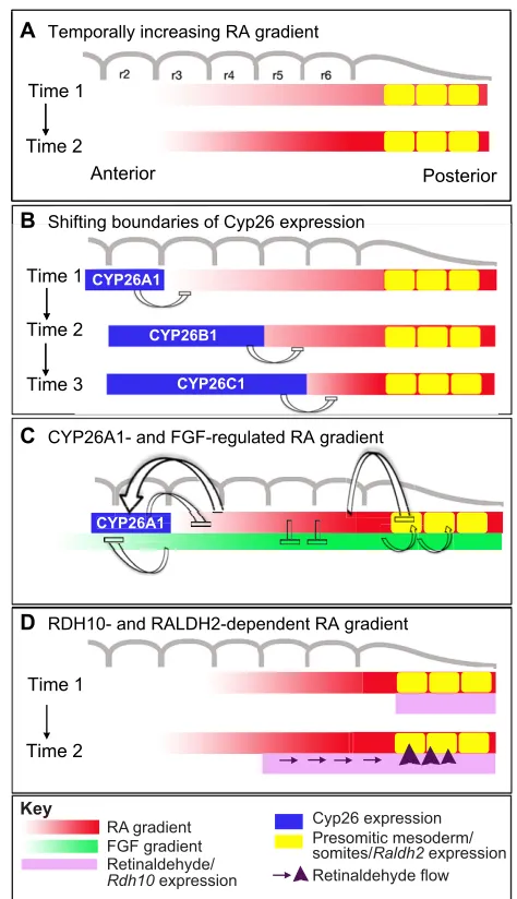
Many in-depth studies have investigated the functions of RA during elongation of the embryonic body axis, where it controls several events relating to mesodermal segmentation and neurogenesis in the caudal neural tube (the future spinal cord). Somites are segmented epithelial structures that are formed sequentially along the left and right paraxial mesoderm. They are the precursors of various tissues: their dorsal portion, the dermomyotome, will differentiate into muscle and dermis, whereas their ventral part, the sclerotome, gives rise to skeletal elements (vertebral column and ribs). Somite formation is a rhythmic process that relies on a ‘clock and wavefront’ mechanism, in which a molecular oscillator driven by Wnt and Notch signalling generates cyclic waves of gene expression that progress rostrally along the PSM (reviewed by Gibb et al., 2010). This oscillator interacts with a system of Box 3. Retinoids in the adult brain
Retinoid functions are likely to persist through postnatal life. Mice mutant for RARand RXR, two receptors specifically expressed in striatal structures including the caudate putamen and nucleus accumbens (NAc), show reduced locomotor activity and impaired motor performance typical of abnormal striatal function. Importantly, Rxrg–/–mice show increased despair behaviour and
another key symptom of depression (anhedonia), which are both reversed by chronic antidepressant treatment. Adenovirus-mediated re-expression of RXR within the NAc also reversed these behaviours, clearly demonstrating a postnatal function for RXR signalling (Krzyzosiak et al., 2010). These findings suggest that altered retinoid signalling could contribute to diseases affecting the nigrostriatal system, such as Parkinson’s and Huntington’s diseases. RA may also act in the hippocampus, a key structure for memory processing and emotion. Neurogenesis occurs throughout life in the hippocampal granular zone, a site of high RA activity. Rarb–/–and
Rxrg–/– mice are deficient in spatial learning and memory, like
vitamin A-deficient (VAD) rats, for which the deficits can be rescued by RA treatment (Bonnet et al., 2008). Altered retinoid signalling may also be involved in the degradation of hippocampal function in aging mice. Some pioneering studies suggest that retinoids might regulate the proliferation and/or differentiation of hippocampal stem cells into functional neurons. Adult neural stem cells are also found in the forebrain subventricular zone and in the olfactory bulb. In vitro, RA can increase neurogenesis by enhancing the proliferation and differentiation of adult forebrain neuroblasts, and in vivo it may regulate the proliferation of slowly dividing astrocytes in the subventricular zone (Haskell and LaMantia, 2005). Further characterisation of these functions might have important implications for the therapy or prevention of neurodegenerative diseases.