0095-1137/97/$04.0010
Copyright © 1997, American Society for Microbiology
Human Isolates of
Staphylococcus caprae: Association with
Bone and Joint Infections
ROGER SHUTTLEWORTH,1* RONALD J. BEHME,1,2ALAN MCNABB,1
ANDW. DAVID COLBY1,2
Division of Microbiology, London Health Sciences Centre,1and the Department of Microbiology and Immunology,2University of Western Ontario, London, Ontario, Canada
Received 10 March 1997/Returned for modification 29 April 1997/Accepted 7 July 1997
Staphylococcus capraeis a coagulase-negative, DNase-positive member of the genusStaphylococcus usually associated with goats, but since 1991 a few laboratories have reported isolating the organism from human clinical specimens. We report on the isolation of 14 strains from human specimens and note that 10 strains were obtained from patients with bone and joint infections. Nine of the 10 infections started with traumatic fractures, and the other was a case of mastoiditis. Seven of these 10 infections were in patients with orthopedic prostheses, which appears to be a risk factor. Three of the 14 strains were from transplant patients. For three
of the patients, S. capraewas the only organism isolated. S. capraemay be misidentified because it is not
represented in the current MicroScan or Vitek identification systems which are in use in many laboratories,
but the organism can be differentiated by a few biochemical tests.S. capraeproduces positive results for DNase,
pyrrolidonyl aminopeptidase, and acid production from mannitol and maltose; it produces negative results for ornithine decarboxylase and tube coagulase.
Coagulase-negative staphylococci are among the most com-mon bacteria isolated in clinical laboratories. Once regarded as harmless commensal organisms, some are now recognized as pathogens capable of causing both nosocomial and communi-ty-acquired infections, particularly in immunosuppressed pa-tients or in individuals who use prosthetic devices (9). Infection withStaphylococcus epidermidisin such patients is well docu-mented, and there are reports implicating other coagulase-negative species in serious infections. In the last two decades a number of new species of staphylococci have been described. We report here on the isolation from human clinical samples of 14 strains of S. caprae, a species usually associated with goats. A large proportion of the strains were isolated from patients with bone and joint infections associated with ortho-pedic prostheses. We also present relevant laboratory data which may be useful for the recognition of strains of this species in human material.
MATERIALS AND METHODS
Bacterial strains.The sources of the 14 strains ofS. caprae, together with
relevant clinical information, are presented in Table 1. The strains were isolated during the period from November 1990 to April 1996. All strains were catalase-positive, gram-positive cocci appearing in clusters, and all grew well on P agar containing 6.5% (wt/vol) sodium chloride. They were identified asS. capraeby means of a self-educating system which has been described in detail elsewhere (1). Briefly, isolates were subjected to cellular fatty acid analysis with the Micro-bial Identification System (MicroMicro-bial ID, Inc., Newark, Del.), as well as 35 biochemical tests. They were assigned to the species only if both identification methods gave the same result.
Cultures of staphylococci were suspended in glycerol citrate solution at270°C for long-term storage and were routinely subcultured to 5% horse blood agar and incubated in air at 35°C.
The type strain ofS. caprae, ATCC 35538, was obtained from the American Type Culture Collection, Rockville, Md.
Colonial morphology.All the strains were grown on P-agar plates (11)
incu-bated at 35°C for 72 h, followed by further incubation for 48 h at room temper-ature. Cultures on brain heart Infusion agar (4311065; BBL), Columbia agar
(CM 331; Oxoid, Basingstoke, United Kingdom) with 5% horse blood, and Columbia agar with 5% sheep blood were incubated at 35°C for 48 h.
Biochemical tests.Sensitivity to desferrioxamine (13) was tested on P-agar
plates with disks impregnated with 1 mg of Desferal (Ciba-Geigy, Dorval, Que-bec, Canada). The disk was placed on the second quadrant of a streaked P-agar plate, and the plate was incubated overnight at 35°C in air.
Tests for bound coagulase and protein A were performed with the Slidex Staph Plus kit (bioMerieux, Marcy l’Etoile, France), according to the manufacturer’s instructions. Tests for heat-stable nuclease were performed by the microslide method of Lachica et al. (12).
The strains were also examined by using MicroScan Pos Combo 6 panels (Dade International, West Sacramento, Calif.) and Vitek GPI cards (bio-Merieux-Vitek, Hazelwood, Mo.), according to the manufacturers’ instructions.
Cellular fatty acid analysis.Organisms taken from storage were subcultured at
least twice on blood agar before analysis. For fatty acid analysis, organisms were grown on Trypticase soy agar (11043; BBL) containing 5% sheep blood, and the plates were incubated in air at 35°C for 2461 h. Analysis of cellular fatty acids was performed as described previously (1) by using the Microbial Identification System. Fatty acid profiles were compared with those in a profile library com-piled in the Division of Microbiology laboratory and containing entries for all the recognized aerobic species and subspecies of staphylococci with the exception of the recently described speciesS. saprophyticussubsp.bovis(6).
Antimicrobial susceptibility testing.Disk diffusion tests were performed by
the standard method of the National Committee for Clinical Laboratory Stan-dards (17). MICs were determined with MicroScan Pos Combo 6 panels and Vitek GPS SB cards (bioMerieux-Vitek) according to the manufacturers’ rec-ommendations.
The type strain ofS. caprae, ATCC 35538, was tested in parallel with all the clinical isolates.
RESULTS
Colonial morphology. Colonies on horse and sheep blood
agars at 35°C were white, circular, entire, raised, 1 to 2 mm in diameter after 24 h and approximately 4 mm in diameter after 48 h. Some strains showed a narrow zone of beta-hemolysis extending less than 1 mm from the edge of the colony after 48 h of incubation. On brain heart infusion agar after 48 h colonies were 4 to 5 mm in diameter, circular, raised and often with a flat top, and shiny but often with a matte center. On P agar after 5 days colony diameters were 6 to 10 mm, and the colonies were usually white, circular, entire, opaque, and raised. Colonies of most strains had a flat, shiny top with a broad ring of radial striations leading to a transparent periph-ery. In some cases tiny microcolonies were visible at the pe-* Corresponding author. Mailing address: Division of Microbiology,
London Health Sciences Centre, 339 Windermere Rd., London, On-tario, Canada, N6A 5A5. Phone: (519) 663-3899. Fax: (519) 663-3880.
2537
on May 15, 2020 by guest
http://jcm.asm.org/
riphery. One strain showed pale yellow pigmentation on P agar.
Biochemical tests.All strains gave negative results in tests
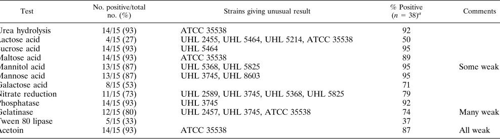
for oxidase, free coagulase, ornithine decarboxylase, esculin hydrolysis, and acid production from salicin, sorbitol, arabi-nose, raffiarabi-nose, cellobiose, xylitol, xylose, and melezitose. None of the strains grew on plates containing novobiocin (1.6 mg/ liter) or furazolidone (0.02%), and none was sensitive to des-ferrioxamine. All strains gave negative reactions when tested for bound coagulase or protein A in the Slidex Staph Plus kit. All strains fermented glucose within 24 h and gave positive reactions in the following tests: arginine hydrolysis, production of DNase (which was not heat stable) and pyrrolidonyl amino-peptidase, and acid production from glucose, trehalose, and fructose. Other tests gave variable results, which are presented in Table 2.
Positive reactions in the gelatinase, Tween 80 lipase, and Voges-Proskauer tests were always weak, whereas DNase tests always gave reactions which were at least as strong as those obtained with anS. aureuscontrol strain. Tests for heat-stable nuclease were negative. This is in contrast to results published previously (23), which stated that the DNase was heat stable but usually gave weak reactions. The type strain, ATCC 35538 (a goat strain), was dissimilar to the clinical isolates, giving negative results in the urea, maltose, and Voges-Proskauer tests. Strain UHL 3745 was also unusual in that it was mannose and phosphatase negative. The test for acid production from sucrose was positive for all strains (including the type strain) except UHL 5464. A previous report (4) stated thatS. caprae strains are negative in this test; this discrepancy is probably due to our use of a more sensitive test.
Cellular fatty acid analysis.All the strains were identified as
S. capraeby analysis of cellular fatty acids and gave similarity indices of.0.6 against library entries developed in the Divi-sion of Microbiology laboratory (1). A similarity index of 0.6 or more represents a good level of confidence in the identification (14). However, the fatty acid profiles were very similar to those for S. capitisand S. haemolyticus. This means that fatty acid analysis is unable to distinguish reliably between these groups and must be supported by other methods.
Commercially available identification methods.S. capraeis
not represented in the MicroScan or Vitek databases. The results obtained with both systems are given in Table 3.
Antimicrobial susceptibility tests. The results obtained by
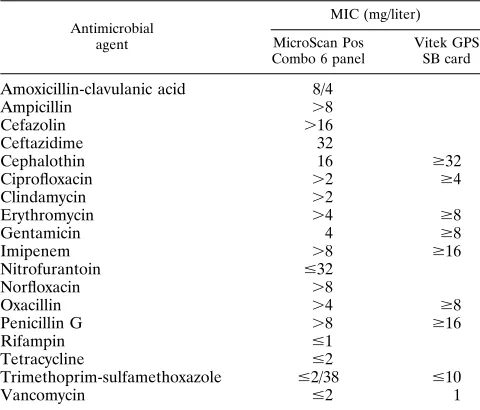
the three methods of antimicrobial susceptibility testing were almost completely consistent. However, UHL 3745 fell in the intermediate category when tested against gentamicin by the disk diffusion method but was reported to be sensitive by the commercially available systems. Three strains (strains UHL 3741 and UHL 5214 and the type strain) were sensitive to penicillin G and did not produce beta-lactamase. The other 12 strains were beta-lactamase positive. UHL 3745 was found to be resistant to several of the antimicrobial agents tested, in-cluding oxacillin (Table 4). Except for the penicillin resistance noted above, all the other strains were sensitive to all the agents tested. Two penicillin-resistant strains (strains UHL 6953 and UHL 8603) showed small zones of inhibition around the penicillin disk, with small colonies within the zone.
DISCUSSION
S. capraewas first described in 1983 in milk samples taken from healthy goats in France (4). It has since been reported as a commensal organism of goats on several occasions (3, 18, 21). Some strains produce toxic shock syndrome toxin type 1 (22),
TABLE 1. Source of strains and relevant clinical information a Isolate no. Patient age (yr) Diagnosis Orthopedic prosthesis Immuno-compromised Site of S. caprae isolation No. of specimens yielding S. caprae / no. examined
Microscopic findings (PC/GPC)
Other isolates (no. of specimens) Outcome UHL 2455 36 Fracture; tibial osteomyelitis, septic arthritis Y N Ankle synovium 1/6 Few/ 2 S. aureus (5) Degenerative arthritis UHL 2457 54 Fracture; knee sepsis, olecranon swelling Y N Bursa fluid from elbow 1/1 Few/ 2 None Cured UHL 2589 52 Bilateral otitis externa N N Left and right ear drainage 2/2 2 / 11 Pseudomonas aeruginosa (2), coliforms (1) Cured UHL 3288 44 Fracture; osteomyelitis of calcaneus N N Calcaneus 8/18 1 /Few Anaerobic GPC (11), coliforms (6), S. aureus (5) Unknown UHL 3741 71 Infected toenail N N Great toe nail 1/1 11 / 11 None Unknown UHL 3745 60 Renal transplant, cytomegalovirus sepsis N Y Nephrectomy site 3/3 111 /Few Klebsiella pneumoniae (1), enterococci (2) Died UHL 5214 69 Fracture of malleolus, wound break-down Y N Ankle 7/11 1 /Few Coliforms (11), Stenotrophomonas maltophilia (4) Cured UHL 5368 44 Renal transplant N Y Nose 1/1 2 / 2 S. aureus (1) Died UHL 5464 33 Fracture of distal tibia-fibula Y N Ankle 5/9 Few/ 2 Staphylococcus epidermidis (2) Unknown UHL 5825 69 Fracture; osteomyelitis of ankle Y N Ankle tissue 1/11 Few/ 2 Streptococcus anginosus group (3), anaero-bic GPC (4) Cured UHL 6259 60 Liver transplant, mastoiditis N Y Left and right ear drainage 2/2 11 / 111 None Died UHL 6451 68 Medial arthrosis, meniscectomy N N Knee 5/6 11 /Few S. lugdunensis (1) Chronic UHL 6953 18 Fracture of patella, necrosis Y N Synovial fluid 5/16 111 / 2 S. epidermidis (5), Streptococcus mitis (1) Unknown UHL 8603 25 Fracture, tibial osteomyelitis Y N Tibia 5/5 Few/ 1 Group G streptococci (5), P. aeruginosa (5), anaerobic GPC (3) Cured aAll patients were males. Abbreviations: N, no; Y, yes; PC, pus cells; GPC, gram-positive cocci.
on May 15, 2020 by guest
http://jcm.asm.org/
the toxin associated with menstruation-related cases of toxic shock syndrome caused byS. aureus(2).
The first report of isolation ofS. capraefrom human sources involved two patients, both of whom had received antibiotics for at least 3 weeks (7). One of the strains was resistant to methicillin (MIC,.100mg/ml). The investigators stated that in 1990 they isolated 13 additional strains from patients in the same hospital and that this species accounted for 6% of their clinical coagulase-negative staphylococcal isolates. It is now known that some strains possess themecAgene (7, 16, 19, 20), which also determines heterotypic methicillin resistance in S. aureus.
Vandenesch et al. (23) reported five cases of human infec-tion withS. caprae, including two cases of urinary tract infec-tion, one case of mitral valve endocarditis, and two cases of bacteremia associated with vascular catheters. A 1996 abstract by Morvan and El Solh (15) describes four cases ofS. caprae infection. Three of these strains were from synovial fluid or tissue from patients with orthopedic prostheses, and the re-maining one was from a patient with endocarditis following the implantation of a prosthetic valve.
We report here on the isolation ofS. caprae from 14 pa-tients, 7 of whom had orthopedic prostheses and one of whom had mastoiditis (Table 1). The 14 strains were isolated during the period from November 1990 to April 1996.
Our first strain ofS. caprae(strain UHL 2455) was at first not identified because it produced acid from sucrose, mannitol,
and fructose, contrary to the original description of the species (4). Subsequent examination of the type strain, strain ATCC 35538, showed that it too gave positive results in these tests by the methods that we used. Valle et al. (21) reported that 16 of 18 strains isolated from goats were fructose positive and that 7 of 18 were mannitol positive. All their strains failed to produce acid from sucrose. Strain UHL 2455 was later included in the study by George and Kloos (5), in which seven strains of S. caprae, including UHL 2455, were subjected to DNA-DNA hybridization and their identities were confirmed.
There are no colonial or morphologic features which allow for a rapid distinction betweenS. capraeand many other spe-cies of coagulase-negative staphylococci. However, it may be recognized by a combination of biochemical tests, including its strongly positive DNase and pyrrolidonyl aminopeptidase re-actions and acid production from maltose. Relatively few hu-man staphylococci give strong reactions in the DNase test, and data for differentiation among them are given in Table 5. Further information may be found in our previous publication (1). Tests for free and bound coagulase and for protein A allow for the easy distinction betweenS. capraeand most strains ofS. aureus.
The low incidence ofS. capraeisolation which is suggested by the literature is not due to difficulty in its biochemical identification. It is more likely due to low awareness in clinical laboratories and to its absence from the databases of several of the more commonly used commercially available systems. Lab-TABLE 2. Biochemical tests in which results for 15 strains ofS. capraewere variable
Test No. positive/total
no. (%) Strains giving unusual result
% Positive
(n538)a Comments
Urea hydrolysis 14/15 (93) ATCC 35538 92
Lactose acid 4/15 (27) UHL 2455, UHL 5464, UHL 5214, ATCC 35538 50
Sucrose acid 14/15 (93) UHL 5464 95
Maltose acid 14/15 (93) ATCC 35538 89
Mannitol acid 13/15 (87) UHL 5368, UHL 5825 95 Some weak
Mannose acid 13/15 (87) UHL 3745, UHL 8603 95
Galactose acid 8/15 (53) 71
Nitrate reduction 11/15 (73) UHL 2589, UHL 3745, UHL 5368, UHL 5825 79
Phosphatase 14/15 (93) UHL 3745 92
Gelatinase 12/15 (80) UHL 2457, UHL 3745, ATCC 35538 74 Many weak
Tween 80 lipase 5/15 (33) 37
Acetoin 14/15 (93) ATCC 35538 87 All weak
[image:3.612.62.556.82.220.2]aData are from reference 1.
TABLE 3. Results for 15 strains ofS. capraeobtained with two commercially available identification systemsa
Strain MicroScan Pos Combo 6 Panels Vitek GPI
UHL 2455 303360S. carnosusLS 69.87% 77567020010S. simulans87% GCMS
UHL 2457 307360S. carnosusLS 72.37% 73565020010S. warneri87% GCMS
UHL 2589 203320 Rare biotype 73565020010S. warneri87% GCMS
UHL 3288 307362S. haemolyticusLS 38.72% 73525020010S. haemolyticus69% GCMS
UHL 3741 307323S. aureusLS 78.47% 73565060010S. warneri92%
UHL 3745 222302S. haemolyticusLS 80.55% 73565060050S. warneri96%
UHL 5214 303320S. haemolyticusLS 58.85% 77567020010S. simulans87% GCMS
UHL 5368 207302S. haemolyticusGood ID 92.50% 77567020010S. simulans87% GCMS
UHL 5464 307364S. epidermidisVGI 97.35% 77567000010S. simulans90%
UHL 5825 207362S. warneriLS 52.03% 77567020010S. simulans87% GCMS
UHL 6259 303362S. haemolyticusLS 71.59% 73565020010S. warneri87% GCMS
UHL 6451 307320S. epidermidisLS 60.84% 73565020010S. warneri87% GCMS
UHL 6953 307363S. aureusGood ID 94.45% 73565020010S. warneri87% GCMS
UHL 8603 313361S. aureusVGI 99.9% 76515020040S. warneri65% GCMS
ATCC 35538 307300S. carnosusVGI 87.72% 77527020010S. haemolyticus56% GCMS
aLS, low selectivity; VGI, very good identification; GCMS, good confidence, marginal separation.
on May 15, 2020 by guest
http://jcm.asm.org/
[image:3.612.61.557.559.717.2]oratories which use such systems may find the profile numbers in Table 3 useful and may confirm an identification ofS. caprae with a few extra tests.
MicroScan Pos Combo 6 panels often gave low selectivity results (Table 3), but 5 of 14 strains were wrongly assigned to other species. The Vitek system usually gave a “good confi-dence-low selectivity” comment, but it identified three strains asS. simulansorS. warneri. Kloos and George (10) included 11 strains (presumably goat strains) in an evaluation of MicroScan Pos ID and Rapid Pos ID panels. They reported correct iden-tifications for 10 strains with the Rapid Pos ID panels but stated that the databases had been modified by the addition of extra data, including data obtained in the study. Obviously, these were not the standard, generally available databases.
Apart from the 14 patients described in Table 1, during the period covered by this study we recognized only one further
strain of S. caprae, from an external environmental sample. Other isolates ofS. capraemay not have been identified to the species level because they were not considered significant. In any case, our isolation rate seems lower than that of Kanda et al. (7), who found thatS. capraeaccounted for about 6% of their isolates of coagulase-negative staphylococci.
The most striking feature of our series of strains is that 10 of 14 (71%) were associated with bone and joint infections (Table 1). Seven of nine fracture patients were initially treated by open reduction and internal fixation, and fixation hardware appears to be a significant risk factor. One patient had bilateral mastoiditis, with positive radiological findings; the culture yielded onlyS. caprae. All 14 patients in our series were male. This possibly reflects lifestyle risk factors for bone and joint injury. Three patients (21%) were transplant patients. The mortality rate for the immunocompromised patients was 100%, but it is unlikely, on the basis of chart review, that these deaths were directly attributable toS. capraeinfection.
Table 1 indicates the number of specimens examined from each patient and the number of samples that yieldedS. caprae. Of 11 patients with multiple specimens, for 9 patientsS. caprae was isolated from more than one specimen, indicating that the organism was present in the lesion over a period of time. Of eight patients whose direct smears showed gram-positive cocci, four yieldedS. capraeas the only gram-positive coccus isolated. S. caprae was isolated along with well-established pathogens from some patients, and it is impossible to determine its sig-nificance in these patients. However, S. caprae was the only organism isolated from three patients. Its presence along with numerous pus cells indicates that it is capable of causing in-fection. For one patient (infected with strain UHL 6451), while S. lugdunensis was isolated from one sample, five samples yieldedS. caprae.
The variety of biochemical and antimicrobial susceptibility patterns among our strains rules out a point source or cross-infection problem within the hospital. While we have no infor-mation regarding contact of any of these patients with goats, our strains are biochemically similar to human isolates re-ported in the literature. Further studies are required to estab-lish the prevalence and distribution of this species in the hu-man body, but it seems likely that the organisms were part of the skin flora of the patients.
The high proportion of patients with orthopedic prostheses strongly suggests an association of this species with bone and joint infections, either as a sole pathogen or in company with other species. It is not known whyS. capraeshould have such a predilection for these sites, and possible virulence factors should be the subject of future studies.
For the reasons stated above, it is quite likely that strains of S. capraeare overlooked or misidentified in the clinical labo-ratory. This possibility was pointed out by Kawamura et al. (8), who identified by DNA-DNA hybridization 80 strains of S. capraewhich had been earlier misidentified asS. haemolyticus, S. warneri,S. hominis, orS. epidermidis. With greater aware-ness, more case reports of human infection withS. capraewill likely arise, and this will permit a better analysis of risk factors. The recognition and identification ofS. capraeare not difficult, and only by general recognition in routine laboratories will its true pathogenic potential be understood.
ACKNOWLEDGMENTS
[image:4.612.58.298.100.305.2]We thank Leona W. Ayers, Ohio State University Hospitals, for helpful advice on the identification of isolates and Shaunalee Garrow for technical assistance.
TABLE 4. MICs of various antimicrobial agents for strain UHL 3745 obtained with MicroScan Pos Combo 6
panels and Vitek GPS SB cards
Antimicrobial agent
MIC (mg/liter)
MicroScan Pos Combo 6 panel
Vitek GPS SB card
Amoxicillin-clavulanic acid 8/4
Ampicillin .8
Cefazolin .16
Ceftazidime 32
Cephalothin 16 $32
Ciprofloxacin .2 $4
Clindamycin .2
Erythromycin .4 $8
Gentamicin 4 $8
Imipenem .8 $16
Nitrofurantoin #32
Norfloxacin .8
Oxacillin .4 $8
Penicillin G .8 $16
Rifampin #1
Tetracycline #2
Trimethoprim-sulfamethoxazole #2/38 #10
Vancomycin #2 1
TABLE 5. Differentiation betweenS. capraeand other DNase-positive staphylococci found
in human samplesa
Staphylococcal species
% Positive
Tube
coagu-lase
Ornithine decarbox-ylase
Mannitol Maltose PYRb
S. caprae(n538) 0 0 95 89 100
S. aureussubsp.aureus
(n5133)
99 0 94 100 0
S. capitissubsp.capitis
(n539)
0 0 97 3 0
S. capitissubsp.ureolyticus
(n536)
0 0 97 100 0
S. intermedius(n586)c 94 0 13 100 99
S. lugdunensis(n5109) 0 100 1 100 99
S. schleiferisubsp. schleiferi(n531)
6 (weak) 0 0 0 100
aData are from reference 1. bPYR, pyrrolidonyl aminopeptidase.
cMainly canine strains; data for strains from other animal hosts may differ.
on May 15, 2020 by guest
http://jcm.asm.org/
[image:4.612.57.298.543.702.2]REFERENCES
1.Behme, R. J., R. Shuttleworth, A. McNabb, and W. D. Colby.1996.
Identi-fication of staphylococci with a self-educating system using fatty acid analysis and biochemical tests. J. Clin. Microbiol.34:3075–3084.
2.Bergdoll, M. S.1985. The toxic shock syndrome toxin-one (TSST-1) and
toxic shock syndrome. Zentralbl. Bakteriol. Mikrobiol. Hyg. I Abt. Suppl.
14:91–94.
3.Deinhofer, M., and A. Pernthaner. 1993. Differenzierung von
Staphy-lokokken aus Schaf- und Ziegenmilchproben. Dtsch. Tiera¨rztl. Wochenschr.
100:234–236.
4.Devriese, L. A., B. Poutrel, R. Kilpper-Ba¨lz, and K. H. Schleifer.1983.
Staphylococcus gallinarumandStaphylococcus caprae, two new species from animals. Int. J. Syst. Bacteriol.33:480–486.
5.George, C. G., and W. E. Kloos.1994. Comparison of theSmaI-digested
chromosomes ofStaphylococcus epidermidisand the closely related species Staphylococcus capitisandStaphylococcus caprae. Int. J. Syst. Bacteriol.44:
404–409.
6.Hajek, V., H. Meugnier, M. Bes, Y. Brun, F. Fiedler, Z. Chmela, Y. Lasne, J.
Fleurette, and J. Freney.1996.Staphylococcus saprophyticussubsp.bovis
subsp. nov., isolated from bovine nostrils. Int. J. Syst. Bacteriol.46:792–796.
7. Kanda, K., E. Suzuki, K. Hiramatsu, T. Oguri, H. Miura, T. Ezaki, and T.
Yokota.1991. Identification of a methicillin-resistant strain ofStaphylococcus
capraefrom a human clinical specimen. Antimicrob. Agents Chemother.
35:174–176.
8. Kawamura, H., S. Adnan, N. Li, H. Miura, Y. Hashimoto, H. Yamamoto, and
T. Ezaki.1992. Identification of staphylococci by quantitative hybridization
and selection of 18 biochemical tests useful for their rapid identification. Conference on Taxonomy and Automated Identification of Bacteria, Prague, Czech Republic.
9. Kloos, W. E., and T. L. Bannerman.1994. Update on clinical significance of
coagulase-negative staphylococci. Clin. Microbiol. Rev.7:117–140.
10. Kloos, W. E., and C. G. George.1991. Identification ofStaphylococcus
spe-cies and subspespe-cies with the MicroScan Pos ID and Rapid Pos ID panel systems. J. Clin. Microbiol.29:738–744.
11. Kloos, W. E., T. G. Tornabene, and K. H. Schleifer.1974. Isolation and
characterization of micrococci from human skin, including two new species: Micrococcus lylaeandMicrococcus kristinae. Int. J. Syst. Bacteriol.24:79–101.
12. Lachica, R. V. F., C. Genigeorgis, and P. D. Hoeprich.1971. Metachromatic
agar-diffusion methods for detecting staphylococcal nuclease activity. Appl. Microbiol.21:585–587.
13. Lindsay, J. A., and T. V. Riley.1991. Susceptibility to desferrioxamine: a new
test for the identification ofStaphylococcus epidermidis. J. Med. Microbiol.
35:45–48.
14. Microbial ID, Inc.1992. Microbial Identification System operating manual,
version 4. Microbial ID, Inc., Newark, Del.
15. Morvan, A., and N. El Solh.1996. Human infections associated with
Staph-ylococcus capraefollowing implantation of prostheses, abstr. 46.InAbstracts of the 8th International Symposium on Staphylococci and Staphylococcal Infections. Societe´ Francaise de Microbiologie, Paris, France.
16. Murakami, K., W. Minamide, K. Wada, E. Nakamura, H. Teraoka, and S.
Watanabe.1991. Identification of methicillin-resistant strains of
staphylo-cocci by polymerase chain reaction. J. Clin. Microbiol.29:2240–2244.
17. National Committee for Clinical Laboratory Standards.1990. Performance
standards for antimicrobial disk susceptibility tests, 4th ed. Approved stan-dard M2-A4, vol. 10. National Committee for Clinical Laboratory Stanstan-dards, Villanova, Pa.
18. Poutrel, B.1984. Udder infection of goats by coagulase-negative
staphylo-cocci. Vet. Microbiol.9:131–137.
19. Suzuki, E., K. Hiramatsu, and T. Yokota.1992. Survey of
methicillin-resis-tant clinical strains of coagulase-negative staphylococci formecAgene dis-tribution. Antimicrob. Agents Chemother.36:429–434.
20. Suzuki, E., K. Kuwahara-Arai, J. F. Richardson, and K. Hiramatsu.1993.
Distribution ofmecregulator genes in methicillin-resistantStaphylococcus clinical strains. Antimicrob. Agents Chemother.37:1219–1226.
21. Valle, J., S. Piriz, R. de la Fuente, and S. Vadillo.1991. Staphylococci
isolated from healthy goats. Zentralbl. Veterinaermed. B38:81–89.
22. Valle, J., S. Vadillo, S. Piriz, and E. Gomez-Lucia.1991. Toxic shock
syn-drome toxin 1 (TSST-1) production by staphylococci isolated from goats and presence of specific antibodies to TSST-1 in serum and milk. Appl. Environ. Microbiol.57:889–891.
23. Vandenesch, F., S. J. Eykyn, M. Bes, H. Meugnier, J. Fleurette, and J.
Etienne.1995. Identification and ribotypes ofStaphylococcus capraeisolates
isolated as human pathogens and from goat milk. J. Clin. Microbiol.33:888– 892.