Original Article
Prokaryotic expression of partial E gene of DENV strain
and its effect on HUVEC permeability
Jing Yuan1,2*, Bingle Lv1*, Li Zuo1
1Department of Immunology, Guizhou Medical University, Guiyang, P R China; 2Department of Nephrology, The
Affiliated People’s Hospital of Guizhou Medical University, Guiyang, P R China. *Equal contributors.
Received February 17, 2017; Accepted March 20, 2017; Epub June 1, 2017; Published June 15, 2017
Abstract: Dengue virus (DENV) has become the one of the major public health problems worldwide, especially in Tropical and subtropical regions. E protein is the biggest structural proteins and the major envelope protein of the virion, which can induce protective immune response and subsequently immune pathological damage. The pathol-ogy of DENV infection is characterized as increased vascular permeability. However, the effect of Protein E on the vascular permeability is not well understood. We isolated DENV2 (M strain) from Aedes albopictus in 2002 and sequenced 1-476 bp of its E gene (GenBank No. AY278226). Here we prepared fusion protein of E gene 1-476 sequence of DENV-2 M strain and NGC strain, named M476 and N476 respectively, and their respective polyclonal antibody. In co-culture system of human umbilical vein endothelial cell (HUVEC) and recombinant protein, indirect
immunofluorescence staining showed that the recombinant protein of M and NGC can bind to HUVEC. Through detecting FITC fluorescence change, it was found that M476 protein had significant effect on the permeability of
HUVEC during the period of 0.5~24 h and 32~48 h, 12 h at the peak. As for N476 protein, it functioned at 0.5~48 h, 3 h at the peak. Compared with untreated cells, the morphology of HUVEC cells treated with M476 or N476 protein showed shrinking cell, wider width between cells and disrupted cell connection. Taken together, our work
strongly confirmed the effect of protein E of DENV-2 on vascular permeability, which might help understanding the
pathogenesis of DENV in depth.
Keywords: DENV strain, HUVEC, prokaryotic expression
Introduction
Dengue virus (DENV), a member of flaviviridae Flavivirus, has four serotypes (DENV1-4). DENV distributes widely in tropical and subtropical regions. The gene encoding DENV is about 11 kb. DENV is single-strand RNA virus wrapped by membrane virion, with a diameter of about 50 nm, constituted by capsid protein C, membrane protein M and envelope protein E [1-4]. After DENV infection there comes out a series of clin-ical manifestations. The patients may be asy- mptomatic at the early stage, and then develop dengue fever (DF), dengue hemorrhagic fever (DHF) and dengue shock syndrome (DSS). In severe cases, the symptoms could be plasma leakage and even life-threatening shock. The harm of DENV on human health is getting worse in the past 10 years [5], and it is the most cru-cial disease influencing humans among mos-quito-borne, with 50~100 million people infect-ed with DENV [6]. DENV has become the one of the major public health problems that the world
temic vascular damage [17]. However, the effect of protein E on the vascular permeability has never been explored.
We isolated the virus (M strain, the same below) from Aedes albopictus in the nature of Ma Wei town, Dushan County, Guizhou Province of Aedes albopictus in 2002. The virus was identi-fied as DENV-2 by indirect immunofluorescence with anti DENV1-4 monoclonal antibodies and digested genotyping of RT-PCR products. Compared with DENV-2 NGC strain, there are a base insertion and five mutations in the virus sequence. The virus sequence was recorded by GenBank, and the number is AY278226 [11, 18]. The sequence studied in the present work is 1-476 bp of DENV-2 E gene, a core area formed by folded monomer [19]. Our targeted sequence located in E protein EDI domain, an important domain involved in by protein E medi-ating membrane fusion process [15, 20]. Based on the work described as above, we pre-pared fusion protein of E gene 1-476 sequence of DENV-2 M strain and NGC strain, named M476 and N476 respectively. We used the pro-karyotic expression system pET28a+ to express protein, purified the protein with the affinity arm of the 6xHis-tag at the N or C-terminus of the target protein. To explore the effect of DENV E recombinant protein on the permeability of cultured human umbilical vein endothelial cell (HUVEC), polyclonal antibody of M-476 and N-476 were prepared. Our probe of the effect of protein E on the vascular permeability would benefit the understanding of the mechanism of DHF/DSS development.
Materials and methods
Reagents, cells and animals
E gene 1-476 fragment of DENV M strain and NGC strain, E-coli DH5-alpha (α) were kept in
purchased from Shanghai Experimental Animal Center of Chinese Academy of Sciences. Prokaryotic expression vector pET28a+ was purchased from Generay (Shanghai, China). BL21 (DE3) was purchased from TIANGEN (Beijing, China). Restriction endonucleases Nde I and Sal I, T4 DNA ligase were purchased from Fermentas (Canada).
DNA gel extraction kit, FITC labeled dextran 4000 Da were purchased from Sigma. Biowest Agarose, ethidium bromide (EB), HEPS were the products of the Sino-American Biotechnology Company. NC membrane was from the Takara Bio. Glycine, Acr, Bis, SDS, TEMED, Triton-100, EDTA and Tris were the products from Amresco (USA). Prime STAR Hot Start DNA Polymerase, IPTG, Kan, HiS-Bind Purification Kit, DTT, and BSA were the products of Merck. Pierce and BCA Protein Assay Kit were purchased from Thermo Electron Corporation. Reap Miniprep Kit, His tag monoclonal antibody, goat anti-mouse IgG antibody-HRP, DAB staining kit and blocking buffer was purchased from TIANGEN (Germany). FITC-conjugated goat anti-mouse IgG secondary antibody was purchased from Zhongshan Golden Bridge (Beijing, China). Enhanced Chemiluminescence was from Santa Cruz (USA).
Preparation of recombinant fusion protein M476, N476
The primers for E gene 1-476 fragment of DENV-2 M strain and NGC strain were synthe-sized by Takara (Dalian, China), which con-tained the NdeI, SalI restriction point. The prim-er sequences are listed in Table 1.
[image:2.612.90.392.97.175.2]Target gene fragment of M476 and N476 were respectively cloned into prokaryotic expression vector pET28a(+). The plasmid then was used to transfect E.coli BL21 to generate M476 and
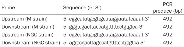
Table 1. Prokaryotic expression primers sequences of M and NGC strains
Prime Sequence (5’-3’) produce (bp)PCR
Upstream (M strain) 5’-cggcatatgcgttgcataggaatatcaaat-3’ 492 Downstream (M strain) 5’-gggtcgacttaccatgtttttcctgtgtca-3’ 492 Upstream (NGC strain) 5’-cggcatatgcgttgcataggaatatcaaat-3’ 492 Downstream (NGC strain) 5’-aggtcgacttagccatgttttcctgtgtca-3’ 492
N476-expressed strain. His-tag M476 and N476 fusion protein were expressed after IPTG induction. Western Blot was performed to iden-tify fusion protein with anti-His tag mouse monoclonal antibody as primary antibody and goat anti-mouse IgG antibody-HRP antibody as the second antibody. Purification of His-tag M476 and N476 fusion protein were performed by His Bind Purification Kit and the purity was assayed by SDS-PAGE. Because the fusion pro-tein was expressed by inclusion bodies, refold-ing of the protein was performed usrefold-ing gradient dialysis.
Preparation and identification of polyclonal antibody of M476 and N476 fusion protein
SDS-PAGE was used to isolate the M476 and N476 fusion protein. The target bands contain-ing M476 and N476 fusion protein were cut under sterile conditions and put mortar to dis-solve it. 22 BALB/c mice of 6-8 wk old were divided into M group, NGC group and control group. There were 10 mice in M group and NGC group, respectively, and two in control group. The prepared M476 and N476 antigen were inoculated subcutaneously at multiple points of the mice in M and NGC group respectively. Mice in control group were given PBS alternatively. The amount of the initial antigen immunization was 50 μg each mouse. One week later, 25 μg of antigen was appended to each mouse once a week for 4 times. Seven days after the last immunization, blood serum was collected and separated to aliquots to store at -80°C. All stud-ies were performed according to internationally recognized guidelines for animal care.
Identification of antiserum was performed by Western blot. Briefly, M476 and N476 protein used for immunization was isolated by SDS-PAGE and then transferred onto PVDF mem-brane. The membranes were incubated with mouse antiserum (1:100) for 1 h. Then the PVDF membranes were incubated with the horse-radish peroxidase-conjugated secondary antibody goat anti-rabbit lgG (1:100) for 1 hour at room temperature. The proteins bands were visualized using DAB staining kit according to the instruction of the manufacturer.
Binding M476 and N476 fusion protein to HUVEC
HUVEC cells were seeded onto sterile cover-slips placed in wells of six-well plates and
cul-tured. Cell seeded was coincubated M476 and N476 protein (10 μg/well) respectively for 12 hours. After that, removed cell medium, washed three times with PBS, and then fixed with 1 ml 4% paraformaldehyde for 10 min. Indirect im- munofluorescence staining was used to deter-mine the binding of M-476 and N-476 protein to HUVEC. In brief, washed the wells with PBST 3 times, 5 min/time, dried coldly followed by add-ing 2% BSA was added into each well, to block for 30 min. aspirated the residual blocking solu-tion 30 min later, then M-476 and N-476 anti-serum was respectively added (diluted 1:100, 200 μl/well) onto the coverslip slowly and incu-bated in a moist chamber at 37°C for 30 min. Then FITC-labeled goat anti-mouse IgG second-ary antibody (diluted 1:1000, 100 μl/well) was added, and incubated in dark for 30 min. After washing with PBS, coverclips were observed under fluorescence microscope.
The effect of M476 and N476 protein on the permeability HUVEC monolayer
The Transwell was placed into 24-well plates, 600 μl of L-DMEM working solution was added into the lower chamber. 200 μl of HUVEC cell was seeded in the upper chamber at a density of 105/ml. After about 12 hours, cells were grown into monolayer.
M476 and N476 protein was added to each transwell of the upper chamber with the final concentration of 10 μg/ml. The permeability was detected after 0.5, 1, 3, 6, 12, 24, 30, 36, 42, 48 and 72 h of incubation, respectively. 10 μl/well of FITC-labeled dextran was added into the upper chamber of transwell in dark. After 15 min of culture, fluorescence value was detected by full wavelength microplate reader. LPS and PBS were used as control. Meanwhile, the morphology of monolayer was observed under inverted microscope and ultra-structure was observed under Transmission electron microscope.
Results
Identification of the M476 and N476 prokary-otic expression product by SDS-PAGE and Western blot
precipita-tion from centrifugaprecipita-tion were analyzed at SDS-PAGE (Figure 1). The molecular weight of our target protein was about 28 KDa, consistent with the theoretical value. Western blot was performed to identify the expression product. The results were as shown in Figure 2, two clear bands appeared, which was consistent with the expected results, suggesting the recombinant protein was the destined protein.
After purified by His-Bind Purification Kit, the recombinant protein was analyzed by SDA-PAGE and gel image instrument. It was shown that the purity of recombinant protein reached to 90%.
Identification of antiserum of M476 and N476 protein
We then prepared the antiserum of M476 and N476. Western blot showed two clear bands which was consistent with the expected results (Figure 3).
M476 and N476 protein bond with HUVEC
To test the effect of M476 and N476 protein on HUVEC cells, we firstly demonstrated the bind-ing of these two recombination fusion protein with HUVEC cells. Indirect immunofluorescence staining was shown that in both of HUVEC cells cultures co-incubated with M476 or N476 pro-tein, FITC specific fluorescence label could be found at the surface of the cell. Cell control was set in order to distinguish the nonspecific adsorption of fluorescence label, using PBS instead of protein (Figure 4).
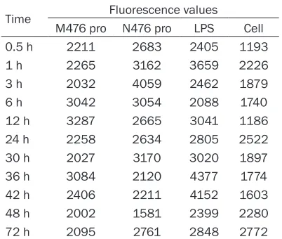
The effect of the M476 and N476 protein on the permeability of HUVEC monolayer
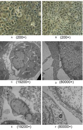
We firstly determined the timing that M476 and N476 protein has the greatest impact on the permeability by detecting FITC fluorescence change at full range microplate reader. It was showed that, M476 protein had significant effect on the permeability of HUVEC during the period of 0.5~24 h and 32~48 h, 12 h at the peak. As for N476 protein, it functioned at 0.5~48 h, 3 h at the peak. During the period of 0.5~10 h and 24~36 h, the effect of N476 pro-tein was stronger than M476, while during the period of 10~24 h and 36~48 h, M476 protein had the higher effect (Table 2; Figure 5). Then we observed the morphology of HUVEC cells at the peak timing that the effect of M476 and N476 protein was most significant under inverted microscope and TEM. For M476 pro-tein, we observed at 12 h after protein treat-ment. It was found that the width between cells became wider compared with untreated cells, cell connection was disrupted and cell was shrinking (Figure 6A). In cultures of HUVEC coincubated with N476 protein, we observed at 3 h after N476 protein treatment. The similar changes were seen and in some field, cell fusion was seen. (Figure 6B) At the respective Figure 1. SDS-PAGE analysis of M476 and N476
fusion protein. Lane 1. Supernatant of pET28a(+) -M476-BL21 induced by IPTG; lane 2. Supernatant of pET28a(+)-N476-BL21 induced by IPTG; lane 3. Precipitation of pET28a(+)-M476 -BL21 induced by IPTG; lane 4. Precipitation of pET28a(+)-N476-BL21
induced by IPTG; lane 5. Purified fusion protein of pET28a(+)-M476; lane 6. Purified fusion protein of
pET28a(+)-N476; M. Protein Marker.
Figure 2. Western blot of His-M476 and His-N476 fusion protein lane 1. Western blot of His-M476 fu-sion protein; lane 2. Western blot of His-N476 fufu-sion protein.
timing, TEM found significant ultra-structural changes as well (Figure 6C-F).
The result from M group was shown in Figure 6C and 6D. The width of part of nuclear mem-brane became wider, part of the mitochondrial ridge disappeared and even was of great vacu-olization, microvillus dropped and envelope was not complete. The result from NGC group was shown in Figure 6E and 6F. The cell mem-brane was broken, cell microvillus disappeared, a large amount of mitochondrial was heavily vacuolization, and mitochondrial ridge disap- peared.
Discussion
Protein E contains three domains, EDI, EDII and EDIII, respectively. Among them, the antigen from EDI and EDII region can bind to the DENV specific antibody [20], and the mono-antibody
which has strong neutralized ability and serum specificity mainly bind to EDIII region [16, 21-23].
The gene in our current research is at the 1-476 bp of the full length E gene of DENV-2. It mainly locate at the EDI region of protein E. EDI is a monomer state core region [14] and the fusion peptide located at EDII region form an exten-sive finger fold, which may be related to the dimerization and membrane fusion of protein E [15, 16]. The processes of protein E allosteric induced membrane fusion go through EDI region. Therefore it is of great significance to study this region.
[image:5.612.95.522.73.207.2]The study of the effect of Protein E to the per-meability of HUVEC makes us better under-stand the pathogenesis of DHF/DSS. Vascular endothelial cells as the initial barrier of the cir-culatory system could regulate cell adhesion molecules and impact the inter-cells communi-cation then mediate the increase of the perme-ability and cause plasma leakage through regu-lating cytokines, chemokine, and cell receptors after DENV infection. Infected cells can pro-duce monocytes to affect the growth and per-meability of endothelial cells in vitro. Other immune cells, such as lymphocytes, could in- teract with endothelial cells to cause plasma leakage. DENV could also directly infect HUVEC to induce cell death or apoptosis to destroy vas-cular wall. It was considered that protein E was participated in the interaction between virus and specific receptors on targeted cell surface and then mediated virus infection directly; actually, the envelope protein of the DENV was the protein E [11].
Figure 4. The image of HUVEC cells under fluorescence microscope (200×. A. The combination of HUVEC with M476
protein; B. The combination of HUVEC with N476 protein; C. HUVEC cells in control.
Table 2. Dynamic level of fluorescence values in out-chamber
Time Fluorescence values
M476 pro N476 pro LPS Cell
0.5 h 2211 2683 2405 1193
1 h 2265 3162 3659 2226
3 h 2032 4059 2462 1879
6 h 3042 3054 2088 1740
12 h 3287 2665 3041 1186
24 h 2258 2634 2805 2522
30 h 2027 3170 3020 1897
36 h 3084 2120 4377 1774
42 h 2406 2211 4152 1603
48 h 2002 1581 2399 2280
[image:5.612.89.290.285.455.2]DENV can infect cells in two ways, one is binding with the Fc receptor of sensitive cell through the IgG then to infect cell, which is called ADE ef- fect; the other one is directly interact with the sensitive cells but not the Fc receptor to infect cells. Protein E as an envelope protein is believed to involve in this process [21, 24]. In current study, the bind-ing of the fragment of 1-476 in gene E with HUVEC was simi-lar with the second mecha-nism, which means it directly binds with the HUVEC but not Fc receptor. We also approved HUVEC is the sensitive cell type to DENV-2. More impor-tantly, this protein significant-ly increases the permeability of single layer HUVEC and obviously affects the cell shape and ultra-structure in our study.
[image:6.612.90.376.73.200.2]Cell membrane plays a pivotal role for cell function. Microvi- llus increases the surface of the cell, which is good for cell uptake. Mitochondrial where oxidation reaction takes place is the energy factory, which could produce energy for cell [20]. The cell in physiological condition interacts with other cell or the basal membrane through cytoskeleton. The ch- ange of cytoskeleton function can alter cell shape and the status of cell-cell contact and the cell to basal membrane connection. Meanwhile, the change of the status of cell-cell contact and the cell-cell to basal membrane connection can cause the rearrangement of cytoskeleton protein thro- ugh cell signal transduction then finally change the perme-ability of endothelial cells. The change in the constringency of endothelial cells was th- ought to be the final common Figure 5. Dynamic level of fluorescence in values in out-chamber.
Figure 6. Transmission electron microscope of HUVEC. A. HUVEC inoculate
with M476 protein (200×); B. HUVEC inoculate with N476 protein (200×); C
[image:6.612.90.378.237.687.2]alteration of permeability induced from differ-ent signal and mechanisms.
The interaction of virus protein with receptor is the initial step of virus infection. Protein E of DENV binding with the receptor in the mem-brane of target cell is of great importance for the DENV infection to cell. This study pave the road for the better understand the pathogene-sis and prevention of DENV infection. For instance design some drug to block the binding site for the virus in the surface of target cell or use some protein containing similar pattern with the virus protein to block the receptor in the surface of sensitive cell. This study will pro-vide some hint to the development of new anti-virus method.
Acknowledgements
This work was supported by grants from National Natural Science Foundation of China (No. 31260224 and No. 81560263), “125” major scientific and technological projects from Guizhou Province Department of Education and Talents funding from Guizhou province gov-ernor, Science and technology fund project of GuiZhou provincial health and family planning commission (gzwjkj2016-1-005).
Disclosure of conflict of interest
None.
Address correspondence to: Dr. Li Zuo, Department of Immunology, Guizhou Medical University, Guiyang, P R China. Tel: 86908108; Fax: +86-851-86908108; E-mail: [email protected]
References
[1] de Wispelaere M, Yang PL. Mutagenesis of the DI/DIII linker in dengue virus envelope protein impairs viral particle assembly. J Virol 2012; 86: 7072-7083.
[2] Li XQ, Qiu LW, Chen Y, Wen K, Cai JP, Chen J, Pan YX, Li J, Hu DM, Huang YF, Liu LD, Ding XX, Guo YH, Che XY. Dengue virus envelope do-main III immunization elicits predominantly cross-reactive, poorly neutralizing antibodies localized to the AB loop: implications for den-gue vaccine design. J Gen Virol 2013; 94: 2191-2201.
[3] Watterson D, Kobe B, Young PR. Residues in domain III of the dengue virus envelope glyco-protein involved in cell-surface glycosaminogly-can binding. J Gen Virol 2012; 93: 72-82. [4] Whitehorn J, Farrar J. Dengue. Br Med Bull
2010; 95: 161-173.
[5] Allicock OM, Lemey P, Tatem AJ, Pybus OG, Bennett SN, Mueller BA, Suchard MA, Foster JE, Rambaut A, Carrington CV. Phylogeography and population dynamics of dengue viruses in the Americas. Mol Biol Evol 2012; 29: 1533-1543.
[6] Hang VT, Nguyet NM, Trung DT, Tricou V, Yok-san S, Dung NM, Van Ngoc T, Hien TT, Farrar J, Wills B, Simmons CP. Diagnostic accuracy of
NS1 ELISA and lateral flow rapid tests for den
-gue sensitivity, specificity and relationship to
viraemia and antibody responses. PLoS Negl Trop Dis 2009; 3: e360.
[7] Rodenhuis-Zybert IA, van der Schaar HM, da Silva Voorham JM, van der Ende-Metselaar H, Lei HY, Wilschut J, Smit JM. Immature dengue virus: a veiled pathogen? PLoS Pathog 2010; 6: e1000718.
[8] Khadka S, Vangeloff AD, Zhang C, Siddavatam P, Heaton NS, Wang L, Sengupta R, Sahas-rabudhe S, Randall G, Gribskov M, Kuhn RJ, Perera R, LaCount DJ. A physical interaction network of dengue virus and human proteins. Mol Cell Proteomics 2011; 10: M111.012187. [9] St John AL, Rathore AP, Raghavan B, Ng ML,
Abraham SN. Contributions of mast cells and vasoactive products, leukotrienes and chy-mase, to dengue virus-induced vascular leak-age. Elife 2013; 2: e00481.
[10] Hung JJ, Hsieh MT, Young MJ, Kao CL, King CC, Chang W. An external loop region of domain III of dengue virus type 2 envelope protein is
in-volved in serotype-specific binding to mosquito
but not mammalian cells. J Virol 2004; 78: 378-388.
[11] Crill WD, Roehrig JT. Monoclonal antibodies that bind to domain III of dengue virus E
glyco-protein are the most efficient blockers of virus
adsorption to Vero cells. J Virol 2001; 75: 7769-7773.
[12] Carr JM, Hocking H, Bunting K, Wright PJ, Da-vidson A, Gamble J, Burrell CJ, Li P. Superna-tants from dengue virus type-2 infected macro-phages induce permeability changes in endothelial cell monolayers. J Med Virol 2003; 69: 521-528.
[13] Garcia-Bates TM, Cordeiro MT, Nascimento EJ, Smith AP, Soares de Melo KM, McBurney SP, Evans JD, Marques ET Jr, Barratt-Boyes SM. As-sociation between magnitude of the
virus-spe-cific plasmablast response and disease sever -ity in dengue patients. J Immunol 2013; 190: 80-87.
[14] Modis Y, Ogata S, Clements D, Harrison SC. A ligand-binding pocket in the dengue virus en-velope glycoprotein. Proc Natl Acad Sci U S A 2003; 100: 6986-6991.
dur-ing repeated dengue infection are virus
glyco-protein specific and bind to multiple virus sero -types. J Immunol 2012; 189: 5877-5885. [16] Allison SL, Schalich J, Stiasny K, Mandl CW,
Heinz FX. Mutational evidence for an internal
fusion peptide in flavivirus envelope protein E.
J Virol 2001; 75: 4268-4275.
[17] Stiasny K, Kiermayr S, Holzmann H, Heinz FX.
Cryptic properties of a cluster of dominant fla -vivirus cross-reactive antigenic sites. J Virol 2006; 80: 9557-9568.
[18] Zuo L, Shu LP. Isolation identification and phy -logenetic analysis of a dengue virus strain
from field aedes albopictus in mawei town
(Guizhou). Chin Med J (Engl) 2004; 117: 1847-1849.
[19] Rothman AL, Ennis FA. Immunopathogenesis of dengue hemorrhagic fever. Virology 1999; 257: 1-6.
[20] Lee E, Leang SK, Davidson A, Lobigs M. Both E protein glycans adversely affect dengue virus
infectivity but are beneficial for virion release.
J Virol 2010; 84: 5171-5180.
[21] Luplertlop N, Missé D, Bray D, Deleuze V, Gon-zalez JP, Leardkamolkarn V, Yssel H, Veas F. Dengue-virus-infected dendritic cells trigger vascular leakage through metalloproteinase overproduction. EMBO Rep 2006; 7: 1176-1181.
[22] Wahala WM, Huang C, Butrapet S, White LJ, de Silva AM. Recombinant dengue type 2 viruses with altered e protein domain III epitopes are
efficiently neutralized by human immune sera.
J Virol 2012; 86: 4019-4023.
[23] Bhardwaj S, Holbrook M, Shope RE, Barrett AD, Watowich SJ. Biophysical characterization
and vector-specific antagonist activity of do
-main III of the tick-borne flavivirus envelope
protein. J Virol 2001; 75: 4002-4007.