COMPUTER DATABASES FOR
TWO-DIMENSIONAL ELECTROPHORESIS
T. Toda, Tokyo Metropolitan Institute of Gerontology, Tokyo, Japan
Copyright^ 2000 Academic Press
Two-dimensional (2-D) electrophoresis is an unri-valled technique of the highest capacity to separate many thousands of proteins in complex specimens such as a crude extract of cells. The development of computer systems for 2-D gel image analysis has promoted the construction of comprehensive databases for 2-D electrophoresis. The recent ad-vance in the Internet computer network has allowed us to share useful pieces of information located in many 2-D electrophoresis databases on the world-wide web (WWW) via the Internet. And a newReld of research named proteomics has been growing based on 2-D electrophoresis databases.
2-D Electrophoresis
2-D electrophoresis, established by O’Farrell in 1975, is a combined technique of isoelectric focusing (IEF) and sodium dodecyl sulfate (SDS)-polyacrylamide gel electrophoresis (PAGE) for achieving the Rnest res-olution of proteins in crude specimens. Protein mol-ecules of different isoelectric points (pI) are separated at theRrst stage of IEF, and components of different molecular masses are resolved in the second-dimen-sional SDS-PAGE.
The IEF in a mobile pH gradient, which is auto-matically generated by lining up carrier ampholytes in the order of pI, is performed in the original protocol of O’Farrell’s 2-D electrophoresis. However, there is still a problem with the conventional IEF method in the drift of the pH gradient toward the cathode. Righetti and colleagues have devised an improved method of IEF which runs on an immo-bilized pH gradient (IPG) to prevent cathodic drift. The IPG-IEF is adopted in most current protocols of 2-D electrophoresis for constructing computer databases.
Protein Detection
Proteins separated on a gel plate are detected by autoradiography if they are labelled with a radioiso-tope. Nonradioactive proteins are visualized by silver or dye staining depending on the protein amount on
the gel. The protein proRle on the 2-D gel reports useful information about qualitative and quantitative states of proteins in the specimen. Most computer databases for 2-D electrophoresis have generally been constructed on such visualized 2-D gel images.
2-D Gel Image Analysis
Computer analysis of the 2-D gel images was de-veloped after O’Farrell’s paper because 2-D gel im-ages were too complicated to be analysed manually. Lipkin and Lemkin of the National Institutes of Health in the USA made their GELLAB system on a DEC minicomputer in 1980, and independently of this, Andersonet al. constructed their original TY-CHO system in 1981. Garrels and Franza prepared a prototype software for 2-D gel image analysis on a compact Hewlett Packard desktop computer in 1979, and then developed it into the QUEST system for an UNIX-based minicomputer in 1983. The PDQUEST, which is a commercially available soft-ware package for SUN UNIX workstations, was a re-vision of the QUEST system. Many other software packages, such as Melanie II and GELLAB II, have been made commercially available. Those Macin-tosh- and Windows-based software packages have allowed 2-D gel image analysis to be performed easily for database construction.
Western Blotting
Western blotting is an immunochemical technique for detecting speciRc proteins. Protein spots separated by 2-D electrophoresis are transferred on to a nitrocel-lulose or polyvinylidene diSuoride (PVDF) membrane by electrotransfer blotting. After blocking, the mem-branes are treated with a speciRc antibody and fol-lowed by a second enzyme-linked antibody. Localiza-tion of speciRc protein on the membrane is visualized by an enzyme reaction. The results of spot protein identiRcation by Western blotting are also included in 2-D electrophoresis databases.
Microsequencing
Figure 1 Proteomic analysis tool PetIdent in the ExPASy Molecular Biology server. The URL is http://www.expasy.ch/tools/ peptident.html.
Proteins that are blocked at the N-terminus are often cleaved with endopeptidase. The digests are separ-ated by reversed-phase high performance liquid chromatography (HPLC) and then subjected to sequencing. Protein databases on the Internet may answer a query on the homology of the amino acid sequence for identiRcation of the protein if the data-base includes an entry of the protein itself or family gene products. The sequence data and the results of homology search are both valuable information for constructing 2-D electrophoresis databases.
Mass Spectrometry
Mass spectrometry is another powerful technique for identiRcation of proteins separated by 2-D electrophoresis. The peptide mass Rngerprinting of endopeptidase digests is extensively used for primary assignment of the protein. The ExPASy Molecular Biology server (http://www.expasy.ch/) offers useful proteomic tools such as PeptIdent, which helps us in peptide massRngerprinting works for protein
Figure 2 TheSWISS-2DPAGEtop page in the ExPASy Molecular Biology server of the Swiss Geneva University Hospital. The URL is http://www.expasy.ch/ch2d/.
History of 2-D Electrophoresis
Databases
The 2-D electrophoresis database, reported by Lipkin and Lemkin in 1980, was only for multiple 2-D gel image analyses on a stand-alone minicomputer. In their GELLAB system, a composite gel (CGEL) database was made by extracting spot data from multiple gels and merging them into a representative gel. The primary database consisted of the lists of corresponding spots, their associated properties and interrelations. The human serum protein 2-D elec-trophoresis database for clinical use, reported by
Anderson and his co-workers in 1981, was also only for personal use on an ofSine computer. Human keratinocyte 2-D electrophoresis databases, made by Celis et al. on a UNIX workstaion with PDQUEST software, were offered to many research groups commercially, but not made accessible on the Internet.
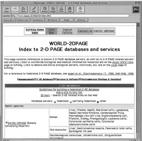
Figure 3 TheWORLD-2DPAGEhome page for indexing to2-D PAGEdatabases and services throughout the world. The URL is http://www.expasy.ch/ch2d/2d-index.html.
WORLD-2DPAGE home page (Figure 3) present-ed at http://www.expasy.ch/ch2d/2d-index.html in the ExPASy server offers a convenient link to many 2-DE databases in the world.
The databases listed in Table 1 were indexed in the WORLD-2DPAGE page for link as of 15 June, 1999.
Some of the databases have been built follow-ing the guidelines for the federated 2-D electro-phoresis database recommended by Appelet al. Rules for the database federation are summarized in
Table 2.
World-Wide Web for
2-D Electrophoresis Database
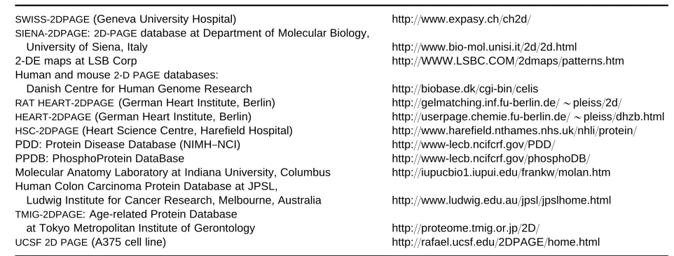
Table 1 A partial list of 2-D electrophoresis databases indexed in the WORLD-2DPAGE home page
SWISS-2DPAGE(Geneva University Hospital) http://www.expasy.ch/ch2d/
SIENA-2DPAGE:2D-PAGEdatabase at Department of Molecular Biology,
University of Siena, Italy http://www.bio-mol.unisi.it/2d/2d.html
2-DE maps at LSB Corp http://WWW.LSBC.COM/2dmaps/patterns.htm
Human and mouse2-D PAGEdatabases:
Danish Centre for Human Genome Research http://biobase.dk/cgi-bin/celis
RAT HEART-2DPAGE(German Heart Institute, Berlin) http://gelmatching.inf.fu-berlin.de/&pleiss/2d/
HEART-2DPAGE(German Heart Institute, Berlin) http://userpage.chemie.fu-berlin.de/&pleiss/dhzb.html
HSC-2DPAGE(Heart Science Centre, Harefield Hospital) http://www.harefield.nthames.nhs.uk/nhli/protein/
PDD: Protein Disease Database (NIMH}NCI) http://www-lecb.ncifcrf.gov/PDD/
PPDB: PhosphoProtein DataBase http://www-lecb.ncifcrf.gov/phosphoDB/
Molecular Anatomy Laboratory at Indiana University, Columbus http://iupucbio1.iupui.edu/frankw/molan.htm Human Colon Carcinoma Protein Database at JPSL,
Ludwig Institute for Cancer Research, Melbourne, Australia http://www.ludwig.edu.au/jpsl/jpslhome.html
TMIG-2DPAGE: Age-related Protein Database
at Tokyo Metropolitan Institute of Gerontology http://proteome.tmig.or.jp/2D/
UCSF 2D PAGE(A375 cell line) http://rafael.ucsf.edu/2DPAGE/home.html
NIMH-NCI, National Institute of Mental Health and National Cancer Institute.
Table 2 Guidelines for building a federated 2-DE database
1. Individual entries in the database must be accessible from remote by keyword search
2. The database must be linked to other databases through active hypertext cross-references
3. A main index has to be supplied that provides a means of querying all databases through one unique entry point 4. Individual protein entries must be accessible through clickable
images
5. 2-DE analysis software, been designed for use with federated 2-DE database, must be able to access directly individual entries in any federated 2-DE database
For full details, see Appel RDet al. (1996) in Electrophoresis 17: 540}546.
Hypertext Cross-Reference
Links from the 2-D electrophoresis database to re-lated databases, such as GenBank DNA database in NCBI Entrez, SWISS-PROT protein database or SWISS-3DIMAGE three-dimensional structure databases, through hypertext cross-references is a common function of most databases for 2-D elec-trophoresis. The function is achieved with anchor tags in hypertext mark-up language (HTML) and executive scripts for common gateway interface (CGI). For example, in TMIG-2DPAGE, the data entry for human nm-23 carries the following hyper-text cross-reference to the corresponding SWISS-PROT (P15531) and NCBI GenBank (X75598) entries respectively.
SWISS-PROT
(A HREF"`http://www.expasy.ch/cgi-bin/ sprot-search-ac?P15531aTARGET"`}blanka' P15531(/A'
GenBank
(A HREF"`http://www3.ncbi.nlm.nih.gov/ htbin-post/Entrez/query?db"n&form" 6&dopt"g&uid"X75598aTARGET"
`}blanka'X75598(/A'
Clickable Image Map
Clickable image map is a function of CGI installed in the WWW server software for achieving an active link from a location on an image to a corresponding
Rle. All federated 2-D electrophoresis databases on the WWW have 2-D gel images for the clickable image map. The content of each protein data entry is displayed on the monitor screen when the mouse button is clicked on the corresponding spot on the 2-D gel image.
Setting up a 2-D Electrophoresis
Database on the WWW
To set up a 2-D electrophoresis database, software for WWW server, such as Apache httpd, must be working on a UNIX or LINUX server computer con-nected to the Internet. The function of clickable im-age map is included in the Apache modules if they are properly installed. To set up a new 2-D electrophor-esis database, prepare the following four types ofRles
Rrst, according to the Apache Reference Manual, which is available in the Apache directory.
1. A hypertext Rle for the image map top page (***.html).
Figure 4 Human keratinocytes 2-D PAGE database in the Danish Centre for Human Genome Research. The URL is http:// biosun.biobase.dk/cgi-bin/make}gel.start?msau00032#0#0#0#0.
3. A map Rle for directing the jumping destination (***.map).
4. TextRles of protein data entries as destinations for links (***.html).
When theseRles are placed in the appropriate di-rectories in the WWW server, the basal activity of the 2-D electrophoresis database starts running.
2-D Electrophoresis Databases on the
WWW
SWISS-2DPAGE
The SWISS-2DPAGE is a fully implemented 2-DE federated database. The ExPASy WWW server is
Figure 5 TMIG-2DPAGE clickable map of TIG-3 human fibroblast line. The URL is http://proteome.tmig.or.jp/2D/Fibro/ humfb}menu.html.
database of yeast (Saccharomyces cerevisiae) genes coding for proteins (LISTA) are also linked through SWISS-PROT.
Human 2-D PAGE Databases of the Danish Centre For Human Genome Research
The human 2-D PAGE databases at the University of Aarhus, which were developed for functional genome analysis in health and disease, contain data on
pro-teins identiRed on various reference maps. Databases of human keratinocytes (Figure 4) are served for the study of skin biology, and those of transitional cell carcinomas are for the study of bladder cancer.
Heart-2DPAGE
federated 2-D database. It contains data on the hu-man heart ventricle and atrium. The data are access-ible both by keyword search on the protein name and by mouse-clicking on two images of 2-D. Entries provide data related to the isoelectric point, the mo-lecular mass and the amino acid sequence of each spot protein.
PDD Protein Disease Database
The PDD Protein Disease Database on the WWW server is a part of the National Institute of Mental Health and National Cancer Institute (NIMH-NCI) Protein}Disease Database Project for correlating dis-eases with proteins observed in serum, cerebrospinal
Suid, urine and other common human body Suids based on biomedical literature. The PDD database includes the data of quantitative and qualitative pro-tein variations with disease states, answering ques-tions on protein patterns found in common body
Suids with respect to disease conditions in the litera-ture.
TMIG-2DPAGE
TMIG-2DPAGE is a database of human proteins in-volved in the mechanisms of cell ageing. It includes two clickable image maps for data entries of proteins in normal cell ageing, and for those in the disease state of Werner’s syndrome patients. On the TMIG-2DPAGE clickable map of TIG-3 human Rbroblast line, quantitative variations of proteins observed in the process of replicative cell ageing are demonstrated with coloured crosses (Figure 5).
As a mouse button is clicked on a protein spot, the corresponding data entry is displayed on the browser. Each data entry contains information on age-related protein variation, physicochemical properties, refer-ences and active links to both SWISS-PROT and NCBI Entrez nucleotide sequence databases.
Keyword Search of
2-D Electrophoresis Databases
The 2DHunt on the ExPASY server (http://www. expasy.ch/ch2d/2DHunt/) is a convenient search en-gine for WWW sites of 2-D electrophoresis databases. The site list for 2DHunt search is periodically created by the site retrieval robot supplied from the Marvin
(Multi-Agent Retrieval Vagabond on Information Networks). Although some irrelevant sites may some-times be hit, the search engine is helpful for many researchers to Rnd speciRc 2-D electrophoresis databases of their own interests.
Further Reading
Anderson NL, Taylor J, Scandore A et al. (1981) The TYCHO system for computer analysis of two-dimen-sional gel electrophoresis patterns. Clinical Chemistry 27(11): 1807}1820.
Appel RD, Hoogland C, Bairoch A and Hochstrasser DF (1999) Constructing a 2-D database for the World Wide Web.Methods in Molecular Biology112: 411}416. Celis JE, Gromov P, Stergaard Met al. (1996) Human 2-D
PAGE databases for proteome analysis in health and disease: http://biobase.dk/cgi-bin/celis. FEBS Letters 398(2}3): 129}134.
Garrels JI and Franza Jr BRF (1989) The REF52 protein
database. Journal of Biological Chemistry 264(9):
5283}5298.
Hoogland C, Sanchez JC, Tonella Let al. (1998) Current status of the SWISS-2DPAGE database.Nucleic Acids Research26(1): 332}333.
Lemkin PF (1997) The 2DWG meta-database of two-di-mensional electrophoretic gel images on the Internet. Electrophoresis18(15): 2759}2773.
Lipkin LE and Lemkin PF (1980) Data-base techniques for multiple two-dimensional polyacrylamide gel
elec-trophoresis analyses. Clinical Chemistry 26(10):
1403}1412.
O’Farrell PH (1975) High resolution two-dimensional elec-trophoresis of proteins.Journal of Biological Chemistry 250(10): 4007}4021.
Righetti PG, Gianazza E and Bjellqvist B (1983) Modern aspects of isoelectric focusing: two-dimensional maps and immobilized pH gradients. Biochemical and Bio-physical Methods8(2): 89}108.
Toda T and Kimura N (1998) TMIG-2DPAGE: a new concept of two-dimensional gel protein database for research on aging.Electrophoresis18(2): 344}348. Toda T and Ohashi M (1995) Review: Current status and
perspectives of computer-aided two-dimensional
densitometry. Journal of Chromatography A 698(1):
41}54.
Vesterberg O and Svensson H (1966) Isoelectric fractiona-tion, analysis, and characterization of ampholytes in natural pH gradients. IV. Further studies on the resolv-ing power in connection with separation of myoglobins. Acta Chemica Scandinavica20(3): 820}834.