Breast Cancer Detection from Digital Mammogram
using Deep Learning Method
Kaveramma C L
1, Mrs. Rajasree P M
2 1, 2RV college of Engineering, Bengaluru, India
Abstract: Breast cancer is the leading cause of cancer death in elderly women. prior detection and diagnosis is the best and most effective approach to decrease the tumour growth as well as the date rate.
At the moment Digital Mammography is put forward as imaging method for early diagnosis of breast cancer. Classification of masses in mammograms is still a challenge and play crucial role in providing assistance to radiologist for accurate diagnosis. In this paper, a patch based convolutional neural network (CNN) based classification technique which is one of the deep learning technique is proposed. The architectural models of patch based CNN are used for classification of mammogram images into benign and malignant. A comparative study on CNN model has been discussed in this paper.
Keywords: CNN, Deep learning, Mammogram
I. INTRODUCTION
Breast cancer is world’s second most occurring cancer among all types of cancer and it is most common cancer among women worldwide.
The important stages of breast cancer are stage 0, stage 1-3 and stage 4. The first clear visible symptom of breast cancer is a lump that is different from the rest of the breast tissue. Each year an estimate of 1.5 lakh new cases of breast cancer are diagnosed in India, making it leading type of cancer among women and replacing cervical cancer. According to the World Health Organization (WHO), the number of cancer cases expected in 2025 will be 19.3 million cases.
Computer-aided detection and diagnosis (CAD) systems are already being used to offer assistance in the decision-making process of radiologists. Such systems may significantly reduce the amount of effort, while minimizing the number of false positives that lead to unnecessary and discomforting biopsies. Machine learning is the scientific study of algorithms and statistical models that computer systems use to progressively improve their performances on a specific task. Machine learning algorithms build a mathematical model of sample data, known as “training data”, in order to make predications or decisions without being explicitly programmed to perform the task.
The main challenge of machine learning technique is building computer-aided detection systems used in many cases of mammography’s diagnostic such as screening radiology, malignant or benign tissue discrimination , and digital pathology that can assist radiologists to be more productive, objective and consistent in diagnosis. Deep learning is considered a significant breakthrough technology of recent years as it has exhibited performance beyond the state-of-the-art in various machine learning tasks including object detection and classification.
Contrary to conventional machine learning methods, which require a hand-crafted feature extraction stage, which is challenging as it relies on domain knowledge, deep learning methods adaptively learn the appropriate feature extraction process from the input data with respect to the target output. Deep learning is a currently developing field which explores areas of artificial intelligence and machine learning to learn features directly from the data, using multiple nonlinear processing layers. Deep learning with Convolutional Neural Networks (CNN) have achieved impressive results on computer visions tasks such as classification, object detection , and segmentation .
II. METHODOLOGY
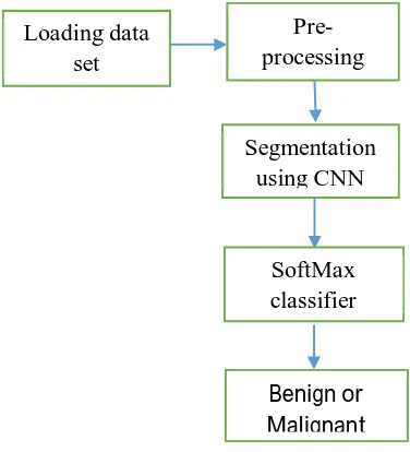
In order to predict breast cancer image as malignant or benign first the images are loaded and pre-processed where they are reduced to size 128x128. Then the image are segmented using convolutional neural network (CNN) and classified using softmax. The dataset consists of training set and testing set ,during training set the images are read and converted into blocks and feature vector is used to train CNN the trained CNN is used for segmentation and segmented output is used to train CNN for classification. In the testing phase the image is read and converted into blocks and output of feature vector is given to deep neural net for segmentation and the output is classified as benign and malignant
[image:2.612.204.392.174.381.2]
Fig 1 B.D of the proposed model
[image:2.612.152.471.409.589.2]CNN Architecture
Fig 2 CNN Architecture
Convolutional neural network are the leading architecture in deep learning that are used to solve an image classification problem. The goal of this paper is to tell which class the input image belongs to. The process of building a convolutional neural network always involves 4 major steps
1) Convolution
2) Pooling
3) Flattening
4) Fully connected layer
Loading data set
Pre-processing
Segmentation using CNN
SoftMax classifier
A. Types of layers
All neurons in one layer, do similar kind of mathematical operations that is how that layer gets its name.
1) Convolution layer: Convolution is the mathematical operation that is used in image processing to filter signal, find pattern in signal etc. each and every neurons in this layer perform convolution on inputs. The most important parameter in a convolutional neuron is the filter size. convolution filter is slither over whole input image to calculate the output across the image and here window is slither by 1 pixel at time this number is called Stride Typically more than 1 filter in one convolution layer is us
Pooling layer is used immediately after the convolutional layer to reduce the spatial size(only width and height, not depth). This reduces the number of parameters, hence computation is reduced. Also, less number of parameters avoid over fitting. The most common form of pooling is Max pooling where we take a filter of size 3X3 and apply the maximum operation over the 3X3 sized part of the image.
2) Fully Connected Layer: If each neuron in a layer get input from all the neurons in the previous layer, then this layer is called fully connected layer. The output of this layer is computed by matrix multiplication followed by bias offset.
III. RESULTS
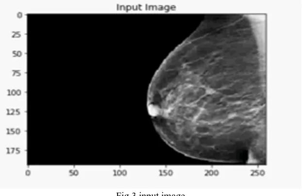
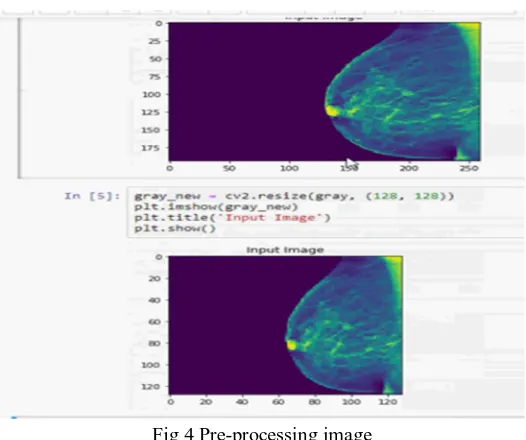
[image:3.612.151.462.469.671.2]The mammogram images obtained are pre-processed in order to resize the image into 128x128. Dataset was pre-processed where the images are of size 250x250 which were resized to 128x128.
Fig 3 input image
Fig 4 Pre-processing image
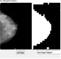
[image:4.612.203.408.373.498.2]Satisfactory results have been obtained using the CNN based proposed breast cancer detection method .The train data contained 80 images as benign and 89 images as malignant . the CNN was trained using these images and the test data contained 10 benign and 10 malignant images output obtained was displayed as benign or malignant in graphical user interface. An accuracy of 96% is obtained from the proposed method.
Fig 5 GUI to display classified output
The GUI is really very user friendly. The GUI of the proposed methodology describes clearly about the outcome of entire process. The GUI of the proposed methodology describes clearly
about the outcome of entire process
[image:4.612.181.430.558.722.2]Fig 7 classified image as malignant
IV. CONCLUSION
In this paper a computer–aided diagnosis (CAD) system termed as convolution neural network is proposed. This convolution neural networks are used on digital mammogram images for detection whether the cancer is benign or malignant . It is best to detect cancer at early stage and advise the patient to treat it. By using CNN architecture breast cancer with 96% accuracy has been obtained.
REFRENCES
[1] M. G. Ertosun, D. L. Rubin, “Probabilistic visual search for masses within mammography images using deep learning,” in Bioinformatics and Biomedicine (BIBM), 2015 IEEE International Conference, pp. 1310–1315,November 2015.
[2] J. Arevalo, F. A. Gonzalez, R. Ramos-Pollan, J. L. Oliveira, M. A. G. Lopez, “Convolutional neural networks for mammography mass lesion classification,” in 2015 37th Annual International Conference of the IEEE Engineering in Medicine and Biology Society (EMBC), pp. 797– 800, August 2015.
[3] syed jamal safdar Gardezi, muhammd awais, ibrahima faye, “mammogram classification using deep learning features”, in 2017 IEEE international conference on signal and image processing applications(IEEE ICSIPA 2017).
[4] Gabriele Valvano, daniele della latta , Nicola martini, dante chiappino “Evaluation of deep convolutional neural network method for the segmentation of breast microcalcifications in mammography imaging. Springer, 2017 European medical and biological engineering conference.
[5] Zhang, Y., et al. DeepSplice: Deep classification of novel splice junctions revealed by RNA-seq. in Bioinformatics and Biomedicine (BIBM), 2016 IEEE International Conference on. 2016. IEEE
[6] Hua, K.-L., et al., Computer-aided classification of lung nodules on computed tomography images via deep learning technique. OncoTargets and therapy, 2015. [7] Pravin S. Mane, Indra Gupta, M. K. Vasantha , “Implementation of RISC Processor on FPGA”, Electrical Engineering Department, Indian Institute of
Technology Roorkee, 2006 IEEE.
[8] Kui YI, Yue-Hua DING, “32-bit RISC CPU Based on MIPS”, International Joint Conference on Artificial Intelligence 2009.