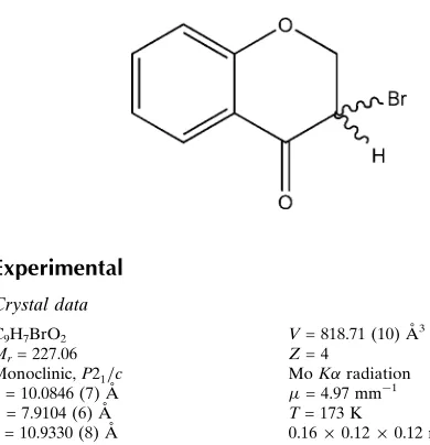
3-Bromochroman-4-one
Mahidansha M. Shaikh,aNeil A. Koorbanally,a* Karen Du Toit,bDeresh Ramjugernathcand Johannes Bodensteinb
a
School of Chemistry and Physics, University of Kwazulu-Natal, Private Bag X54001, Durban 4000, South Africa,bDiscipline of Pharmaceutical Science, University of KwaZulu-Natal, Private Bag X54001, Durban 4000, South Africa, andcSchool of
Engineering, University of KwaZulu-Natal, Durban 4041, South Africa Correspondence e-mail: [email protected]
Received 18 February 2013; accepted 25 February 2013
Key indicators: single-crystal X-ray study;T= 173 K; mean(C–C) = 0.004 A˚; Rfactor = 0.025;wRfactor = 0.061; data-to-parameter ratio = 15.2.
The heterocyclic ring of the title compound, C9H7BrO2,
obtained by bromination of 4-chromanone with copper bromide, adopts a half-chair conformation. The supramol-ecular structure is governed by a weak C—H O hydrogen bond. There is also –stacking between symmetry-related benzene rings; the centroid–centroid distance is 3.9464 (18), the perpendicular distance between the rings is 3.4703 (11) and the offset is 1.879 A˚ .
Related literature
For similar structures, see: Schollmeyeret al.(2005); Pielet al. (2011); Betzet al.(2011). For synthesis involving chromanone intermediates, see: Simaset al.(2002); Zhanget al.(2008). For the biological activity of chromanone derivatives, see: Choet al.(1996); Xuet al.(1998); Shaikhet al.(2012, 2013a,b).
Experimental
Crystal data
C9H7BrO2 Mr= 227.06 Monoclinic,P21=c a= 10.0846 (7) A˚
˚
V= 818.71 (10) A˚3 Z= 4
MoKradiation = 4.97 mm1
Data collection
Bruker Kappa DUO APEXII diffractometer
Absorption correction: multi-scan (SADABS; Sheldrick, 1997)
Tmin= 0.504,Tmax= 0.587
5434 measured reflections 1659 independent reflections 1392 reflections withI> 2(I)
Rint= 0.026
Refinement
R[F2> 2(F2)] = 0.025 wR(F2) = 0.061
S= 1.05 1659 reflections
109 parameters
H-atom parameters constrained
max= 0.39 e A˚
3
min=0.39 e A˚
[image:1.610.49.244.517.719.2]3
Table 1
Hydrogen-bond geometry (A˚ ,).
D—H A D—H H A D A D—H A
C2—H2A O2i 0.99 2.44 3.311 (3) 146
Symmetry code: (i)x;yþ1;z.
Data collection:APEX2(Bruker, 2006); cell refinement:SAINT (Bruker, 2006); data reduction:SAINT; program(s) used to solve structure:SHELXS97(Sheldrick, 2008); program(s) used to refine structure: SHELXL97 (Sheldrick, 2008); molecular graphics: ORTEP-3 for Windows(Farrugia, 2012); software used to prepare material for publication:SHELXL97.
We thank the University of KwaZulu-Natal, the National Research Foundation (NRF) and the South African Research Chairs initiative of the Department of Science and Technology for financial support and Ms Hong Su for the data collection.
Supplementary data and figures for this paper are available from the IUCr electronic archives (Reference: GO2082).
References
Betz, R., McCleland, C. & Marchand, H. (2011).Acta Cryst.E67, o1151. Bruker (2006).APEX2andSAINT. Bruker AXS Inc., Madison, Wisconsin,
USA.
Cho, H., Katoh, S., Sayama, S., Murakami, K., Nakanishi, H., Kajimoto, Y., Ueno, H., Kawasaki, H., Aisaka, K. & Uchida, I. (1996).J. Med. Chem.39, 3797–3805.
Farrugia, L. J. (2012).J. Appl. Cryst.45, 849–854.
Piel, I., Steinmetz, M., Hirano, K., Fro¨hlich, R., Grimme, S. & Glorius, F. (2011).Angew. Chem. Int. Ed.50, 4983–4987.
Schollmeyer, D., Kammerer, B., Peifer, C. & Laufer, S. (2005).Acta Cryst.E61, o868–o869.
Shaikh, M. M., Kruger, H. G., Bodenstein, J., Smith, P. & du Toit, K. (2012).
Nat. Prod. Res.26, 1473–1483.
Shaikh, M. M., Kruger, H. G., Smith, P., Bodenstein, J. & du Toit, K. (2013a).J. Pharm. Res.6, 21–25.
Shaikh, M. M., Kruger, H. G., Smith, P., Munro, O. Q., Bodenstein, J. & du Toit, K. (2013b).J. Pharm. Res.6, 1–5.
Sheldrick, G. M. (1997).SADABS. University of Go¨ttingen, Germany. Sheldrick, G. M. (2008).Acta Cryst.A64, 112–122.
Simas, A. B. C., Furtado, L. F. O. & Costa, P. R. R. (2002).Tetrahedron Lett.43, 6893–6895.
Xu, Z.-Q., Buckheit Jnr, R. W., Stup, T. L., Flavin, M. T., Khilevich, A., Rizzo, J. D., Lin, L. & Zembower, D. E. (1998).Bioorg. Med. Chem. Lett.8, 2179– 2184.
Zhang, L., Zhang, W.-G., Kang, J., Bao, K., Dai, Y. & Yao, X.-S. (2008).J. Asian
Acta Crystallographica Section E
Structure Reports Online
supporting information
Acta Cryst. (2013). E69, o473 [doi:10.1107/S1600536813005394]
3-Bromochroman-4-one
Mahidansha M. Shaikh, Neil A. Koorbanally, Karen Du Toit, Deresh Ramjugernath and Johannes
Bodenstein
S1. Comment
Many chromanone derivatives are used as versatile intermediates in the synthesis of natural products such as flavanone,
isoflavanone and homoisoflavanones (Simas et al., 2002, Zhang et al., 2008). These derivatives possess anticancer and
antibiotic properties (Cho et al., 1996.). Chromanone derivatives also possess antiviral activities against HIV and the
simian immunodeficiency virus (SIV) (Xu et al., 1998). We recently reported the synthesis of several homoisoflavanone
analogues from their corresponding chromanone derivatives with antiinflammatory (Shaikh et al., 2012; Shaikh et al.,
2013a) and antifungal activities (Shaikh et al., 2013b).
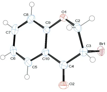
In the title compound, the pyranone moiety is fused with the benzene ring and adopts a half chair conformation. The
dihedral angle between the benzene ring and the (C3—C2—O1) of the pyranone moiety is 43.03 (17)° and C2 flips out of
the plane of the benzene ring by 0.5734 (31) Å (Fig. 1).
The supramolecular structure is governed by a weak C-H···O hydrogen bond, C2 –H2A···.O2 (-x,1-y,-z) with an H···O
distance of 2.44 Å, a C···O distance of 3.311 (3)Å and an angle at H of 146°.
There is also π–π stacking between the two benzene rings across the centre-of-symmetry at (1/2,1/2,0), the centroid to
centroid distance is 3.9464 (18)Å, the perpendicular distance between the rings is 3.4703 (11)Å and the offset is 1.879Å.
S2. Experimental
To a mixture of copper bromide (II) (11.351 g, 50.673 mmol) in ethyl acetate, chloroform (20:20 ml) was stirred under
inert atmosphere at room temperature. Into this mixture, chroman-4-one (5 g, 33.783 mmol) in chloroform (20 ml) was
added and the reaction mixture refluxed vigorously under inert atmosphere at 70 °C for 6 h. Completion of the reaction
was monitored by thin layer chromatography. Upon completion, the reaction mixture was cooled, filtered and washed
with chloroform (20 ml). The filtrate solution was evaporated under reduced pressure to get the pure title compound with
a yield of 86%.
1H NMR (400 MHz, CDCl
3): δ (p.p.m.): 4.53–4.65 (3H, m, H-2a, H-2 b & H-3), 6.98–7.06 (2H, m, H-6 & H-8), 7.48–
7.52 (1H, m, H-7), 7.89 (1H, dd, J = 1.60, 7.92 Hz, H-5).
13C NMR (400 MHz, CDCl
3): δ (p.p.m.): 45.43 (C-3), 71.26 (C-2), 117.95 (C-8), 11877 (C-10), 122.33 (C-6), 128.24
(C-7), 136.74 (C-5), 160.65 (C-9), 185.21 (C-4).
S3. Refinement
All non-hydrogen atoms were refined anisotropically. All hydrogen atoms were placed in idealized positions and refined
Figure 1
The molecular structure of the title compound, with atom labels and anisotropic displacement ellipsoids (drawn at 50%
probability level).
3-Bromochroman-4-one
Crystal data
C9H7BrO2
Mr = 227.06
Monoclinic, P21/c
Hall symbol: -p 2ybc a = 10.0846 (7) Å b = 7.9104 (6) Å c = 10.9330 (8) Å β = 110.164 (2)° V = 818.71 (10) Å3
Z = 4
F(000) = 448 Dx = 1.842 Mg m−3
Mo Kα radiation, λ = 0.71073 Å Cell parameters from 5434 reflections θ = 2.2–26.4°
µ = 4.97 mm−1
T = 173 K Block, colourless 0.16 × 0.12 × 0.12 mm
Data collection
Bruker Kappa DUO APEXII diffractometer
Radiation source: fine-focus sealed tube Graphite monochromator
0.5° φ scans and ω scans
Absorption correction: multi-scan (SADABS; Sheldrick, 1997)
5434 measured reflections 1659 independent reflections 1392 reflections with I > 2σ(I) Rint = 0.026
θmax = 26.4°, θmin = 2.2°
Refinement
Refinement on F2
Least-squares matrix: full R[F2 > 2σ(F2)] = 0.025
wR(F2) = 0.061
S = 1.05 1659 reflections 109 parameters 0 restraints
Primary atom site location: structure-invariant direct methods
Secondary atom site location: difference Fourier map
Hydrogen site location: inferred from neighbouring sites
H-atom parameters constrained w = 1/[σ2(F
o2) + (0.0298P)2 + 0.3551P]
where P = (Fo2 + 2Fc2)/3
(Δ/σ)max < 0.001
Δρmax = 0.39 e Å−3
Δρmin = −0.39 e Å−3
Special details
Experimental. 1H NMR (400 MHz, CDCl3): δ (p.p.m.): 4.53–4.65 (3H, m, H-2a, H-2b & H-3), 6.98–7.06 (2H, m, H-6 &
H-8), 7.48–7.52 (1H, m, H-7), 7.89 (1H, dd, J = 1.60, 7.92 Hz, H-5). 13C NMR (400 MHz, CDCl3): δ (p.p.m.): 45.43 (C-3), 71.26 (C-2), 117.95 (C-8), 118.77 (C-10), 122.33 (C-6), 128.24 (C-7), 136.74 (C-5), 160.65 (C-9), 185.21 (C-4).
Geometry. All e.s.d.'s (except the e.s.d. in the dihedral angle between two l.s. planes) are estimated using the full
covariance matrix. The cell e.s.d.'s are taken into account individually in the estimation of e.s.d.'s in distances, angles and torsion angles; correlations between e.s.d.'s in cell parameters are only used when they are defined by crystal symmetry. An approximate (isotropic) treatment of cell e.s.d.'s is used for estimating e.s.d.'s involving l.s. planes.
Refinement. Refinement of F2 against ALL reflections. The weighted R-factor wR and goodness of fit S are based on F2,
conventional R-factors R are based on F, with F set to zero for negative F2. The threshold expression of F2 > σ(F2) is used
only for calculating R-factors(gt) etc. and is not relevant to the choice of reflections for refinement. R-factors based on F2
are statistically about twice as large as those based on F, and R- factors based on ALL data will be even larger.
Fractional atomic coordinates and isotropic or equivalent isotropic displacement parameters (Å2)
x y z Uiso*/Ueq
Br1 0.22559 (3) 0.04339 (3) 0.04673 (3) 0.03040 (11)
O1 0.33588 (18) 0.3999 (2) 0.20473 (16) 0.0258 (4)
O2 0.00306 (19) 0.3386 (2) −0.13799 (18) 0.0348 (5)
C2 0.2062 (3) 0.3261 (3) 0.2053 (2) 0.0261 (6)
H2A 0.1470 0.4155 0.2238 0.031*
H2B 0.2276 0.2418 0.2764 0.031*
C3 0.1239 (3) 0.2415 (3) 0.0786 (2) 0.0251 (6)
H3 0.0297 0.2053 0.0807 0.030*
C4 0.1031 (3) 0.3566 (3) −0.0374 (2) 0.0242 (5)
C5 0.2106 (3) 0.5942 (3) −0.1218 (3) 0.0274 (6)
H5 0.1348 0.5872 −0.2026 0.033*
C6 0.3170 (3) 0.7098 (3) −0.1066 (3) 0.0330 (7)
H6 0.3148 0.7825 −0.1764 0.040*
C7 0.4279 (3) 0.7196 (3) 0.0121 (3) 0.0339 (7)
H7 0.5018 0.7987 0.0223 0.041*
C8 0.4323 (3) 0.6165 (3) 0.1148 (3) 0.0284 (6)
H8 0.5080 0.6253 0.1955 0.034*
C9 0.3249 (3) 0.4994 (3) 0.0995 (2) 0.0212 (5)
Atomic displacement parameters (Å2)
U11 U22 U33 U12 U13 U23
Br1 0.04127 (18) 0.01971 (15) 0.03327 (17) 0.00253 (11) 0.01674 (13) 0.00006 (12) O1 0.0294 (10) 0.0252 (9) 0.0199 (9) −0.0012 (8) 0.0046 (8) 0.0022 (8) O2 0.0315 (11) 0.0358 (11) 0.0289 (11) 0.0004 (8) −0.0002 (9) −0.0033 (9) C2 0.0339 (15) 0.0240 (13) 0.0218 (13) 0.0027 (11) 0.0116 (12) 0.0023 (11) C3 0.0265 (13) 0.0225 (13) 0.0294 (14) 0.0016 (11) 0.0137 (12) −0.0006 (11) C4 0.0259 (13) 0.0233 (13) 0.0242 (13) 0.0058 (11) 0.0095 (12) −0.0027 (11) C5 0.0392 (15) 0.0233 (13) 0.0217 (13) 0.0094 (12) 0.0129 (12) 0.0008 (11) C6 0.0521 (18) 0.0196 (13) 0.0364 (16) 0.0075 (12) 0.0269 (15) 0.0071 (12) C7 0.0394 (16) 0.0199 (14) 0.0504 (18) −0.0021 (12) 0.0255 (15) −0.0019 (13) C8 0.0276 (14) 0.0236 (13) 0.0348 (15) −0.0006 (11) 0.0116 (12) −0.0066 (12) C9 0.0263 (13) 0.0175 (12) 0.0209 (12) 0.0033 (9) 0.0097 (11) −0.0014 (9) C10 0.0267 (13) 0.0172 (12) 0.0221 (13) 0.0032 (10) 0.0122 (11) −0.0027 (10)
Geometric parameters (Å, º)
Br1—C3 1.969 (2) C5—C6 1.375 (4)
O1—C9 1.367 (3) C5—C10 1.401 (4)
O1—C2 1.434 (3) C5—H5 0.9500
O2—C4 1.218 (3) C6—C7 1.393 (4)
C2—C3 1.505 (3) C6—H6 0.9500
C2—H2A 0.9900 C7—C8 1.376 (4)
C2—H2B 0.9900 C7—H7 0.9500
C3—C4 1.515 (3) C8—C9 1.390 (4)
C3—H3 1.0000 C8—H8 0.9500
C4—C10 1.476 (4) C9—C10 1.399 (4)
C9—O1—C2 115.40 (19) C6—C5—H5 119.7
O1—C2—C3 113.01 (19) C10—C5—H5 119.7
O1—C2—H2A 109.0 C5—C6—C7 119.5 (2)
C3—C2—H2A 109.0 C5—C6—H6 120.2
O1—C2—H2B 109.0 C7—C6—H6 120.2
C3—C2—H2B 109.0 C8—C7—C6 121.1 (3)
H2A—C2—H2B 107.8 C8—C7—H7 119.5
C2—C3—C4 112.1 (2) C6—C7—H7 119.5
C2—C3—Br1 111.18 (17) C7—C8—C9 119.5 (3)
C4—C3—Br1 105.11 (15) C7—C8—H8 120.3
C2—C3—H3 109.4 C9—C8—H8 120.3
C4—C3—H3 109.4 O1—C9—C8 116.7 (2)
Br1—C3—H3 109.4 O1—C9—C10 123.0 (2)
O2—C4—C10 123.6 (2) C8—C9—C10 120.3 (2)
O2—C4—C3 121.3 (2) C9—C10—C5 119.0 (2)
C10—C4—C3 115.2 (2) C9—C10—C4 120.2 (2)
O1—C2—C3—C4 −51.4 (3) C7—C8—C9—C10 −0.1 (4)
O1—C2—C3—Br1 66.0 (2) O1—C9—C10—C5 179.9 (2)
C2—C3—C4—O2 −153.8 (2) C8—C9—C10—C5 −0.5 (3)
Br1—C3—C4—O2 85.3 (2) O1—C9—C10—C4 −2.5 (3)
C2—C3—C4—C10 27.4 (3) C8—C9—C10—C4 177.1 (2)
Br1—C3—C4—C10 −93.5 (2) C6—C5—C10—C9 0.6 (3)
C10—C5—C6—C7 0.0 (4) C6—C5—C10—C4 −177.0 (2)
C5—C6—C7—C8 −0.6 (4) O2—C4—C10—C9 179.9 (2)
C6—C7—C8—C9 0.7 (4) C3—C4—C10—C9 −1.4 (3)
C2—O1—C9—C8 158.5 (2) O2—C4—C10—C5 −2.5 (4)
C2—O1—C9—C10 −21.8 (3) C3—C4—C10—C5 176.2 (2)
Hydrogen-bond geometry (Å, º)
D—H···A D—H H···A D···A D—H···A
C2—H2A···O2i 0.99 2.44 3.311 (3) 146