Crystal structure of
5-(1,3-dithian-2-yl)-2
H
-1,3-benzodioxole
Julio Zukerman-Schpector,a* Ignez Caracelli,bHe´lio A. Stefani,cOlga Gozhinacand Edward R. T. Tiekinkd
aDepartmento de Quı´mica, Universidade Federal de Sa
˜o Carlos, 13565-905 Sa˜o Carlos, SP, Brazil,bDepartmento de Fı´sica, Universidade Federal de Sa
˜o Carlos, 13565-905 Sa˜o Carlos, SP, Brazil,cDepartamento de Farma´cia, Faculdade de Cieˆncias Farmaceˆuticas, Universidade de Sa˜o Paulo, 05508-900 Sa˜o Paulo-SP, Brazil, anddDepartment of Chemistry, University of Malaya, 50603 Kuala Lumpur, Malaysia. *Correspondence e-mail: [email protected]
Received 4 February 2015; accepted 5 February 2015
Edited by W. T. A. Harrison, University of Aberdeen, Scotland
In the title compound, C11H12O2S2, two independent but
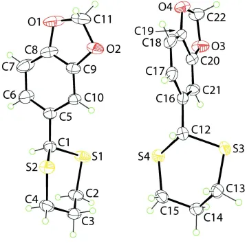
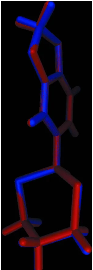
virtually superimposable molecules, A and B, comprise the asymmetric unit. In each molecule, the 1,3-dithiane ring has a chair conformation with the 1,4-disposed C atoms being above and below the plane through the remaining four atoms. The substituted benzene ring occupies an equatorial position in each case and forms dihedral angles of 85.62 (9) (moleculeA) and 85.69 (8) (molecule B) with the least-squares plane
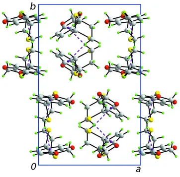
through the 1,3-dithiane ring. The difference between the molecules rests in the conformation of the five-membered 1,3-dioxole ring which is an envelope in molecule A (the methylene C atom is the flap) and almost planar in molecule B(r.m.s. deviation = 0.046 A˚ ). In the crystal, molecules ofA self-associate into supramolecular zigzag chains (generated by glide symmetry along the c axis) via methylene C—H interactions. Molecules of Bform similar chains. The chains pack with no specific directional intermolecular interactions between them.
Keywords:crystal structure; 1,3-dithiane; conformation; 1,3-benzo-dioxole; C—H interactions.
CCDC reference:1047484
1. Related literature
The title compound has been prepared previously, see: Ballesteros et al. (2005). For the structure of a related compound containing the same molecular skeleton as in the title compound,i.e. (19R,21R,25S)-( )-2-(2-menthyloxycarb-onyl-3,4-methylenedioxyphenyl)1,3-dithiane, see: Ratajczak-Sitarzet al.(1996).
2. Experimental
2.1. Crystal data
C11H12O2S2 Mr= 240.33 Monoclinic,P21=c a= 11.4765 (3) A˚
b= 17.5504 (4) A˚
c= 11.6397 (2) A˚ = 104.275 (1)
V= 2272.05 (9) A˚3
Z= 8
MoKradiation = 0.45 mm1 T= 296 K
0.590.400.26 mm
2.2. Data collection
Bruker APEXII CCD diffractometer
Absorption correction: multi-scan (SADABS; Sheldrick, 1996)
Tmin= 0.702,Tmax= 0.745
14839 measured reflections 4164 independent reflections 3759 reflections withI> 2(I)
Rint= 0.020
2.3. Refinement
R[F2> 2(F2)] = 0.035 wR(F2) = 0.092 S= 1.03 4164 reflections
271 parameters
H-atom parameters constrained max= 0.29 e A˚
3
min=0.44 e A˚
3
Table 1
Hydrogen-bond geometry (A˚ ,).
Cg1 andCg2 are the centroids of the C5–C11 and C16–C21 rings, respectively.
D—H A D—H H A D A D—H A
C4—H4b Cg1i
0.97 2.77 3.731 (2) 170 C13—H13a Cg2ii
0.97 2.54 3.4841 (19) 165
Symmetry codes: (i)x;yþ1 2;z
1
2; (ii)x;y 1 2;z
3 2.
Data collection: APEX2 (Bruker, 2009); cell refinement: SAINT (Bruker, 2009); data reduction:SAINT; program(s) used to solve structure: SIR2014 (Burla et al., 2015); program(s) used to refine structure: SHELXL2014 (Sheldrick, 2015); molecular graphics: ORTEP-3 for Windows(Farrugia, 2012),QMOL(Gans & Shalloway, 2001) and DIAMOND (Brandenburg, 2006); software used to prepare material for publication:MarvinSketch (ChemAxon, 2010) andpublCIF(Westrip, 2010).
Acknowledgements
(305626/2013-2 to JZS, 306121/2013-2 to IC and 308320/2010-7 to HAS), FAPESP (2012/00424-2 and 2013/21925-2) and CAPES are acknowledged for financial support.
Supporting information for this paper is available from the IUCr electronic archives (Reference: HB7361).
References
Ballesteros, L., Noguez, O., Arroyo, G., Velasco, B., Delgado, F. & Miranda, R. (2005).J. Mex. Chem. Soc.49, 302–306.
Brandenburg, K. (2006).DIAMOND. Crystal Impact GbR, Bonn, Germany.
Bruker (2009).APEX2andSAINT. Bruker AXS Inc., Madison, Wisconsin, USA.
Burla, M. C., Caliandro, R., Carrozzini, B., Cascarano, G. L., Cuocci, C., Giacovazzo, C., Mallamo, M., Mazzone, A. & Polidori, G. (2015).J. Appl. Cryst.48, 306–309.
ChemAxon (2010).Marvinsketch.http://www.chemaxon.com. Farrugia, L. J. (2012).J. Appl. Cryst.45, 849–854.
Gans, J. & Shalloway, D. (2001).J. Mol. Graph. Model.19, 557–559. Ratajczak-Sitarz, M., Naruszewicz, M., Kosturkiewicz, Z., Jozkowiak, J. &
Rozwadowska, M. D. (1996).Pol. J. Chem.70, 310–319.
Sheldrick, G. M. (1996).SADABS. University of Go¨ttingen, Germany. Sheldrick, G. M. (2015).Acta Cryst.C71, 3–8.
supporting information
Acta Cryst. (2015). E71, o167–o168 [doi:10.1107/S2056989015002455]
Crystal structure of 5-(1,3-dithian-2-yl)-2
H
-1,3-benzodioxole
Julio Zukerman-Schpector, Ignez Caracelli, H
é
lio A. Stefani, Olga Gozhina and Edward R. T.
Tiekink
S1. Experimental
A solution of the corresponding 2H-1,3-benzodioxole-5-carbaldehyde (0.037 mol, 1 equiv.) in chloroform (20 ml) was combined with an equimolar amount of propane-1,3-dithiol (3.7 ml, 0.037 mol) at room temperature. The solution was stirred for 1 h at this temperature, then cooled to -20 °C after which BF3 etherate (0.46 ml, 0.0037 mol, 0.1 equiv.) was
added drop-wise. The reaction solution was allowed to warm to room temperature and stirred overnight. After this time, the solution was washed three times each with water, 10% aqueous KOH, then water followed by drying over MgSO4.
Evaporation of the solvent furnishes a pure product as colourless crystals in 97% yield. To obtain crystals suitable for X-ray analysis, the product was crystallized from CH3OH. The spectroscopic data matched those reported in the literature
(Ballesteros et al., 2005).
S2. Refinement
Figure 1
Figure 2
Figure 3
A view of the zigzag supramolecular chain comprising molecules of A along the c axis (glide symmetry) mediated by C— H···π interactions are shown as purple dashed lines.
Figure 4
A view in projection down the c axis of the unit-cell contents. The C—H···π interactions are shown as purple dashed lines.
5-(1,3-Dithian-2-yl)-2H-1,3-benzodioxole
Crystal data
C11H12O2S2
Mr = 240.33
Monoclinic, P21/c
[image:6.610.125.487.232.589.2]b = 17.5504 (4) Å c = 11.6397 (2) Å β = 104.275 (1)° V = 2272.05 (9) Å3
Z = 8
F(000) = 1008 Dx = 1.405 Mg m−3
Mo Kα radiation, λ = 0.71073 Å Cell parameters from 9068 reflections θ = 2.9–25.4°
µ = 0.45 mm−1
T = 296 K Prism, colourless 0.59 × 0.40 × 0.26 mm
Data collection
Bruker APEXII CCD diffractometer φ and ω scans
Absorption correction: multi-scan (SADABS; Sheldrick, 1996) Tmin = 0.702, Tmax = 0.745
14839 measured reflections
4164 independent reflections 3759 reflections with I > 2σ(I) Rint = 0.020
θmax = 25.4°, θmin = 1.8°
h = −13→13 k = −21→19 l = −14→8
Refinement
Refinement on F2
Least-squares matrix: full R[F2 > 2σ(F2)] = 0.035
wR(F2) = 0.092
S = 1.03 4164 reflections 271 parameters 0 restraints
Hydrogen site location: inferred from neighbouring sites
H-atom parameters constrained w = 1/[σ2(F
o2) + (0.0459P)2 + 0.9363P]
where P = (Fo2 + 2Fc2)/3
(Δ/σ)max = 0.001
Δρmax = 0.29 e Å−3
Δρmin = −0.44 e Å−3
Special details
Geometry. All e.s.d.'s (except the e.s.d. in the dihedral angle between two l.s. planes) are estimated using the full covariance matrix. The cell e.s.d.'s are taken into account individually in the estimation of e.s.d.'s in distances, angles and torsion angles; correlations between e.s.d.'s in cell parameters are only used when they are defined by crystal symmetry. An approximate (isotropic) treatment of cell e.s.d.'s is used for estimating e.s.d.'s involving l.s. planes.
Fractional atomic coordinates and isotropic or equivalent isotropic displacement parameters (Å2)
x y z Uiso*/Ueq
C6 0.22667 (18) 0.69163 (15) 0.55466 (19) 0.0640 (6) H6 0.1835 0.7230 0.5937 0.077* C7 0.1889 (2) 0.68440 (18) 0.4319 (2) 0.0779 (7) H7 0.1215 0.7100 0.3882 0.094* C8 0.25501 (19) 0.63842 (14) 0.37937 (16) 0.0602 (6) C9 0.35527 (17) 0.60054 (11) 0.44322 (15) 0.0464 (4) C10 0.39414 (16) 0.60659 (10) 0.56338 (15) 0.0426 (4) H10 0.4621 0.5809 0.6056 0.051* C11 0.3389 (3) 0.57881 (16) 0.25229 (18) 0.0757 (7) H11A 0.3891 0.6094 0.2143 0.091* H11B 0.3145 0.5335 0.2047 0.091* S3 0.09983 (5) 0.28175 (2) 0.03890 (4) 0.04927 (14) S4 0.16401 (4) 0.44631 (2) 0.00810 (4) 0.04651 (14) O3 0.33099 (13) 0.39219 (10) 0.48171 (12) 0.0623 (4) O4 0.17354 (14) 0.42693 (9) 0.56131 (12) 0.0626 (4) C12 0.07298 (15) 0.38075 (9) 0.06991 (15) 0.0393 (4) H12 −0.0118 0.3922 0.0342 0.047* C13 0.05738 (19) 0.28499 (10) −0.12132 (16) 0.0496 (4) H13A 0.0628 0.2340 −0.1517 0.060* H13B −0.0258 0.3011 −0.1470 0.060* C14 0.13363 (18) 0.33772 (11) −0.17442 (16) 0.0484 (4) H14A 0.1138 0.3306 −0.2597 0.058* H14B 0.2176 0.3245 −0.1438 0.058* C15 0.11586 (19) 0.42070 (10) −0.14730 (16) 0.0504 (5) H15A 0.0312 0.4330 −0.1756 0.060* H15B 0.1596 0.4518 −0.1914 0.060* C16 0.09597 (16) 0.39253 (9) 0.20158 (15) 0.0401 (4) C17 0.00141 (17) 0.41118 (10) 0.25062 (18) 0.0460 (4) H17 −0.0757 0.4149 0.2014 0.055* C18 0.01841 (18) 0.42465 (11) 0.37213 (18) 0.0521 (5) H18 −0.0452 0.4381 0.4044 0.063* C19 0.13232 (18) 0.41716 (10) 0.44076 (16) 0.0451 (4) C20 0.22716 (16) 0.39691 (10) 0.39325 (16) 0.0435 (4) C21 0.21260 (16) 0.38462 (11) 0.27471 (15) 0.0441 (4) H21 0.2772 0.3716 0.2437 0.053* C22 0.2965 (2) 0.40546 (14) 0.58912 (18) 0.0616 (5) H22A 0.3083 0.3596 0.6371 0.074* H22B 0.3455 0.4457 0.6338 0.074*
Atomic displacement parameters (Å2)
U11 U22 U33 U12 U13 U23
C3 0.0580 (12) 0.0621 (12) 0.0359 (9) −0.0002 (9) 0.0092 (8) −0.0045 (8) C4 0.0662 (13) 0.0545 (12) 0.0459 (11) −0.0100 (10) 0.0068 (9) −0.0122 (9) C5 0.0380 (9) 0.0554 (11) 0.0370 (9) −0.0024 (8) 0.0130 (7) 0.0002 (8) C6 0.0459 (11) 0.0932 (17) 0.0539 (12) 0.0180 (11) 0.0142 (9) 0.0020 (11) C7 0.0512 (12) 0.124 (2) 0.0516 (12) 0.0217 (14) −0.0001 (10) 0.0118 (13) C8 0.0507 (11) 0.0919 (16) 0.0343 (10) −0.0106 (11) 0.0032 (8) 0.0042 (10) C9 0.0516 (10) 0.0543 (11) 0.0358 (9) −0.0091 (8) 0.0155 (8) −0.0023 (8) C10 0.0439 (9) 0.0509 (10) 0.0335 (8) 0.0003 (8) 0.0104 (7) 0.0015 (7) C11 0.0981 (19) 0.0934 (18) 0.0356 (11) −0.0223 (15) 0.0166 (11) −0.0127 (11) S3 0.0712 (3) 0.0321 (2) 0.0429 (3) −0.0009 (2) 0.0108 (2) 0.00570 (17) S4 0.0588 (3) 0.0368 (2) 0.0442 (3) −0.01144 (19) 0.0132 (2) −0.00229 (18) O3 0.0544 (8) 0.0930 (11) 0.0390 (7) 0.0080 (7) 0.0104 (6) −0.0048 (7) O4 0.0798 (10) 0.0687 (9) 0.0452 (8) 0.0103 (8) 0.0265 (7) −0.0080 (7) C12 0.0401 (9) 0.0347 (8) 0.0426 (9) 0.0011 (7) 0.0091 (7) 0.0036 (7) C13 0.0643 (12) 0.0356 (9) 0.0443 (10) −0.0069 (8) 0.0046 (9) −0.0028 (7) C14 0.0598 (11) 0.0477 (10) 0.0362 (9) −0.0050 (8) 0.0092 (8) −0.0039 (8) C15 0.0677 (12) 0.0404 (10) 0.0420 (10) −0.0090 (9) 0.0111 (9) 0.0061 (8) C16 0.0447 (9) 0.0330 (8) 0.0448 (9) 0.0036 (7) 0.0155 (8) 0.0024 (7) C17 0.0434 (9) 0.0388 (9) 0.0578 (11) 0.0092 (7) 0.0161 (8) 0.0032 (8) C18 0.0550 (11) 0.0453 (10) 0.0654 (13) 0.0117 (8) 0.0325 (10) −0.0021 (9) C19 0.0623 (12) 0.0348 (9) 0.0440 (10) 0.0037 (8) 0.0240 (9) −0.0034 (7) C20 0.0460 (10) 0.0436 (10) 0.0424 (9) 0.0032 (8) 0.0136 (8) 0.0000 (7) C21 0.0411 (9) 0.0535 (11) 0.0412 (9) 0.0062 (8) 0.0169 (8) −0.0004 (8) C22 0.0757 (15) 0.0689 (14) 0.0411 (10) −0.0015 (11) 0.0162 (10) −0.0090 (10)
Geometric parameters (Å, º)
C6—H6 0.9300 C17—H17 0.9300 C7—C8 1.353 (3) C18—C19 1.360 (3) C7—H7 0.9300 C18—H18 0.9300 C8—C9 1.377 (3) C19—C20 1.383 (2) C9—C10 1.363 (2) C20—C21 1.365 (2) C10—H10 0.9300 C21—H21 0.9300 C11—H11A 0.9700 C22—H22A 0.9700 C11—H11B 0.9700 C22—H22B 0.9700
C9—C8—O1 109.6 (2) O4—C19—C20 109.45 (17) C10—C9—O2 127.83 (18) C21—C20—O3 128.03 (16) C10—C9—C8 122.31 (18) C21—C20—C19 122.16 (17) O2—C9—C8 109.86 (16) O3—C20—C19 109.81 (16) C9—C10—C5 116.72 (17) C20—C21—C16 117.23 (16) C9—C10—H10 121.6 C20—C21—H21 121.4 C5—C10—H10 121.6 C16—C21—H21 121.4 O1—C11—O2 109.06 (18) O4—C22—O3 108.66 (16) O1—C11—H11A 109.9 O4—C22—H22A 110.0 O2—C11—H11A 109.9 O3—C22—H22A 110.0 O1—C11—H11B 109.9 O4—C22—H22B 110.0 O2—C11—H11B 109.9 O3—C22—H22B 110.0 H11A—C11—H11B 108.3 H22A—C22—H22B 108.3
Hydrogen-bond geometry (Å, º)
Cg1 and Cg2 are the centroids of the C5–C11 and C16–C21 rings, respectively.
D—H···A D—H H···A D···A D—H···A C4—H4b···Cg1i 0.97 2.77 3.731 (2) 170
C13—H13a···Cg2ii 0.97 2.54 3.4841 (19) 165