C-MYC and BCL2 Expression in Normal Tissue Around Proliferative Breast
Conditions in Relation to ER, PR in a Sample of Iraqi Women
Ahmed F. Hameed
1*, Mustafa M. Ibraheem
2, Basim Sh. Ahmed
31
Assist. Lecturer, M.Sc. Anatomy; Histology & Embryology, Department of Anatomy, Histology and Embryology, College of Medicine, Mustansiriyah University, Baghdad-00964, Iraq
2Assist. Prof., Histology & Embryology, Department of Anatomy, Histology and Embryology, College of Medicine, Mustansiriyah
University, Baghdad-00964 Iraq
3
Assist. Prof, Department of Pathology & Forensic Medicine, Mustansiriyah University, Baghdad-00964, Iraq
Article Information Received 15 April 2018
Received in revised form 16 May 2018 Accepted 18 May 2018
Abstract
Breast cancer describes several subtypes of cancer of the breast that differs in clinical
presentation, which reveals different gene expression and different molecular characteristics.
As new advances in diagnosis and treatment emerge with an already prevalent but still
curable disease, more research is required for such advanced diagnostic and prognostic
parameters. A Total of 120 tissue samples were included in the current prospective study.
Normal breast tissue taken from reduction mammoplasty (40 samples), normal tissue around
a breast primary ductal carcinoma (40 samples) and normal tissue around Fibroadenoma
(40 samples) were enrolled in the study. Tissue samples were immunohistochemically
stained for four markers: BCL2, C-MYC, ER & PR and the score results of the markers were
statistically examined and correlated. There was a highly statistically significant expression of
BCL2 & C-MYC in normal tissue around breast carcinoma more than other proliferative
conditions, with high significant correlation with ER, PR. Though there was overexpression of
C-MYC &BCL2 in all three proliferative conditions, it was more pronounced in breast cancer.
Keywords:
C-MYC, BCL2, ER, PR,
Proliferative breast disease, Immunohistochemistry Corresponding Author:
E-mail: [email protected] Mob.: 009647701702443
1 Introduction
The umbrella term of the breast cancer describes several
subtypes of cancer of the breast that differ in clinical
presentation, which reveals different gene expression and
different molecular characteristics1,2. Breast cancer is regarded
as the most frequent cancer diagnosed in females where it
occurs 1 in 4 of women diagnosed in all cancer. It is the second
cause of cancer-related death in women aged 40-59 years3. So
with multiple subtypes and different molecular characteristics,
breast cancer is a heterogeneous disease that requires proper
and reliable biomarker to diagnose and treated 4.
MYC is proto-oncogene and nuclear transcription factor that
deregulated in different cancer, in breast cancer the MYC is
over expressed in 30-50 % of high-grade cancer5. MYC show
site specific DNA binding activity as a transcription factor with its
binding associated factor X (MAX), this MYC-MAX site is rate
limiting in cell cycle progression during G1 phase and this
process is regulated partly by cyclin dependent kinase. The
oncogene C-MYC play important role in cellular pathway that
encourage the cancer cells proliferation and anticancer drug
resistance6
The proto-oncogene BCL2, is a member of BCL-family that
involved in apoptosis and first described in translocation (14:18)
of human follicular lymphoma7.The role of proto-oncogene it
quickly discovered but its role as anti-apoptotic gene discovered
some years later8. Due to the influence of BCL2-family in
survival and death of the cells, the cells had constricted balance
on expression of these proto-oncogenes. This is particularly true
for a number of pro-apoptosis molecules like PUMA, Noxa, Bid,
and Bad in a p53- dependent manner9.
Compared to breast cancer, fibroadenomas are the commonest
benign tumors in adulthood of the female that arise from TDLU
UK Journal of Pharmaceutical and Biosciences
Available at www.ukjpb.com
of the breast10. Therefore, a similar and comprehensive study of
their nature is as important as studying their malignant
counterpart.
The current prospective study was designed to study the
immunohistochemical detection and expression of C-MYC and
BCL2 in in relation to the concurrent expression of ER & PR in
normal breast tissue surround proliferative breast disorders
(benifn and malignant) and compare such expression to healthy
normal breast without proliferative properties, in an attempt to
evaluate the diagnostic and prognostic values of such markers.
2 Material and Methods
2.1 Sample collection and grouping
A Total of 120 tissue samples included in this prospective study.
Were obtained from normal tissue of breast either: from totally
normal breast tissue that took from reduction mammoplasty (40
samples), or from normal tissue around breast primary ductal
carcinoma (40 samples) in addition to (40 sample) from normal
tissue around Fibroadenoma lump, from the period of
December 2016 until October 2017, which collected from Al
Yarmouk teaching hospital and 2 private laboratory.
Breast biopsies with benign results initially were categorized
into 1 of the following 3 categories using the criteria outlined by
Dupont and Page11: (1) nonproliferative (normal breast tissue),
(2) proliferative without atypia (Fibroadenoma), or (3) atypical
hyperplasia (ductal)
The age of patients was divided into three groups; group1 totally
normal tissue that aged <40, group2 normal tissue around
proliferative Fibroadenoma that aged <40 years and 40-60
years, and normal tissue around proliferative ductal carcinoma
that divided into 3 levels <40—40-60 -- >60 years.
For each sample, 5 serial sections of 4µm were taken, one
representative section stained with H&E, the other four sections
were stained immunohistochemically with C-MYC, BCL2, ER &
PR markers.
2.2 Immunohistochemistry
For maximum staining performance after Deparaffinizing, the
slides were heated in the antigen retrieval buffer in a domestic
microwave oven at 800W for 20min. Slides were stained using
manual method. The standard immuno-peroxidase protocol of
Abcam Company was used for detection. Dilutions for the
primary antibodies were 1:50 for C-MYC (H00004609-M02 MYC
monoclonal antibody), 1:100 for bcl2 (Abcam Clone [Bcl2/100]
Code: ab117115). Following counterstain the slide immediately
analyze using light microscope (Micros Austria)
All tissues were assessed blindly by two observers. For
assessment; six normal lobules were identified for each sample
and graded according to the extent and intensity of staining.
The positive control for each batch of staining was used as the
reference point for assessing intensity. Intensity was scored as;
0 (no staining), 1 (weak), 2 (moderate) and 3 (strong). Extent
of staining was categorized by proportion; 0–25% (0.25) of cell stained positively, 26–50% (0.50), 51–75% (0.75), 76–100%
(1.00)
An index for each lobule, between 0 and 3, was generated by
multiplying these two scores, and the mean of the 6 lobules
gave the index for that tissue.
2.3 Statistical analysis
By using analysis of variance (SPSS Software, version 24.0
2017), the results were evaluated statistically, and whenever
there was a difference between the correlated groups, student
t-test was applied to estimate the degree of significance by
comparing the mean of data and standard deviation of each
group. Therefore, data are presented as measures of mean ±
standard deviation, at 95% confidence interval.
3 Results
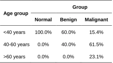
3.1 Age distribution and differences
In normal breast tissue 100% of the cases were <40 years. In
normal tissue around benign disorders; 60% of the cases were
<40 years and 40% of the cases 40-60 years.
In normal tissue around malignant disorders 15.4% of the cases
<40 years, 61.5% from 40-60 years and 23.1% >60 years
(Table 1 and Fig 1).
Table 1: Frequency distribution of age according to the
conditions of breast tissue
Age group
Group
Normal Benign Malignant
<40 years 100.0% 60.0% 15.4%
40-60 years 0.0% 40.0% 61.5%
>60 years 0.0% 0.0% 23.1%
The normal control group had more expression of ER when
compared to normal tissue around Benign and Malignant
conditions, followed by normal tissue around malignant
condition. On the other hand, the normal tissue around benign
condition had the least expression than other samples with
marked statistical significant.
The BCL2 protein was expressed in all conditions of normal
breast tissue but it exhibit more expression in benign and
malignant conditions than in normal control group. The C-MYC expression didn’t differ in control and benign group but it
increased in malignant condition especially in age level of 40-60
years.
As shown in figure 2, there was no statistical difference in
percentage of staining when comparing different sample from
control, benign and malignant for all markers used in this study.
The markers also showed no statistical difference after
calculating the final IHS of expression in the three samples of
the breast where P value >0.05 (Fig 3).
C-MYC expression showed significant direct correlation with PR
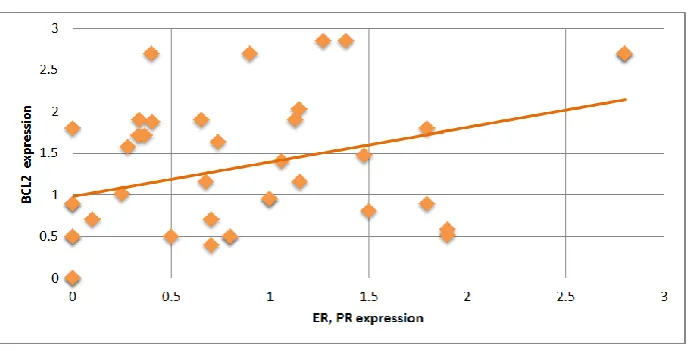
not with ER (Fig 4). On the other hand, BCL2 expression
correlated directly positively with statistical significance with
both ER & PR (Fig 5).
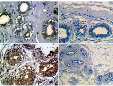
Fig 1: Immunohitochemical expression of different markers in normal breast tissue. (A: positive BCL2 around breast
cancer, B: positive ER in normal breast, C: positive C-MYC around cancer, D: Negative ER around cancer)
Fig 2:Bar chart showing the percentages of the staining of ER, PR, BCL2 and C-MYC in Control, Benign and malignant
groups of breast tissue. (Bars represent mean, error bars represent standard deviation)
This study was designed to detect presence of ER, PR, BCL2
and C-MYC in normal breast tissue of three proliferative
conditions and it revealed that there was presence of ER and
PR expression in normal tissue of the breast in any condition
that affirm by series of study12,13 that reported detection of ER,
PR in any amount in normal breast tissue.
Fig 3: Bar chart showing the immunohistochemical score of ER, PR, BCL2 and C-MYC in Control, Benign and malignant
groups of breast tissue. (Bars represent mean, error bars represent standard deviation)
Fig 4: Scatter diagram showing a direct positive correlation of expression between PR & C-MYC markers in breast tissue
Fig 5:Scatter diagram showing a direct positive correlation of expression between ER & PR with BCL2 markers in breast
tissue
Also this study revealed detection of BCL2 in all conditions but
it more in normal tissue around malignant that run in same line
with other reports14-16, they all demonstrated there is a
significantly higher level of expression of anti-apoptotic protein
(BCL2) in the normal tissue around breast carcinoma in
cancer. In same manner there was an expression of C-MYC in
all conditions but it more in malignant condition this agreed with
most of studies that mention; C-MYC detected in normal tissue
and cancerous from 1-94% of the cases17-19.
Regarding the correlation, this study found statistically
significant direct correlation between BCL2 and ER, PR
expression this in concordance of other study20,21 that reported
direct correlation of BCL2 and ER, PR expression in cancer
condition than benign. In same line result revealed, there were
60.5% of the cases ER+ had C-MYC overexpression with no
significant correlation between them, also there was no effect of
ER overexpression that confined with recent study22 that
reported 70.9% of ER+ in C-MYC over expression.
A PR result in this study was; 70% of cases were PR+ with
weak direct correlation between them, which confined with other
reported of23 while others, reported negative correlation with PR
or didn’t find any correlation between C-MYC and PR24
.
5 Conclusion:
C-MYC & BCL2 expressed in all normal breast tissue around
proliferative conditions but it more around carcinoma with direct
correlation with hormonal receptors.
6 Acknowledgments
The authors are grateful to Department of Anatomy, Histology
and Embryology, College of Medicine, Mustansiriyah University,
Iraq for providing the facilities needed to complete this work.
7 Competing Interests
Authors have declared that no competing interests exist.
8 Author’s contributions
AFH carried out literature review and histological staining. MMI
reviewed the statistical data and finalized the manuscript. BSA
gave provided histopathological reviews and helped finalize the
manuscript and statistical analysis. All authors read and
approved the final manuscript.
9 References
1. Angahar LT. An Overview of Breast Cancer Epidemiology,
Risk Factors, Pathophysiology, and Cancer Risks
Reduction. MOJ Biol Med. 2017; 1(4): 00019.
2. Curtis C, Shah SP, Chin SF, Turashvili G, Rueda OM. The
genomic and transcriptomic architecture of 2,000 breast
tumours reveals novel subgroups. Nature. 2012;
486(7403): 346-352.
3. Ferlay J, Soerjomataram I, Dikshit R, Eser S, Mathers C,
Rebelo M, Parkin DM, Forman D, Bray F. Cancer
incidence and mortality worldwide: sources, methods and
major patterns in GLOBOCAN 2012. Int J Cancer. 2015;
136(5): 359-86.
4. Schnipper L. Clinical implications of tumor-cell heterogeneity. N. Engl. J. Med. 1986; 314: 1423–1431.
5. Prall OW, Rogan EM, Musgrove EA, Watts CK, Sutherland
RL. C-Myc or cyclin D1 mimics estrogen effects on cyclin
E-Cdk2 activation and cell cycle reentry. Mol. Cell. Biol. 1998; 18: 4499–4508.
6. Hartl, M. The Quest for Targets Executing
MYC-Dependent Cell Transformation. Front. Oncol. 2016; 6,
132.
7. Cleary ML, Sklar J. Nucleotide sequence of a t(14;18)
chromosomal breakpoint in follicular lymphoma and
demonstration of a breakpoint-cluster region near a
transcriptionally active locus on chromosome 18. Proc Natl
Acad Sci USA. 1985; 82(21):7439–43.
8. Nunez G, London L, Hockenbery D, Alexander M,
McKearn JP, Korsmeyer SJ. Deregulated Bcl-2gene
expression selectively prolongs survival of growth
factor-deprived hemopoietic cell lines. J Immunol. 1990; 144(9):3602–10.
9. Jiang P, Du W, Heese K, Wu M. The Bad guy cooperates
with good cop p53: Bad is transcriptionally up-regulated by
p53 and forms a Bad/p53 complex at the mitochondria to
induce apoptosis. Mol Cell Biol. 2006; 26(23):9071–82.
10. Rosen PP. Fibroepithelial Neoplasms. In Rosen PP.
Rosen‟s Breast Pathology, 3rd edition. Lippincott Williams
and Wilkins. 2009; 187-229.
11. Dupont WD, Page DL. Risk factors for breast cancer in
women with proliferative breast disease. N Engl J Med.
1985; 312:146-151.
12. Ricketts D, Turnbull L, Ryall G. Estrogen and
Progesterone Receptors in the Normal Female Breast.
Cancer res. 1991; 51: 1817-1822.
13. Khan SA, Rogers MAM, Khurana KK. Estrogen Receptor
Expression in Benign Breast Epithelium and Breast Cancer
Risk. J. of the National Cancer Inst. 1998; 90(1):37-42.
14. Hassan HI, Walker RA. Decreased apoptosis in
non-involved tissue from cancer-containing breasts. J
Pathology. 1998; 184:258–264
15. Adams JM, Cory S. The Bcl-2 apoptotic switch in cancer
development and therapy. Oncogene.2007;26:1324–1337
16. Batchelder AJ,Gordon-Weeks AN, Walker RA. Altered
expression of anti-apoptotic proteins in non-involved tissue
from cancer-containing breasts. Springer Science+Business Media. 2008; 114:63–69
17. Fallah Y, Brundage J, Allegakoen P, Shajahan-Haq AN.
MYC-Driven Pathways in Breast Cancer Subtypes.
Biomolecules.2017; 7: 53.
18. Blancato J, Singh B, Liu A, Liao DJ, Dickson RB.
Correlation of amplification and overexpression of the
c-myc oncogene in high-grade breast cancer: FISH, in situ
hybridisation and immunohistochemical analyses. British J.
19. Liao DJ, Dickson RB. C-Myc in breast cancer. Endocr Relat
Cancer. 2000;7:143-64.
20. Honma N, Takubo K, Akiyama F et al. Expression of
GCDFP-15 and AR decreases in larger or node-positive
apocrine carcinomas of the breast. Histopathology. 2005; 47: 195–201.
21. Moinfar F, Okcu M, Tsybrovskyy O. Androgen receptors
frequently are expressed in breast carcinomas: potential
relevance to new therapeutic strategies. Cancer. 2003; 98:
703–711.
22. Sarrafzadeh Sh, Geranpayeh L, Tasharrofi B. Expression
Study and Clinical Correlations of MYC and CCAT2 in
Breast Cancer Patients. Iranian Biomed J. 2017; 21(5):
303-311
23. Rummukainen JK, Salminen T, Lundin J, Kytola S,
Joensuu H, Isola JJ. Amplification of c-myc by fluorescence
in situ hybridization in a population-based breast cancer
tissue array. Mod Pathol. 2001; 14: 1030–5.
24. Dueck AC, Reinholz M.M, Geiger XJ. Impact of c-MYC
Protein Expression on Outcome of Patients with
Early-Stage HER2+ Breast Cancer Treated with Adjuvant
![Speech by Mr. J. Rey, Member of the Commission of the European Economic Community, delivered at the plenary meeting [GATT-thirteenth session] of the contracting parties. 17 October 1958](data:image/gif;base64,R0lGODlhAQABAIAAAP///wAAACH5BAEAAAAALAAAAAABAAEAAAICRAEAOw==)