Mass Value Assignment of Total and Subclass Immunoglobulin G in a
Human Standard Anthrax Reference Serum
V. A. Semenova,* E. Steward-Clark, K. L. Stamey, T. H. Taylor, Jr., D. S. Schmidt, S. K. Martin,
N. Marano, and C. P. Quinn
Meningitis and Special Pathogens Branch, Division of Bacterial and Mycotic Diseases, Centers for Disease Control and
Prevention, Atlanta, Georgia
Received 19 April 2004/Returned for modification 28 May 2004/Accepted 22 June 2004
An anti-Anthrax Vaccine Adsorbed (anti-AVA) standard human reference serum pool, AVR414, has been
prepared, and the total and protective antigen (PA)-specific immunoglobulin G (IgG) were quantified. AVR414
was prepared by plasmapheresis of healthy adults who had received a minimum of four subcutaneous
injections of AVA. Mass values (in milligrams per milliliter) for total IgG and IgG subclasses 1 to 4 were
determined by radial immunodiffusion. Anti-PA-specific IgG assignment (in micrograms per milliliter) was
done by consensus of two complementary approaches: homologous enzyme-linked immunosorbent assay
(ELISA) with affinity-purified anti-PA IgG as a calibrator and summation of mean PA-specific IgG subclass
concentrations determined by IgG subclass-specific ELISA using the United States National Reference
Prep-aration for Human Serum Proteins as a standard. The total IgG concentration assigned to AVR414 reference
serum was 8.33 mg/ml. IgG subclass concentrations were the following: for IgG1, 4.48 mg/ml; for IgG2, 3.35
mg/ml; for IgG3, 0.37 mg/ml; and for IgG4, 0.30 mg/ml. The assigned mass value for total anti-PA-specific IgG
was 141.2
g/ml. Anti-PA-specific IgG subclass concentrations were the following: for IgG1, 79.6
g/ml; for
IgG2, 35.3
g/ml; for IgG3, 3.2
g/ml; and for IgG4, 25.3
g/ml. Human reference serum pool AVR414 will have
direct application in the standardization of anthrax serological assays, in reagent qualification, and as a
standard for quantification of PA-specific IgG in humans who have been vaccinated with or otherwise exposed
to
Bacillus anthracis
PA.
The immune response to anthrax toxin protective antigen
(PA) is central to protection against anthrax (19, 20).
Immu-noglobulin G (IgG) is the most abundant immuImmu-noglobulin in
human serum and provides the dominant immune response to
protein antigens after vaccination with multiple injections (16,
16a). Measurement of anti-PA IgG antibody is therefore an
appropriate marker of human immune responses to
Bacillus
anthracis
infection and anthrax vaccines. A lack of assay
stan-dardization and qualified reagents has been a major obstacle to
the comparative analysis of human serological responses to
clinical anthrax and anthrax vaccines. Compounding this
prob-lem are variations in antigen selection, preparation, and purity;
variations in assay methodology and end point determination
between laboratories; the diversity of antibodies in polyclonal
serum; and the absence of a suitable standard reference serum
(32). In 2001, the Centers for Disease Control and Prevention
(CDC; Atlanta, Ga.) initiated the Anthrax Vaccine Research
Program to determine the feasibility of reducing the number of
priming series doses of the licensed Anthrax Vaccine
Ad-sorbed (AVA or BioThrax; BioPort Corp., Lansing, Mich.)
(17, 26, 27) from six to three and changing the route of
ad-ministration from subcutaneous (s.c.) to intramuscular (28)
without reducing the vaccine’s immunogenicity. The Anthrax
Vaccine Research Program required the development of
pre-cise, accurate, specific, and sensitive serological assays for the
quantification of anti-PA IgG responses in humans (32).
Fun-damental to the consistency of such assays is the availability of
a standard reference serum and qualified control reagents
to-gether with standardized assay technologies and methods for
end point determination (29). In the present study, we report
the preparation and assignment of mass values for total and
PA-specific IgG and IgG subclasses for an anti-AVA human
reference serum, AVR414. The performance characteristics of
AVR414 as a standard reference reagent for quantification of
anti-PA IgG responses in human serum and the assignment of
PA-specific IgG mass values to positive quality control (QC)
sera and standards (AVR801) for use in anthrax serological
assays are also demonstrated.
MATERIALS AND METHODS
Preparation of anti-AVA human standard reference serum.The anti-AVA
human reference serum AVR414 (CDC standard anthrax reference sera AVR414 and AVR801 may be obtained free of charge under a suitable materials transfer agreement by application to C. P. Quinn, CDC) was prepared by pooling equal volumes of serum from each of three healthy adult CDC volunteers who had received a minimum of four s.c. injections of AVA with the licensed regimen (at 0, 2, and 4 weeks and 6, 12, and 18 months with two yearly boosters). Serum selection was based on anti-PA IgG titers in the range of 3,200 to 6,400 as determined by an anti-PA IgG enzyme-linked immunosorbent assay (ELISA) (32). Plasmapheresis of selected donors and subsequent serum conversion were done at the Emory Transfusion Medicine Program, Emory University School of Medicine (Atlanta, Ga.) and the Scientific Resource Program at the CDC, respectively, by TPE DUAL-NEEDLE operation using a Spectra apheresis sys-tem as described by the manufacturer (Cobe BCT, Inc., Blood Component Technology, Lakewood, Colo.). The plasma units were stored frozen at⫺70°C, thawed overnight at 4°C prior to use, and converted to serum by the injection of 4.0 ml of sterile glass microbeads (B. Braun Instruments, Burlingame, Calif.) suspended in 1.5 M CaCl2–2.0 Mε-amino-caproic acid (Sigma, St. Louis, Mo.).
Clots were allowed to form overnight at 4°C and were then removed by
centrif-* Corresponding author. Mailing address: Microbial Pathogenesis &
Immune Response Laboratory, Meningitis and Special Pathogens
Branch, Division of Bacterial and Mycotic Diseases, Centers for
Dis-ease Control and Prevention, Mail Stop D-01, Atlanta, GA 30333.
Phone: (404) 639-4390. Fax: (404) 639-4550. E-mail: [email protected].
919
on August 17, 2020 by guest
http://cvi.asm.org/
ugation at 2,200⫻gfor 15 min at 4°C. The serum from each unit was recovered by aspiration and stored separately in 500-ml sterile polycarbonate containers (Nalge Nunc International, Rochester, N.Y.). The level of residual anticoagu-lants was not determined (32). The anti-AVA human standard reference serum AVR414 was stored frozen in 3-ml aliquots at⫺70°C.
Calibration standard for serum immunoglobulins.The U.S. National
Refer-ence Preparation for Human Serum Proteins (USNRP; IS1644, lot 20575L) was obtained in lyophilized form from the CDC (33) and was used as a standard reference in IgG subclass-specific ELISAs for quantification of anti-PA IgG subclasses. The USNRP was reconstituted in 1.0 ml of sterile distilled water to provide final concentrations of 11.28 mg of total IgG/ml, comprising 7.12 mg of IgG1/ml, 2.79 mg of IgG2/ml, 0.60 mg of IgG3/ml, and 0.60 mg of IgG4/ml (33).
Anthrax toxin PA.Purified recombinant PA with an amino acid sequence
concurring with that of theB. anthracisV770-NP1-R anthrax vaccine strain was provided by Stephen H. Leppla (National Institute of Allergies and Infectious Diseases, National Institutes of Health, Bethesda, Md.). Antigen was stored frozen in 100- to 500-l aliquots (4.75 mg/ml) in 5 mM HEPES, pH 7.3, at ⫺70°C. PA was expressed and purified to homogeneity as described previously (18, 25).
Affinity purification of human IgG.Total IgG was purified from the AVR414
serum pool with a HiTrap Protein G column (Amersham Biosciences Corp., Piscataway, N.J.). Bound IgG was eluted with 0.1 M glycine-HCl (Sigma), pH 2.75, in 1.0-ml fraction volumes and immediately neutralized by the addition of 0.1-ml volumes of 1.0 M Tris-HCl, pH 9.0 (Life Technologies, Gaithersburg, Md.). Eluted peak fractions were dialyzed against 0.01 M phosphate-buffered saline (PBS), pH 7.4 (Life Technologies). The concentration of purified IgG was determined by radial immunodiffusion (RID) and the Bio-Rad (Hercules, Calif.) protein assay using purified commercially obtained human IgG as a standard (ICN Biomedicals, Inc., Costa Mesa, Calif.).
Affinity purification of PA-specific IgG.The anti-PA-specific IgG from the
protein G-purified AVR414 total IgG was isolated by affinity adsorption to PA immobilized on CNBr-activated Sepharose 4B according to the manufacturer’s instructions (Amersham Biosciences Corp.). Affinity-purified total IgG was loaded onto the column (2.5-ml bed volume) and eluted under neutral conditions using ActiSep elution medium according to the manufacturer’s instructions (Ste-rogene Bioseparations, Inc., Carlsbad, Calif.) (7). Fractions (0.5 ml) were col-lected, dialyzed against several changes of 100 mM HEPES buffer (Life Tech-nologies), pH 7.4, containing 100 mM sodium chloride (Sigma), and then concentrated using Slide-A-Lyzer concentrating solution (Pierce Chemical Com-pany, Rockford, Ill.). The concentration of purified anti-PA-specific IgG was determined by adsorption at 280 nm using an extinction coefficient of 1.35 (9a).
Quantitative anti-PA IgG ELISA.The quantitative ELISA for human anti-PA
IgG has previously been described in detail (32). Briefly, Immulon 2 HB micro-titer plates (Thermo Labsystems, Franklin, Mass.) were coated with 100l of purified recombinant PA (2g/ml) in 0.01 M PBS, pH 7.4 (Life Technologies). After incubation overnight at 4°C, the plates were washed three times with PBS containing 0.1% Tween 20. Serum samples (and all subsequent antibody added to the plates) were diluted in PBS containing 5% skim milk and 0.5% Tween 20 (pH 7.4), loaded, and transferred in twofold dilutions down the plate. After incubation for 60 min at 37°C, plates were washed as described above. Bound human anti-PA IgG was detected by incubation for 60 min at 37°C with horse-radish peroxidase-conjugated mouse monoclonal anti-human IgG Fc PAN clone HP6043 (Hybridoma Reagent Laboratory, Baldwin, Md.) (12). Plates were washed three times, and ABTS Microwell peroxidase substrate system (Kirkeg-aard and Perry Laboratories, Gaithersburg, Md.) was added for incubation for 30 min at 37°C. ABTS peroxidase stop solution (Kirkegaard and Perry Laborato-ries) was added to stop the assay, and plates were read with an MRX Revelation microtiter plate reader (Thermo Labsystems) at a wavelength of 410 nm with a 610-nm reference filter. The affinity-purified anti-PA IgG (139.0g/ml) was used as a calibrator to generate a 7-point standard curve in triplicate over a twofold dilution series in an anti-PA ELISA to assign a PA-specific IgG concentration to AVR414. Experiments to assign mass values were repeated a minimum of three times on three different assay plates by three different operators. The anti-PA IgG concentration for AVR414 was calculated by interpolation from the cali-bration curve using a four-parameter logistic log (4-PL) model and ELISA for Windows software (version 2.0) (31).
Quantitative IgG subclass-specific ELISA.Monoclonal antibodies to human
IgG1 (HP6069␥1 Fc), IgG2 (HP6002␥1 Fc), IgG3 (HP6047, anti-hinge region), and IgG4 (HP6025␥1 Fc) (12, 34) were purchased from the Hybridoma Reagent Laboratory. For determination of a specific IgG subclass concentration, Immu-lon 2 HB microtiter plates (Thermo Labsystems) were coated with the appro-priate monoclonal anti-subclass capture antibody at a concentration of 2g/ml
in 0.01 M PBS (Life Technologies), pH 7.4, at 4°C for 16 to 24 h. The USNRP was used to generate 7-point standard curves in triplicate in a threefold dilution series (1:100 to 1:12,800). Purified anti-PA IgG from the human standard serum AVR414 was diluted in serum diluent, added to the first pair of test wells (100 g/ml in 100l per well), mixed, and serially transferred down the plate in a threefold dilution series. Bound IgG subclasses were detected with polyclonal sheep anti-human IgG–horseradish peroxidase (ICN Biomedicals, Inc.). All sub-sequent reagents, incubation, and washing steps were performed as described above for the human anti-PA IgG ELISA. PA-specific IgG subclass concentra-tions were derived from a minimum of eight separate determinaconcentra-tions. The anti-PA IgG subclass concentrations were calculated as described above (31).
Determination of total IgG and IgG subclasses by RID.Mass values (in
micrograms per milliliter) for total IgG and IgG subclasses in AVR414 were determined by RID using the NL NANORID and BINDARID kits, respectively (The Binding Site, Inc., San Diego, Calif.). Mass values assigned by RID were calculated from a minimum of five separate determinations over three noncon-secutive days using the kit-supplied calibrator as a standard. Concentrations of total IgG and IgG subclasses were interpolated from the standard curves gen-erated on the same plate.
Performance characteristics of AVR414 in anti-PA IgG ELISA.Performance
characteristics established for AVR414 in human anti-PA IgG ELISA included dilution-corrected linearity of the assigned anti-PA IgG value, intermediate precision (repeatability over time) of a 7-point dilution series, and the goodness of fit of these data to a 4-PL model. Evaluation of parallelism between the AVR414 curve and the curves of test serum samples was performed in accor-dance with the guidelines described previously (30). Briefly, if the within-assay coefficient of variation (CV) isⱕ20%, the curves are considered parallel (30). Dilution-corrected linearity of the curve of AVR414 in an anti-PA IgG ELISA was evaluated by comparison of the expected concentrations of anti-PA IgG in AVR414 for seven twofold dilutions (1:100 to 1:6,400; calibration factor, 141.2 g/ml) with computed concentrations for the same dilutions (SAS version 8.0; SAS Institute, Cary, N.C.). Dilutional linearity was determined from triplicate curves in 34 independent assays (204 observations). The reportable IgG concen-trations (in micrograms per milliliter) of three positive QC sera (AVR216, AVR284, and AVR370) were used to evaluate the assay’s intermediate precision using AVR414 as a standard. The positive QC sera were obtained from CDC volunteers who had received at least four injections of AVA. The QC sera were tested in duplicate at single dilutions selected to represent high, medium, and low optical density regions of the reference serum standard curve. Intermediate precision was expressed as the CV of the concentration calculated for the stan-dard curve dilutions between different assay plates over time and performed by different operators. The goodness of fit of the standard was evaluated in 503 different observations and expressed as the approximate multiple correlation coefficient (R2) (31). The utility of AVR414 as a human standard reference
serum in an anti-PA IgG ELISA was evaluated with a separate vaccinee serum pool (AVR801) and a panel of 14 positive control vaccinee sera with a range of 58.0 to 398.4g of anti-PA IgG per ml. AVR801 was prepared by pooling equal volumes of serum from 57 different recipients of AVA. Donors for AVR801 had received a minimum of four s.c. injections of AVA with the licensed regimen. AVR414 was used to assign anti-PA-specific IgG and anti-PA-specific IgG sub-class concentrations to AVR801 using the anti-PA IgG and anti-PA IgG1 to IgG4 subclass-specific ELISAs, respectively, as described above. The anti-PA IgG concentration of AVR801 was determined from the mean of 150 individual test results performed by three operators working independently over a 4-month period of time. Anti-PA IgG1 to IgG4 concentrations were determined from the mean of results from nine individual tests for each IgG subclass performed by three operators working independently using AVR414 as the standard reference serum in anti-PA IgG1 to IgG4 ELISAs. The collection and use of human sera were performed in concordance with the CDC and NIH Institutional Review Boards.
Statistical analysis.The same mathematical and statistical methods were used
for calculations of total and anti-PA-specific IgG and IgG subclasses. The IgG subclass concentrations were assayed separately by ELISA or by RID, and the means were determined, summed, and then compared to the mean concentration of total IgG from the independent assay, ELISA, or RID by using a two-sample
ttest (23). Thettest was computed for various sample size alternatives based on the assumption that sample size might be interpreted as the minimum number for any one subclass, the design number for each subclass, or the total number used for all the subclasses. For the purpose of computing the overall variance (standard deviation) of the subclass sum, the four measurements were assumed to be statistically dependent, with central tendency matched at the mean for the purpose of matching unmatched data.
on August 17, 2020 by guest
http://cvi.asm.org/
RESULTS
Total IgG and IgG subclasses.
The concentrations of total
IgG in AVR414 determined by RID and by summation of IgG
subclasses were 8.33 mg/ml and 8.50 mg/ml, respectively. These
values are not statistically significantly different (
P
⫽
0.284)
(Table 1). The values for individual IgG subclasses in AVR414
determined by RID were the following: for IgG1, 4.48 mg/ml
(52.7% of total IgG); for IgG2, 3.35 mg/ml (39.4%); for IgG3,
0.37 mg/ml (4.4%); and for IgG4, 0.30 mg/ml (3.5%) (Table 1).
PA-specific IgG and IgG subclasses.
The assignment of the
anti-PA-specific IgG concentration in AVR414 was done by
consensus of two complementary approaches: an anti-PA IgG
ELISA using antigen affinity-purified anti-PA IgG as a
calibra-tor and summation of the mean PA-specific IgG subclass
con-centrations determined by the IgG subclass-specific ELISA
with USNRP as a standard. The anti-PA IgG concentration in
AVR414 as determined by ELISA with purified anti-PA IgG
was 139.0
g/ml (Table 2). IgG subclass summation data were
obtained from independent determinations in duplicate of 20,
8, 23, and 29 observations for IgG1, IgG2, IgG3, and IgG4,
respectively (Table 3). The predominant anti-PA IgG subclass
in AVR414 was IgG1, with a concentration of 79.6
g/ml
(55.51% of total IgG). The concentrations of IgG2 and IgG4
were 35.3
g/ml (24.62% of total IgG) and 25.3
g/ml
(17.64%). The concentration of IgG3 in AVR414 was low (3.2
g/ml; 2.2% of total IgG). These data indicate that the anti-PA
IgG subclasses in AVR414 are primarily IgG1, IgG2, and IgG4
(Table 3). The sum of the individual IgG subclass
concentra-tions (143.4
g/ml) was compared with the concentration of
total anti-PA IgG obtained by anti-PA IgG ELISA (139.0
g/
ml) by two-sample
t
test (23). There was no significant
differ-ence between the two mass values calculated (
P
⫽
0.42); a
conservative minimum sample size of eight was used for
cal-culating the sum of IgG subclasses. The overall anti-PA IgG
concentration was computed as an equally weighted mean of
the results from the two methods. The PA-specific IgG
com-ponent of AVR414 was assigned to be 141.2
g/ml and was
1.7% of the total IgG (Table 2).
ELISA performance characteristics of human reference
se-rum AVR414.
An evaluation of parallelism between the
AVR414 curve and the curves of test serum samples showed
that the within-assay CV was
ⱕ
20% and that the
antibody-binding characteristics of standard reference serum AVR414
and the serum samples are similar enough to allow the
deter-mination of antibody concentrations in diluted serum samples
(30). The interassay precision level of the human anti-PA IgG
ELISA was high. The CVs for three positive QC sera were less
then 20%: 13.3% for QC1 (AVR216; 163 tests), 17.8% for
QC2 (AVR284; 201 tests), and 16.4% for QC3 (AVR370; 215
tests). These values are within the accepted values of 20% for
interassay precision (11) and are indicative of a high level of
precision for this type of assay (24). The goodness of fit (mean
R
2) for the AVR414 standard curve calculated over 503
sepa-rate assays was 0.998. The intermediate precision of the
posi-tive control serum panel expressed as the mean CV was 12.7%
(14 separate sera; 378 observations). These values for
interas-say precision levels are also within the accepted values of 20%
for enzyme immunoassays and indicate a high level of
preci-sion for the performance of the AVR414 standard in these
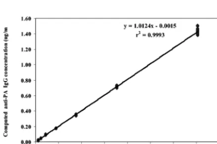
assays (24). Diluting AVR414 over the range from 1:100 to
1:6,400 and evaluating the resultant plot of measured
concen-tration versus expected concenconcen-tration by regression analyses of
interpolated results from the 4-PL model fit indicated a highly
significant linear relationship (mean
R
2⫽
0.999;
P
⬍
0.001),
with a slope of 1.0 and an intercept through the origin (Fig. 1).
Analysis of serum pool AVR801 showed that the anti-PA IgG
concentration (109.4
g/ml) determined by anti-PA IgG
ELISA using AVR414 as the standard was not significantly
different (
P
⫽
0.467) from the anti-PA IgG concentration
obtained by the summation of anti-PA IgG subclasses (111.3
g/ml). The anti-PA IgG subclass concentrations with
AVR801 obtained by anti-PA ELISAs using AVR414 as the
standard were the following: for IgG1, 60.2
g/ml; for IgG2,
25.8
g/ml; for IgG3, 4.5
g/ml; and for IgG4, 20.8
g/ml
(Table 3). The anti-PA IgG subclass distribution in AVR801
was similar to that in AVR414 (54.1% for IgG1, 23.2% for
IgG2, 4.0% for IgG3, and 18.7% for IgG4) (Table 3).
DISCUSSION
A qualified and characterized standard reagent is a critical
component in the development and standardization of
sero-logical assays and provides a benchmark for the comparative
analysis of new assay technologies (1, 15, 21). In this study, we
TABLE 1. Total IgG and IgG subclass concentrations in anti-AVA
standard reference serum AVR414 determined by RID
Total IgG or IgG subclass (mg/ml)Mean (mg/ml)SD (%)CV n % totalIgG by subclassa
IgG1
4.48
0.09
2.0
5
52.7
IgG2
3.35
0.22
6.6
7
39.4
IgG3
0.37
0.02
5.4
7
4.4
IgG4
0.30
0.00
0.0
5
3.5
Independent observation
of total IgG
8.33
0.29
3.5
12
98
Sum of total IgG
subclasses
8.50
0.24
2.8
5
100
aA total IgG concentration of 8.50 mg/ml was used for more-accurate
calcu-lation of percentages of total IgG by subclass and based on consideration of equality of each set of data (P⫽0.284).
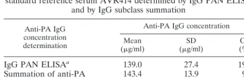
TABLE 2. Anti-PA-specific IgG concentrations in anti-AVA
standard reference serum AVR414 determined by IgG PAN ELISA
and by IgG subclass summation
Anti-PA IgG concentration determination
Anti-PA IgG concentration Mean
(g/ml) (SDg/ml) (%)CV
IgG PAN ELISA
a139.0
27.4
19.7
Summation of anti-PA
IgG subclasses
b143.4
13.9
9.7
Mean of two methods
c141.2
15.5
11.0
aAssignment of total anti-PA IgG concentration was based on 147
indepen-dent determinations in duplicate.
bThe sample size for summation was approximated by the effective degrees of
freedom (n⫽12). Variance-weighted computation was based on the actual sample sizes for each subclass.
cThe variance and corresponding CV were computed as the squared weighted
mean of the observed variances in the repeats of the respective experiments. The effective sample size was calculated as 20.
on August 17, 2020 by guest
http://cvi.asm.org/
report the quantification of total IgG and IgG subclasses and
the assignment of mass values for total and subclass-specific
anti-PA IgG in human standard anthrax reference serum
AVR414. The concentration of total IgG assigned for AVR414
(8.33 mg/ml) was within the established range for human
se-rum IgG in healthy adults (8 to 16 mg/ml) (2, 8, 9, 16). The
concentrations of IgG1 (4.48 mg/ml) and IgG3 (0.37 mg/ml) in
AVR414 were lower than the established range for normal
healthy human adults (5.0 to 12.0 mg/ml and 0.5 to 1.0 mg/ml,
respectively) (2, 8, 16, 22). The percentages of total IgG
rep-resented by IgG1 (52.7%) and IgG3 (4.4%) were also lower
than the range in normal adult sera (60.3 to 71.5% and 5.0 to
8.4%, respectively) (8). The concentration of IgG2 (3.35 mg/
ml) was within the established range for normal adult sera with
a dynamic range of 2.0 to 6.0 mg/ml (2, 8, 22, 36), but the IgG2
percentage of total IgG (39.4%) was higher than the
estab-lished range (19.4 to 31.0%) (8). The AVR414 standard
ref-erence serum pool contains anti-PA-specific IgG of all four
subclasses, with concentrations of PA-specific IgG1, IgG2, and
IgG4 being the highest (55.5, 24.6, and 17.6% of total anti-PA
IgG) and the concentration of IgG3 being the lowest (2.2% of
total anti-PA IgG). A possible explanation for the low
concen-tration of anti-PA IgG3 in AVR414 is affinity maturation and
isotype switching to IgG2 and IgG4 (4) due to the plasma
donors in this study receiving a minimum of four vaccinations
of the licensed AVA regimen.
The anti-PA IgG concentrations assigned by the two
meth-ods were not significantly different (
P
⫽
0.42). This level of
probability from two different approaches for mass values
as-signment is an indication of the robustness and reliability of the
assigned units. The utility of AVR414 has been clearly
dem-onstrated in its application to anti-PA IgG mass value
assign-ments in a panel of QC and positive vaccine control sera and
the assignment of PA-specific values to an additional standard
reference serum pool, AVR801.
It has been our objective to create a standardized platform
technology and reagents that will enable the comparative
anal-yses of human anti-PA IgG responses to clinical anthrax and
anthrax vaccines. The characterization of the anti-PA-specific
total IgG and IgG subclasses of the standard AVR414
de-scribed here provides a strong basis for this standardization.
AVR414 has been successfully applied in quantitative
serolog-ical assays to evaluate human humoral antibody immune
re-sponses to vaccination with AVA (N. Marano, J. Lingappa, P.
Pittman, V. Semenova, S. Leitman, P. Plikaytis, C. Quinn, and
B. Perkins, Abstr. 5th Int. Conf. Anthrax, abstr. O609, 2003),
for analysis of the immune response to PA in individuals with
bioterrorism-associated cutaneous and inhalation anthrax (5,
10, 13, 14, 32, 35, 37), and for evaluation of anti-PA IgG
subclass distribution in anthrax vaccinees and confirmed cases
of clinical anthrax (V. A. Semenova, P. M. Dull, D. S. Schmidt,
T. H. Taylor, E. Steward-Clark, M. M. Ballard, and C. P.
Quinn, Int. J. Infect. Dis. vol. 8, abstr. 35.010, p. S111, 2004).
For each of those studies, the AVR414 standard reference
serum was the pivotal unifying reagent. Qualified reagents,
such as AVR414, will facilitate assay technology transfer and
reduce interassay and interlaboratory variance (3, 6). In
addi-tion, the determination of assay end points using a 4-PL model
to describe the standard curve parameters (29) also makes a
significant contribution to assay standardization by reducing
the number and type of possible errors related to interpolating
antibody concentrations from the standard curve (30).
To-gether these are valuable tools in standardizing the evaluation
of the human anti-PA IgG immune response in recipients of
PA-containing vaccines and in cases of human anthrax.
ACKNOWLEDGMENTS
We acknowledge the contribution of CDC employees and NIH
volunteers for their donation of plasma and serum samples, Emory
Blood Bank for plasmapheresis of CDC volunteers, Wanda Philips for
conversion of plasma to serum, Stephen H. Leppla for provision of
anthrax toxin protective antigen, and Susan Leitman (NIH) for
pro-viding the plasma for AVR801.
FIG. 1. Linear regression of the results of diluting AVR414 from
1:100 to 1:6,400 and evaluating measured concentration versus
ex-pected concentration of anti-PA IgG. Data points are the means of
results from triplicate determinations.
TABLE 3. Comparison of anti-PA-specific IgG subclass distributions in the two anti-AVA standard reference sera, AVR414 and AVR801
IgG subclass
AVR414 AVR801
Mean
(g/ml) (SDg/ml) CV (%) n % totalIgGa (Meang/ml) (SDg/ml) CV (%) n % totalIgGa
IgG1
79.6
9.46
11.7
20
55.5
60.2
5.19
8.62
9
54.1
IgG2
35.3
9.73
27.6
8
24.6
25.8
2.79
10.81
9
23.2
IgG3
3.2
0.70
21.9
23
2.2
4.5
0.53
11.78
9
4.0
IgG4
25.3
2.97
11.7
29
17.6
20.8
3.11
14.95
9
18.7
aThe total anti-PA IgG concentrations obtained by sum of the mean of the anti-PA IgG subclass concentrations (143.4g/ml for AVR414 and 111.3g/ml for
AVR801) were used for a more accurate calculation of the percentage of total IgG by subclass. This approach was based on consideration of equality of each set of data for comparison of means, with aPvalue of 0.785 for AVR414 and aPvalue of 0.467 for AVR801.
on August 17, 2020 by guest
http://cvi.asm.org/
REFERENCES
1. Biagini, R. E., D. L. Sammons, J. P. Smith, B. A. MacKenzie, C. A. F. Striley,
V. Semenova, E. Steward-Clark, K. Stamey, A. E. Freeman, C. P. Quinn, and
J. E. Snawder.2004. Comparison of a multiplexed fluorescent covalent
microsphere immunoassay and an enzyme-linked immunosorbent assay for measurement of human immunoglobulin G antibodies to anthrax toxins. Clin. Diagn. Lab. Immunol.11:50–55.
2. Bradwell, A. R.1995. IgG and IgA subclasses in disease, p. 6. The binding
site, San Diego, Calif.
3. Carlone, G. M., C. E. Frasch, G. R. Siber, S. Quataert, L. L. Gheesling, S. H.
Tuner, B. D. Plikaytis, L. O. Helsel, W. E. DeWitt, W. F. Bibb, B. Swami-nathan, G. Arakere, C. Thompson, D. Phipps, D. Madore, and C. V. Broome.
1992. Multicenter comparison of levels of antibody to theNeisseria menin-gitidisgroup A capsular polysaccharide measured by using an enzyme-linked immunosorbent assay. J. Clin. Microbiol.30:154–159.
4. Devey, M. E.1990. Affinity of IgG subclass antibodies, p. 185–194.InFarouk
Shakib (ed.), The human IgG subclasses: molecular analysis of structure, function and regulation. Pergamon Press, Inc., Elmsford, N.Y.
5. Dewan, P. K., A. M. Fry, K. Laserson, B. C. Tierney, C. P. Quinn, J. A.
Hayslett, L. N. Broyles, A. Shane, K. L. Winthrop, I. Walks, L. Siegel, T. Hales, V. A. Semenova, S. Romero-Steiner, C. Elie, R. Khabbaz, A. S. Khan, R. A. Hajjeh, A. A. Schuchat, and the members of the Washington, D.C.,
Anthrax Response Team.2002. Inhalational anthrax outbreak among postal
workers, Washington, D. C., 2001. Emerg. Infect. Dis.8:1066–1072.
6. Gheesling, L. L., G. M. Carlone, L. B. Pais, P. F. Holder, S. E. Maslanka,
B. D. Plikaytis, M. Achtman, P. Densen, C. E. Frasch, H. Ka¨yhty, J. P. Mays, L. Nencioni, C. Peeters, D. C. Phipps, J. T. Poolman, E. Rosenqvist, G. R. Siber, B. Thiesen, J. Tai, C. M. Thompson, P. P. Vella, and J. D. Wenger.
1994. Multicenter comparison ofNeisseria meningitidisserogroup C capsular polysaccharide antibody levels measured by standardized enzyme-linked im-munosorbent assay. J. Clin. Microbiol.32:1475–1482.
7. Grandics, P., Z. Szathmary, and S. Szathmary.1990. A novel immunoaffinity
chromatography system for the purification of therapeutic proteins. Ann. N. Y. Acad. Sci.589:148–156.
8. Hamilton, R. G.1998. The human IgG subclasses, p. 28.
Calbiochem-Nova-biochem, La Jolla, Calif.
9. Harlow, E., and D. Lane.1988. Antibodies: a laboratory manual, p. 10. Cold
Spring Harbor Laboratory, Cold Spring Harbor, N.Y.
9a.Harlow, E., and D. Lane.1988. Antibodies: a laboratory manual, p. 658–681.
Cold Spring Harbor Laboratory, Cold Spring Harbor, N.Y.
10. Hsu, V. P., S. L. Lukacs, T. Handzel, J. Hayslett, S. Harper, T. Hales, V. A.
Semenova, S. Romero-Steiner, C. Elie, C. P. Quinn, R. Khabbaz, A. S. Khan,
G. Martin, J. Eisold, A. Schuchat, and R. A. Hajjeh.2002. Opening aBacillus
anthracis-containing envelope, Capitol Hill, Washington, D.C.: the public health response. Emerg. Infect. Dis.8:1039–1043.
11. Jacobson, R. H.1998. Validation of serological assays for diagnosis of
infec-tious diseases. Rev. Sci. Tech.17:469–526.
12. Jefferis, R., C. B. Reimer, F. Skvaril, G. de Lange, N. R. Ling, J. Lowe, M. R.
Walker, D. J. Phillips, C. H. Aloisio, T. W. Wells, J. P. Vaerman, C. G. Magnusson, H. Kubagawa, M. Cooper, F. Vartdal, B. Vandvik, J. J. Haaij-man, O. Makela, A. Sarnesto, Z. Lando, Z. J. Gergely, E. Rajnavolgyi, G.
Laszio, J. Radi, and G. A. Molinaro.1985. Evaluation of monoclonal
anti-bodies having specificity for human IgG sub-classes: results of an IUIS/WHO collaborative study. Immunol. Lett.10:223–252.
13. Jernigan, D. B., P. L. Raghunathan, B. P. Bell, R. Brechner, E. A. Bresnitz,
J. C. Butler, M. Cetron, M. Cohen, T. Doyle, M. Fischer, C. Greene, K. S. Griffith, J. Guarner, J. L. Hadler, J. A. Hayslett, R. Meyer, L. R. Petersen, M. Phillips, R. Pinner, T. Popovic, C. P. Quinn, J. Reefhuis, D. Reissman, N. Rosenstein, A. Schuchat, W. J. Shieh, L. Siegal, D. L. Swerdlow, F. C. Tenover, M. Traeger, J. W. Ward, I. Weisfuse, S. Wiersma, K. Yeskey, S. Zaki, D. A. Ashford, B. A. Perkins, S. Ostroff, J. Hughes, D. Fleming, J. P. Koplan, J. L. Gerberding, and the National Anthrax Epidemiologic
Inves-tigation Team.2002. Investigation of bioterrorism-related anthrax, United
States, 2001: epidemiologic findings. Emerg. Infect. Dis.8:1019–1028.
14. Jernigan, J. A., D. S. Stephens, D. A. Ashford, C. Omenaca, M. S. Topiel, M.
Galbraith, M. Tapper, T. L. Fisk, S. Zaki, T. Popovic, R. F. Meyer, C. P. Quinn, S. A. Harper, S. K. Fridkin, J. J. Sejvar, C. W. Shepard, M. McCon-nell, J. Guarner, W.-J. Shieh, J. M. Malecki, J. L. Gerberding, J. M. Hughes, B. A. Perkins, and members of the Anthrax Bioterrorism Investigation
Team.2001. Bioterrorism-related inhalational anthrax: the first 10 cases
reported in the United States. Emerg. Infect. Dis.7:933–944.
15. Johnson, A. M., J. T. Whicher, T. B. Ledue, A. Carlstrom, Y. Itoh, and P. H.
Petersen.2000. Effect of a new international reference preparation for
pro-teins in human serum (certified reference material 470) on results of the College of American Pathologists Surveys for plasma proteins. Arch. Pathol. Lab. Med.10:1496–1501.
16. Kuby, J.1996. Immunology, p. 124–126. W. H. Freeman and Company, New
York, N.Y.
16a.Kuby, J.1996. Immunology, p. 325–326. W. H. Freeman and Company, New
York, N.Y.
17. Lansing, Michigan, Department of Public Health.1978. Anthrax vaccine
adsorbed (package insert). Lansing, Michigan, Department of Public Health, Lansing.
18. Leppla, S. H.1991. Purification and characterization of adenylyl cyclase from
Bacillus anthracis. Methods Enzymol.195:153–168.
19. Little, S. F., B. E. Ivins, P. F. Fellows, and A. M. Friedlander.1997. Passive
protection by polyclonal antibodies againstBacillus anthracisinfection in guinea pigs. Infect. Immun.65:5171–5175.
20. Little, S. F., and B. E. Ivins. 1999. Molecular pathogenesis ofBacillus
anthracisinfection. Microbes Infect.1:131–139.
21. Ma¨kela¨, O., and F. Pe´terfy.1983. Standard sera in solid-phase
immunoas-says. Eur. J. Immunol.13:815–819.
22. Maranon, F., M. Casanovas, L. Berrens, J. M. Olles, and M. A. Dieguez.
1994. A competitive enzyme immunoassay subclass for the determination of total IgG subclass levels in human serum. J. Immunol.15:147–156.
23. Moore, D. S., and G. P. McCabe.1993. Introduction to the practice of
statistics, 2nd ed., p. 499–573. W. H. Freeman and Company, New York, N.Y.
24. O’Connel, M. A., B. A. Belanger, and P. D. Haaland.1993. Calibration and
assay development using the four-parameter logistic model. Chemom. Intell. Lab. Syst.20:97–114.
25. Park, S., and S. H. Leppla.2000. Optimized production and purification of
Bacillus anthracislethal factor. Protein Expr. Purif.18:293–302.
26. Pittman, P. R., J. A. Mangiafico, C. A. Rossi, T. L. Cannon, P. H. Gibbs,
G. W. Parker, and A. M. Friedlander.2000. Anthrax vaccine: increasing
intervals between the first two doses enhances antibody response in humans. Vaccine19:213–216.
27. Pittman, P. R., P. H. Gibbs, T. L. Cannon, and A. M. Friedlander.2001.
Anthrax vaccine: short-term safety experience in humans. Vaccine20:972– 978.
28. Pittman, P. R., G. Kim-Ahn, D. Y. Pifat, K. Coonan, P. Gibbs, S. Little, J. G.
Pace-Templeton, R. Myers, G. W. Parker, and A. M. Friedlander.2002.
Anthrax vaccine: immunogenicity and safety of a dose-reduction, route-change comparison study in humans. Vaccine20:1412–1420.
29. Plikaytis, B. D., S. H. Turner, L. L. Gheesling, and G. M. Carlone.1991.
Comparisons of standard curve-fitting methods to quantitateNeisseria men-ingitidisgroup A polysaccharide antibody levels by enzyme-linked immu-nosorbent assay. J. Clin. Microbiol.29:1439–1446.
30. Plikaytis, B. D., P. F. Holder, L. B. Pais, S. E. Maslanka, L. L. Gheesling,
and G. M. Carlone.1994. Determination of parallelism and nonparallelism
in bioassay dilution curves. J. Clin. Microbiol.32:2441–2447.
31. Plikaytis, B. D., P. F. Holder, and G. M. Carlone.1996. Program ELISA for
Windows user’s manual, version 2.00. Centers for Disease Control and Pre-vention, Atlanta, Ga.
32. Quinn, C. P., V. A. Semenova, C. M. Elie, S. Romero-Steiner, C. Greene, H.
Li, K. L. Stamey, E. Steward-Clark, D. S. Schmidt, E. Mothershed, J. Pruckler, S. Schwartz, R. F. Benson, L. O. Helsel, P. Holder, S. E. Johnson, M. Kellum, T. Messmer, W. L. Thacker, L. Besser, B. D. Plikaytis, T. H. Taylor, Jr., A. E. Freeman, K. J. Wallace, P. Dull, J. Sejvar, E. Bruce, R. Moreno, A. Schuchat, J. R. Lingappa, S. K. Martin, J. Walls, M. Bronsdon, G. M. Carlone, M. Bajani-Ari, D. A. Ashford, D. S. Stephens, and B. A.
Perkins.2002. Specific, sensitive, and quantitative enzyme-linked
immu-nosorbent assay for human immunoglobulin G antibodies to anthrax protec-tive antigen. Emerg. Infect. Dis.8:1103–1110.
33. Reimer, C. B., S. J. Smith, T. W. Wells, R. M. Nakamura, P. W. Keitges, R. F.
Ritchie, G. W. Williams, D. J. Hanson, and D. B. Dorsey.1982. Collaborative
calibration of the U.S. National and the College of American Pathologists reference preparation for specific serum proteins. Am. J. Clin. Pathol.77:
12–19.
34. Reimer, C. B., D. J. Phillips, C. H. Aloisio, D. Moore, G. G. Galland, T. W.
Wells, C. M. Black, and J. S. McDougal.1984. Evaluation of thirty-one
mouse monoclonal antibodies to human IgG epitopes. Hybridoma3:263– 273.
35. Shieh, W. J., J. Guarner, C. Paddock, P. Greer, K. Tatti, M. Fischer, M.
Layton, M. Philips, E. Bresnitz, C. P Quinn, T. Popovic, B. A. Perkins, and
S. R. Zaki.2003. The critical role of pathology in the investigation of
biot-errorism-related cutaneous anthrax. Am. J. Pathol.163:1901–1910.
36. So¨derstrom, T., R. So¨derstrom, A. Avanzini, P. Brandtzaeg, G. Karlsson,
and L. A. Hanson.1987. Immunoglobulin G subclass deficiencies. Int. Arch.
Allergy Appl. Immunol.82:476–480.
37. Traeger, M. S., S. T. Wiersma, N. E. Rosenstein, J. M. Maleckit, C. W.
Shepard, P. L. Raghunathan, S. P. Pillai, T. Popovic, C. P. Quinn, R. F. Meyer, S. R. Zaki, S. C. Tierney, J. D. Jones, B. A. Perkins, and the Florida
Investigation Team.2002. First case of bioterrorism-related international
anthrax in the United States, Palm Beach County, Florida, 2001. Emerg. Infect. Dis.8:1029–1034.