www.impactjournals.com/oncotarget/ Oncotarget, Vol. 5, No. 19
The Functional Characterization of Long Noncoding RNA
SPRY4-IT1
in Human Melanoma Cells
Joseph Mazar1, Wei Zhao1, Ahmad M. Khalil2, Bongyong Lee1, John Shelley1, Subramaniam S. Govindarajan1, Fumiko Yamamoto1, Maya Ratnam2, Muhammad Nauman Aftab1, Sheila Collins1, Brian N. Finck3, Xianlin Han1, John S. Mattick4, Marcel E. Dinger4, and Ranjan J. Perera1*
1 Sanford-Burnham Medical Research Institute, Orlando, FL 32827, USA
2 Department of Genetics and Genome Sciences, Case Western Reserve University School of Medicine, Cleveland, OH 44106,
USA
3 Division of Geriatrics and Nutritional Sciences, Washington University School of Medicine, St. Louis, MO 63110, USA 4 Garvan Institute of Medical Research and St Vincent’s Clinical School, University of New South Wales, Darlinghurst NSW
2010, Australia
Correspondence to: Ranjan J. Perera, email: [email protected]
Keywords: long noncoding RNA, melanoma
Received: January 23, 2014 Accepted: March 24 2014 Published: March 26, 2014
This is an open-access article distributed under the terms of the Creative Commons Attribution License, which permits unrestricted use, distribution, and reproduction in any medium, provided the original author and source are credited.
ABSTRACT:
Expression of the long noncoding RNA (lncRNA) SPRY4-IT1 is low in normal human melanocytes but high in melanoma cells. siRNA knockdown of SPRY4-IT1 blocks melanoma cell invasion and proliferation, and increases apoptosis. To
investigate its function further, we affinity purified SPRY4-IT1 from melanoma
cells and used mass spectrometry to identify the protein lipin 2, an enzyme that converts phosphatidate to diacylglycerol (DAG), as a major binding partner. SPRY4-IT1 knockdown increases the accumulation of lipin2 protein and upregulate the expression of diacylglycerol O-acyltransferase 2 (DGAT2) an enzyme involved in the conversion of DAG to triacylglycerol (TAG). When SPRY4-IT1 knockdown and control melanoma cells were subjected to shotgun lipidomics, an MS-based assay that
permits the quantification of changes in the cellular lipid profile, we found that SPRY4-IT1 knockdown induced significant changes in a number of lipid species, including
increased acyl carnitine, fatty acyl chains, and triacylglycerol (TAG). Together, these results suggest the possibility that SPRY4-IT1 knockdown may induce apoptosis via lipin 2-mediated alterations in lipid metabolism leading to cellular lipotoxicity.
INTRODUCTION
Eukaryotic genomes express a complex repertoire of thousands of RNAs that lack protein-coding capacity [1, 2]. These noncoding RNAs (ncRNAs) are broadly
classified as long or small based on a nucleotide length
of >200 or <200 nucleotides (nt), respectively. Small regulatory ncRNAs are commonly conserved and are involved in transcriptional and posttranscriptional gene
regulation through specific base pairing with their target
genes or transcripts. In contrast, long noncoding RNAs
(lncRNAs) are less well conserved and regulate gene
expression by diverse mechanisms, including epigenetic mechanisms, that remain incompletely understood [3-6].
LncRNAs may be derived from genomic sequences that
are intergenic, intronic, overlapping, or antisense with
respect to nearby protein-coding genes, and although some lncRNAs may be translated into short polypeptides, the vast majority are rarely or never translated [7, 8].
Until recently, lncRNAs were frequently dismissed as non-functional transcriptional ‘‘noise’’ [9]. However, the past few years have seen a rapid increase in our
understanding of the regulatory functions of lncRNAs and their role in human disease and development [10, 11]. LncRNAs exhibit exquisite context-dependency
commensurate with their presumed regulatory role, with cell type-specific expression and localization to discrete
lncRNAs influence target gene expression at specific genomic loci by directly interacting with chromatin
regulatory proteins and/or by modulating the activity of their interacting partners [15-20]. LncRNAs can function as decoys for bound proteins and can also alter the structure and function of the protein [17]. Although lncRNAs play physiological roles during normal cellular development and differentiation [21], changes in their
expression are associated with several diseases, including cancer, heart disease, Alzheimer’s disease, psoriasis,
and spinocerebellar ataxia type 8 [22]. Accumulating evidence suggests that lncRNAs may also play a role in tumorigenesis. For example, increased expression of
HOTAIR is associated with poor prognosis in pancreatic
cancer [23], and increased expression of PCGEM1 and
PCA3/DD3 is associated with a high risk of developing
prostate cancer [24].
Recently, we identified a number of lncRNAs that
are differentially expressed in melanoma cell lines relative to melanocytes and keratinocytes [14, 25]. One of these,
SPRY4-IT1 (Sprouty4-Intron 1; GenBank accession ID AK024556), is highly expressed in melanoma cells
relative to melanocytes, and is localized predominantly in the cytoplasm. Previously we showed that SPRY4-IT1 is derived from the intronic region of the SPRY4 gene and that its predicted secondary structure contains several long
hairpins [14]. Moreover, RNAi-mediated knockdown of SPRY4-IT1 inhibited invasion and proliferation and induced apoptosis of melanoma cells, suggesting an important role for this lncRNA in melanoma biology.
In the present study, we sought to identify SPRY4-IT1-interacting proteins and elucidate this lncRNA’s molecular function. In melanoma cells, SPRY4-IT1
transcripts are processed in the nucleus prior to transport
to the cytoplasm, where they are primarily located in
polysomes. We also identify the phosphatidate lipin 2 as a major SPRY4-IT1-binding protein, and demonstrate the existence of a novel lipid regulatory mechanism involving
SPRY4-IT1 and lipin 2. Our results are consistent with
the possibility that SPRY4-IT1 knockdown may induce
apoptosis via lipin 2-mediated alterations in lipid metabolism leading to cellular lipotoxicity. Together these results provide novel insight into the mechanisms by
which extranuclear processing of lncRNAs contributes to
melanoma biology.
RESULTS
Processing of SPRY4-IT1 Transcripts
SPRY4-IT1 was originally identified as a 706 bp
transcript in adipose tissue as part of a large-scale cDNA sequencing study [26]. The SPRY4-IT1 sequence was shown by PCR to initiate in intron 1 of the SPRY4 gene
and extend to exon 3 (Fig. 1A). Interestingly, the 5′ region
of the full-length SPRY4-IT1 transcript was detected in
samples of nuclear, but not cytoplasmic RNA, suggesting that the processing of SPRY4-IT1 takes place before
nucleocytoplasmic export (Fig. 1B). We confirmed using 5′ RACE that the first 244 nt of the 5′ sequence is missing
from cytoplasmic SPRY4-IT1 (Fig. S1). To validate this
observation and to visualize the subcellular localization
of SPRY4-IT1, we designed several RNA-FISH probes specific for the 3′ or 5′ regions of SPRY4-IT1. This analysis
confirmed the presence of the full-length transcript in the
nucleus but not in the cytoplasm (Fig. S2), in agreement
with PCR results. SPRY4-IT1 staining in melanocytes is
mainly localized to the nucleus and this may be the reason
of intense nuclear staining in melanocytes. Together these data demonstrate that the SPRY4-IT1 transcript undergoes maturation by cleavage of the 5’ region prior to its transport to the cytoplasm. This may suggest that the regulation of the 5’ cleavage controls the export of the transcript to the cytoplasm thus, the overall
SPRY4-IT1 content may depend on the efficiency of the nuclear
excision event.
Although the precise 3′ termination site of the SPRY4-IT1 transcript is not yet known, northern blot analysis shows a SPRY4-IT1 transcript size of approximately 1.8 kb (Fig. 1C), and the 3′ sequence of the
transcript is very similar to that of Exon 3 of the SPRY4 gene. Therefore, we propose that SPRY4-IT1 carries both intronic and exonic sequences and can be considered a noncoding splice variant of the SPRY4 gene.
SPRY4-IT1 and SPRY4 Gene Transcripts Function Independently
We next asked whether the transcription and
function of SPRY4-IT1 and SPRY4 are coordinately or independently regulated. Despite the similar expression patterns of SPRY4 and SPRY4-IT1, siRNA-mediated
knockdown of SPRY4 (siRNA targeting exon 1) in A375 melanoma cells reduced the expression of SPRY4 but not
SPRY4-IT1 (Fig. 1D), demonstrating that transcription of the coding (SPRY4) and noncoding (SPRY4-IT1)
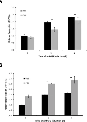
genes occurred independently. To confirm this in a more physiological setting, we treated melanocytes (low expression of SPRY4-IT1) with fibroblast growth factor 2 (FGF2), which is known to induce SPRY4 expression [27]. In serum-containing medium, FGF2 induced expression of SPRY4-IT1 and SPRY4 to similar extents (Fig. 2A).
However, SPRY4-IT1 transcriptionwas further elevated in melanocytes treated with FGF2 in serum-free medium (Fig. 2B), confirming independent transcription of SPRY4-IT1 and SPRY4. These results suggest that SPRY4-IT1 responds to starvation and general stress responses.
To determine whether the stability of SPRY4-IT1 and
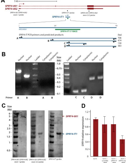
Figure 1:Maturation and Cellular Compartmentalization of SPRY4-IT1 Transcripts. (A) The sequence and location of
SPRY4 and SPRY4-IT1. SPRY4-IT1 is embedded in the SPRY4 parent transcript. SPRY4-IT1 starts in the first intron of the SPRY4 gene and
extends up to exon 3. 5′ RACE shows the maturation and cleavage of a 244 nt transcript in the 5′ region of SPRY4-IT1. (B) Detection of nuclear and cytoplasmic forms of SPRY4-IT1. (C) Northern blot analysis shows the sizes of SPRY4 (4.9 kb) and SPRY4-IT1 (1.8 kb). The
SPRY4 exon 1 probe hybridizes specifically to SPRY4, but the SPRY4 exon 3 probes recognize both SPRY4 and SPRY4-IT. (D) SPRY4 exon
A375 cells with the polymerase II transcriptional inhibitor α-amanitin and measured the transcript levels by RT-PCR. We observed that SPRY4 RNA decayed faster than
SPRY4-IT1 in both the nucleus and cytoplasm, supporting the functional independence of the transcripts (Fig. S3).
Finally, we independently knocked down SPRY4 and
SPRY4-IT1 in A375 cells using transcript-specific siRNAs. Confirming our previous report [14], A375 cell invasion was inhibited ~50% by SPRY4-IT1 knockdown but was
unaffected by SPRY4 silencing (Fig. S4A). Similarly,
SPRY4-IT1 silencing induced apoptosis of A375, as measured by caspase 3 activity, more effectively than did
SPRY4 knockdown (Fig. S4B). Collectively, these data
establish the transcriptional and functional independence of SPRY4-IT1 and its host gene SPRY4.
SPRY4-IT1 Is Localized to the Polysome Fraction and Binds the Lipid Phosphatase Lipin 2
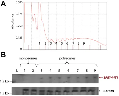
In a recent ribosomal footprint study, Ingolia et al. [28] reported that cytoplasmic lncRNAs are primarily found in ribosomal clusters or polysomes. To determine the location of SPRY4-IT1, we used density gradient
centrifugation to isolate monosomal and polysomal fractions from A375 cells and recovered 13 fractions: 2 monosomal and 11 polysomal (Fig. 3A). Total RNA
was isolated from the individual fractions and analyzed
for the presence of SPRY4-IT1 by northern blotting.
SPRY4-IT1 was detected only in polysomal fractions
3 through 9 (Fig. 3B), indicating that SPRY4-IT1 is
[image:4.612.161.450.273.678.2]indeed associated with polysomes and is not retained in monosomes, consistent with the findings of Ingolia et al. [28]. Sequence analysis further confirmed that SPRY4-IT1
Figure 2: SPRY4 and SPRY4-IT1 Are Coordinately Regulated in Melanocytes. Relative expression of SPRY4-IT1 and SPRY4 following induction by FGF2. Melanocytes were treated with 10 ng/ml FGF2 for 1 or 2 h in the presence or absence of FBS, and the fold
does not show any coding potential; understanding its role
in possible translational regulation is an ongoing study in our laboratory.
We next sought to identify SPRY4-IT1-binding proteins in melanoma cells. For this, we affinity purified
endogenous SPRY4-IT1 from crosslinked A375 lysate (Fig. 4A) and interrogated the SPRY4-IT1-associated proteins by mass spectrometry (MS). A group of candidate
proteins was identified and quantified by spectral counting (Table S1). The most abundant protein candidate was
lipin 2 [29], a phosphatidic acid phosphatase (PAP) that converts phosphatidate into diacylglycerol (DAG). Lipin
2 was pulled down specifically with SPRY4-IT1 but not
with scrambled (control) probes. To verify the physical association between lipin 2 and SPRY4-IT1, lipin 2 was immunoprecipitated, and associated RNAs were isolated
and interrogated for the presence of SPRY4-IT1 by qPCR. As shown in Figure 4B, SPRY4-IT1 transcripts were
enriched in anti-lipin 2 immunoprecipitated compared
with control IgG.
We next investigated the functional significance of the association between SPRY4-IT1 and lipin 2 in
melanoma cells using gene-specific RNAi. Knockdown of lipin 2 with two independent siRNAs led to a concurrent
loss of SPRY4-IT1 (Fig. 4C). This result suggests that the loss of lipin 2 most likely destabilizes SPRY4-IT1. In
contrast, knockdown of SPRY4-IT1 increased both lipin
2 mRNA and protein by ~2.5-fold (Fig. 4D and 4F). PAP enzymatic activity was markedly increased in SPRY4-IT1 knockdown cells (Fig. 4E). Given that SPRY4-IT1 knockdown had no effect on lipin 1 expression (Fig. S5),
these data suggest that the increased PAP activity in these cells is due to the increased abundance of lipin 2. It is
worth noting that knockdown of the SPRY4 gene did not affect the expression of either SPRY4-IT1 or lipin 2 (Fig. 4).
SPRY4-IT1 Knockdown Increases Triacylglycerol Production via Lipin 2
To investigate the effect of SPRY4-IT1 modulation of lipin 2 expression on global lipid metabolism in melanoma
cells, we subjected SPRY4-IT1 knockdown and control
A375 cells to shotgun lipidomics, an MS-based assay that
[image:5.612.109.511.348.673.2]permits the quantification of changes in the cellular lipid profile (Table S2). We found that SPRY4-IT1 knockdown induced significant changes in a number of lipid species,
Figure 3: SPRY4-IT1 Accumulates in Polysomes and Is Absent from Monosomes. (A) Density gradient fractionation of
including increased acyl carnitines (+80.21%), fatty acyl chains (+14.24%), and triacylglycerol (TAG) (+15.02%), as well as decreased phosphatidic acid (-37.72%), phosphatidylcholine (-35.58%), phosphatidylinositol (-27.78%), and phosphatidylserine (-26.40%). Interestingly, DAG content was decreased despite the
increase in TAG levels. Since DAG is required for
TAG synthesis, we hypothesized that the reduction in
DAG levels in SPRY4-IT1 knockdown cellsmay be due
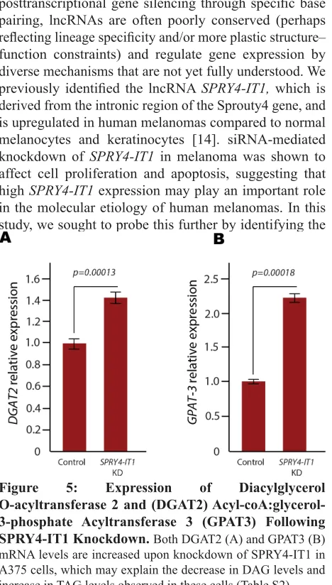
to efficient conversion of DAG to TAG. To test this, we examined the expression of
acyl-CoA:glycerol-3-phosphate acyltransferase 3 (GPAT3), diacylglycerol
O-acyltransferase 1 (DGAT1), and diacylglycerol
O-acyltransferase 2 (DGAT2), since these enzymes play essential roles in DAG to TAG conversion. qPCR analysis showed that both DGAT2 and GPAT3 mRNA levels were increased in SPRY4-IT1 knockdown cells
(Fig. 5), providing a possible mechanism for the changes in DAG and TAG levels in these cells. We postulate that
one mechanism by which SPRY4-IT1 knockdown induces
apoptosis could be through lipin 2-mediated lipotoxicity.
To investigate further, we knocked-down and
over-expressed lipin 2 in A375 cells and measured cell invasion
[image:6.612.149.464.201.641.2]efficiency and doubling time. Interestingly, we observed
Figure 4:SPRY4-IT1 Knockdown Increases Lipin 2 Protein Accumulation in Melanoma Cells. A) Affinity purification
of SPRY4-IT1 from A375 cell lysates with SPRY4-IT1-specific probes followed by qPCR. SPRY4-IT1 is enriched compared to scrambled
(control) probes. U1 RNA was used as endogenous control for pull-downs. (B) qPCR validation showing enrichment of SPRY4-IT1 following immunoprecipitation of A375 cell lysates with lipin 2-specific or control IgG antibodies. (C)Relative expression of SPRY4-IT1 in lipin 2 knock-down cells (D) Relative expression of lipin 2 after siRNA-mediated knockdown of SPRY4-IT1, SPRY4 exon 1 or lipin 2.
(E) Phosphatidic acid phosphatase assay. (F) Western blot analysis of lipin 2 following siRNA-mediated knockdown of SPRY4-IT1 or lipin
that cell doubling time was increased and cell invasion was decreased in both knock-down and force-expressed
cells (Fig. S6A and B). We postulate that lipin 2 levels must be maintained on a steady state level in melanoma cells, and modulating lipin 2 levels (either an increase or decrease) can negatively affect cell physiology. For
comparison, we subjected the primary melanocyte cell
line HEM-l to shotgun lipidomics, and compared cellular
lipid profiles to A375 cells. Interestingly, we found that lipid levels were significantly different in these two
cell types. For an example, phosphatidylethanolamine (PE), Phosphatidylserine (PS), Phosphatidic acid (PA),
Sphingomyelin (SM) were higher in melanocytes,
compared to melanoma cells (Table S3). Studying the role of lipids in melanocyte and melanoma biology is an ongoing study in our laboratory.
DISCUSSION
In contrast to small ncRNAs, which are highly
conserved and play a role in transcriptional and
posttranscriptional gene silencing through specific base
pairing, lncRNAs are often poorly conserved (perhaps
reflecting lineage specificity and/or more plastic structure–
function constraints) and regulate gene expression by diverse mechanisms that are not yet fully understood. We
previously identified the lncRNA SPRY4-IT1, which is
derived from the intronic region of the Sprouty4 gene, and is upregulated in human melanomas compared to normal melanocytes and keratinocytes [14]. siRNA-mediated
knockdown of SPRY4-IT1 in melanoma was shown to
affect cell proliferation and apoptosis, suggesting that high SPRY4-IT1 expression may play an important role in the molecular etiology of human melanomas. In this
study, we sought to probe this further by identifying the
molecular function of SPRY4-IT1 and elucidating its possible molecular function in melanomas.
We found that SPRY4-IT1 is part of a longer noncoding transcript that is processed by cleavage of a
244 nt 5′ fragment prior to transport to the cytoplasm. The fate of the 244 nt fragment is unknown, but it is likely to
be degraded by exo- and endonucleases. The maturation of SPRY4-IT1 is similar to the nuclear processing of
MALAT1 in lung cancer [30]. We also showed that SPRY4
and SPRY4-IT1 are independent transcripts, although they may be regulated by the same transcriptional machinery. A recent study classifying lncRNAs according to their
genomic origin showed they may originate from (a)
a bidirectional promoter adjacent to a protein-coding
gene, (b) the 5′ UTR, (c) a retained intron, (d) within an intron, (e) the 3′ UTR, (f) an intergenic
non-protein-coding region, (g) antisense to protein-non-protein-coding genes, or (f) a combination of the above [31]. According to this
classification, SPRY4-IT1 therefore belongs to the group of intron-retained lncRNAs.
One of the direct targets of SPRY4-IT1 identified
in this study is lipin 2, a member of the lipin family
of phosphatidate phosphatase enzymes [32]. The mouse lipin 2 protein has previously been shown to be post-transcriptionally regulated [33]. We showed that knockdown of SPRY4-IT1 modulates cellular concentrations of lipin 2 substrates, including phosphatidate, and cells expressing high SPRY4-IT1
levels, such as melanomas, might thus be expected to accumulate lipin 2 substrates. Indeed, increased levels of phosphatidate have been linked to certain types of cancers through allosteric activation of the molecular target of rapamycin (mTOR) [34] and to a possible role for lipin 2 in statin resistance of colorectal carcinoma cells in vitro [35]. It would be of interest to determine whether the influence of SPRY4-IT1 on melanocyte and melanoma proliferation involves inhibition of lipin 2 activity and/or activation of mTOR.
Our results are the first to demonstrate a role for
a lncRNA in lipid metabolism in melanoma cells, and collectively, the data support the notion that SPRY4-IT1
may play a critical role in melanocytic transformation.
MATERIAL AND METHODS
Cell Lines
Human epidermal melanocytes (HEM-l) were grown in MelM media containing MelGS growth supplements, 0.5% fetal bovine serum (FBS), penicillin, and streptomycin. Melanoma cells A375 (stage 4) were grown in Complete Tu medium containing a 4:1 mixture of MCDB-153 medium with 1.5 mg/ml sodium bicarbonate and Leibovitz’s L-15 medium with 2 mM L-glutamine, Figure 5: Expression of Diacylglycerol
O-acyltransferase 2 and (DGAT2) Acyl-coA:glycerol-3-phosphate Acyltransferase 3 (GPAT3) Following SPRY4-IT1 Knockdown. Both DGAT2 (A) and GPAT3 (B)
mRNA levels are increased upon knockdown of SPRY4-IT1 in A375 cells, which may explain the decrease in DAG levels and
[image:7.612.59.293.306.718.2]2% FBS, and 1.68 mM CaCl2. Cells were grown at 37°C in a humidified 5% CO2 atmosphere. All cell lines were obtained from ATCC (http://www.atcc.org).
Cloning and Identification of 3′ Spliced Forms of
SPRY4-IT1
Total RNA was isolated from A375 cells with TRIzol (Invitrogen) and reversed transcribed using M-MLV reverse transcriptase. The cDNA was used as a template for PCR amplification with various 5′ primers within SPRY4-IT1 and 3′ primers downstream in the SPRY4 locus. Successful amplification was achieved with two forward primers: 5′ IT1 (gtagagatgggggtttcatcctgttg), which anneals to the first base of SPRY4-IT1, and SPRY4-IT1 qPCR (gctgagctggtggttgaaaggaatc), which is located 305 bp within the SPRY4-IT1 sequence. Successful
amplification at the 3′ end was achieved with the reverse
primer SPRY4 Exon 3 (gtccgctttgggccggtgg), which is
located 229 bp into the third exon of SPRY4. Amplification was unsuccessful using primers 500 bp downstream of this location. PCR products were cloned and sequenced. Sequences were aligned to the genomic template and verified using Vector NTi Advance and AlignX.
Northern Blot Analysis
Total RNA was isolated from each cell line with TRIzol and concentrated. Aliquots of RNA (20 μg per sample) were mixed with equal volumes of 2× NorthernMax-Gly Loading Dye (with ethidium bromide), heated at 50°C for 30 min, and resolved on a 1% glyoxal agarose gel run at 65 V for 2 h 20 min in 1× running buffer. The gel was transferred to a nylon membrane (BrightStar Plus, Ambion) using downward capillary
transfer, crosslinked using a UVP UV crosslinker at
1200 (UVP HL-2000 HybriLinker), prehybridized for 30 min at 42°C in 6 ml ULTRAhyb (Ambion), and then probed overnight at 42°C. Blots were processed with the BrightStar BioDetect kit (Ambion) and exposed to film. The following biotin-labeled DNA probes (IDT) were
used at 166 nM: SPRY4-IT1, ctccactgggcatattctaaaa;
SPRY4 Exon 1, gatgttgcaaccactgcctgg; SPRY4
Exon 3, catggctggtcttcacctggtc; and GAPDH, gggccatgaggtccaccaccc.
Invasion Assay
A375 cells were trypsinized and reverse transfected using RNAiMax (Life Technologies) with 50 μM of the following siRNAs (Life Technologies): SPRY4-IT1 Stealth RNAi 594, SPRY4 Exon 1 Custom Select siRNA, lipin 2
Silencer Select siRNAs s18590 and s18591, and Universal
Negative control (Cat #46-2001).
BD BioCoat™ Matrigel™ Invasion Chambers (24-well plates) were prepared by rehydrating the BD Matrigel™ matrix coating with 0.5 ml of serum-free Complete Tu medium for 2 h at 37°C in a 5% CO2 atmosphere. After rehydration, the solution was removed and 0.5 ml of Complete Tu medium containing chemoattractant (2% FBS) was added to the lower wells, and 0.5 ml of serum-free Complete Tu medium containing 5 × 10⁴ A375 cells transfected with siRNAs was added to the insert wells. Invasion assay plates were incubated for 2 days at 37°C in a humidified 5% CO2 atmosphere.
Non-invading cells were removed by scrubbing the upper surface of the insert, and invading cells on the lower surface of the insert were stained with crystal violet. Each membrane was mounted on a microscope slide for visualization and analysis. Slides were scanned using a
Scanscope digital slide scanner, and the migrated cells
were enumerated using Aperio software. All experiments were performed with biological triplicates.
Apoptosis Assay
A375 cells were trypsinized and reverse transfected with siRNAs as described above. Caspase activity was measured using the Caspase-Glo® 3/7 Assay kit (Promega)
according to the manufacturer’s recommendations, and
samples were read on a GloMax luminometer (Promega).
Polysome Fractionation
Cells grown in 10 cm dishes were gently removed with PBS and collected by centrifugation for 4 min at 100 × g. The cell pellet was resuspended in 500 μl of lysis buffer (100 mM KCl, 50 mM Tris-HCl pH 7.4, 1.5 mM MgCl2, 1 mM dithiothreitol (DTT), 1 mg/ml heparin, 1.5% NP-40, 100 μM cycloheximide, 1% aprotinin, 1 mM
AEBSF, and 100U/ml of RNasin) and incubated on ice
for 15 min. The lysate was centrifuged at 1000 × g and the infranatant lysate was removed from underneath the “fat cake”. The nuclei were pelleted by centrifugation
in a microfuge for 10 min at 12,000 rpm. The resulting
supernatant was carefully removed and applied to the top of a 15–60% sucrose gradient (in 100 mM KCl, 5 mM MgCl2, 20 mM HEPES pH 7.4, and 2 mM DTT). The gradients were centrifuged at 35,000 rpm for 3.5 h at 4°C
in a Beckman SW41 rotor. Samples containing (i) free (unbound) mRNA, ribosomal subunits, and monosomes,
and (ii) polysomes were recovered from the gradients
using a Brandel density gradient fractionator equipped
with an ISCO UA-6 flow cell set to 254 nm (a 70% sucrose solution containing orange G was slowly pumped
into the bottom of the tube to displace the contents from
Bioanalyzer prior to analysis by northern blotting.
Affinity Purification of Endogenous SPRY4-IT1
A375 cells (108 per sample) were grown to 80-90% confluency, the medium was discarded, and the plates were washed with PBS. The cells were trypsinized, pelleted at low speed, washed with PBS, and resuspended in 90 ml PBS (~106 cells/ml). Cells were crosslinked by incubation in 1% formaldehyde at RT with rotation,
and then quenched by the addition of glycine to a
final concentration of 375 mM for 5 min. The cells were centrifuged at low speed, washed in PBS, and recentrifuged. The pellet was resuspended in Buffer A (1% NP-40, 1 mM DTT), homogenized, and spun at low speed. The pellet was resuspended in nuclei lysis buffer (1 mM DTT, 1× protease inhibitor, and 2 μl RNaseOut) at a volume of 1ml per 25 × 106 cell equivalents. The lysate was incubated on ice, sonicated to yield 100-500 bp fragments (Duty Cycle, 20%; intensity, 10; cycles/burst,
200; and time, 8 min), and then centrifuged at high speed.
The supernatant was removed and mixed with 0.5 M LiCl2, then 500 pmol/25 × 106 cell equivalents of a
SPRY4-IT1 probe was added, and the sample was incubated at 37°C for 24 h. The probe consisted of a 25-bp sequence
complementary to SPRY4-IT1 constructed with a locked nucleic acid (LNA) backbone and a 5′-biotin label. Control
samples contained probes complementary to the test probe sequence. A 2 ml aliquot of streptavidin-coated Dynabeads
(Life Technologies) was washed twice with water and once with hybridization buffer, and then blocked with 800 μg yeast tRNA and 800 μg BSA for 1 h at 37°C. The beads were pelleted, washed twice in nuclear lysis buffer, and
resuspended in 2 ml of the same buffer. Prepared beads
were added to the nuclear lysate (125 μl per 500 pmol of probe), rotated at 37°C, and then washed twice in 1 ml nuclear lysis buffer and twice in 1 ml wash buffer (5 mM Tris-HCl pH 7.5, 0.5 mM EDTA, 1M NaCl). Beads from replicate samples were combined, washed twice in PBS, resuspended in PBS, incubated at 75°C for 5 min, and applied to a magnet. The isolated beads were incubated at 65°C overnight to reverse crosslinking, treated with 1 μg/
ml RNase A, vortexed, and incubated at RT. The samples
were then treated with proteinase K, resuspended in Buffer AL, and incubated at 56°C. Finally, RNA in the complexes was purified using the DNeasy Kit, eluted in RNase-free
H2O, and qRT-PCR was performed to quantify SPRY4-IT1. The RNA-protein complexes were then subjected to LTQ
Orbitrap Velos MS for analysis of SPRY4-IT1–associated
proteins. Protein digestion, TiO2-based phosphopeptide enrichment, and ESI-MS/MS was performed at the
proteomics core facility at Sanford-Burnham. Table S1
lists the identified SPRY4-IT1–binding proteins. RNA co-immunoprecipitation (RIP) was performed as previously described [36]. Lipin-2 antibody H-160 was from Santa Cruz (sc-134433).
Real-time qRT-PCR
Quantitative PCR was carried out using
TaqMan mRNA or SYBR Green mRNA assays
with a 7500 Real-Time PCR System (Applied Biosystems/Life Technologies), in accordance with the manufacturer’s protocols. SDS1.2.3 software (Applied Biosystems) was used for comparative Ct analysis, with 18S rRNA serving as the endogenous control. Primers for SYBR qPCR were as follows: SPRY4-IT1 qPCR For, gctgagctggtggttgaaaggaatc; SPRY4-IT1 qPCR Rev, gcttggcccacgatgacttgg; SPRY4 Exon 1 qPCR For, ggcagtggttgcaacatcgccg; SPRY4 Exon 1 qPCR Rev, tagccgccgctgtactcgcagac; LPIN2 qPCR For, cgtctaagcagcccttctatgctgc; LPIN2 qPCR Rev, acatgctccacgagctcactcagc; LPIN1 qPCR
For, ggagagctggtacaggaacatgcaaag; LPIN1 qPCR
Rev, gcagtggctctctccaaaaggtgaag; GPAT3 qPCR
For, caaggaggcctgactgaacttccc; GPAT3 qPCR
Rev, ccgtcctcttagctgagagatccattg; DGAT1 qPCR
For, caacaaggacggagacgccgg; DGAT1 qPCR Rev,
gatgccacggtagttgctgaagcc; DGAT2 qPCR For,
ctgcactgattgctggctcatcg; and DGAT2 qPCR Rev,
gaaagtagcgccacacagcccag.
RNA-FISH Analysis
RNA-FISH was performed using an LNA-modified probe for human SPRY4-IT1 (5′-Texas Red-TCCACTGGGCATATTCTAAAA) and a RiboMap in situ hybridization kit (Ventana Medical Systems, Inc.) on
a Ventana machine. SPRY4-IT1-EE and vector-only cells
were resuspended at 105 cells/ml, and 100 µl was added
to separate cloning rings on an autoclaved glass slide. The
following day, the rings were removed, and the slide was washed in PBS and fixed in 4% paraformaldehyde and 5% acetic acid. After acid treatment using hydrochloride-based RiboClear reagent (Ventana Medical Systems) for 10 min at 37°C, the slide was treated with the
ready-to-use protease 3 reagent, and subjected to a denaturing
pre-hybridization step for 4 min at 80°C. The cells were then hybridized with 40 nM LNA-modified probe in RiboHybe hybridization buffer (Ventana Medical Systems) for 2 h at 58°C. The slide was washed 3 times for 4 min at 60°C; once at low stringency with 0.1× SSC (Ventana Medical Systems) and twice with 1× SSC. The slides were fixed in RiboFix and counterstained with DAPI in an antifade reagent (Ventana Medical Systems). Images were acquired
using a Nikon A1R VAAS laser confocal microscope.
5′ RACE for SPRY4-IT1
(Ambion, Life Technologies). Total RNA was isolated from A375 cells with TRIzol, and 1 μg per sample was ligated to the 5′ RACE adapter using T4 RNA Ligase. This sample was then reverse transcribed using M-MLV reverse transcriptase. The samples were not treated with
either calf intestinal alkaline phosphatase or tobacco acid pyrophosphatase and mRNA molecules thus retained both
the 5′ 7-methyl cap and the 5′ phosphate. Multiple reverse primers were designed within the SPRY4-IT1 sequence to
complement the forward 5′ RLM-RACE primers. PCR reactions were performed using Phusion polymerase (Finnzymes), run out and purified from agarose gels, and cloned into pCR4-TOPO (Life Technologies) for sequence
evaluation (Retrogen). The SPRY4-IT1 RACE product was successfully acquired using the 5′ RACE inner primer (supplied with the FirstChoice® kit) and 5′ RACE SPRY4-IT1 reverse primer, residing 385 bp into SPRY4-IT1 (ctctctggggacgatgcagcatccgatgg).
ACKNOWLEDGMENTS
We thank Drs. Ze’ev Ronai and Fred Levin (both from SBMRI) for their valuable comments and suggestions to improve the manuscript. We also thank Drs. Marc Beal and Arturo Orjalo (both from Biosearch) for Stellaris RNA-FISH probes, Dr. Susan Magdaleno (Life Technologies) for siRNA reagents, Debbie McFadden (SBMRI) for formatting the manuscript, and
Dr. Anne O’Rourke for editorial assistance. This work was supported by National Institutes of Health grants CA165184 and NCI 5P30CA030199 and an Oakstone
Foundation grant to RJP and DK078187 to BNF. MED and
JSM are supported by a Career Development Award and an Australia Fellowship, respectively, from the National Health and Medical Research Council of Australia. The authors have no conflicts of interest to declare.
REFERENCES
1. Carninci P, Kasukawa T, Katayama S, Gough J, Frith MC, Maeda N, Oyama R, Ravasi T, Lenhard B, Wells C, Kodzius R, Shimokawa K, Bajic VB, Brenner SE, Batalov
S, Forrest AR, et al. The transcriptional landscape of the mammalian genome. Science. 2005; 309(5740):1559-1563.
2. Cheng J, Kapranov P, Drenkow J, Dike S, Brubaker S, Patel
S, Long J, Stern D, Tammana H, Helt G, Sementchenko V, Piccolboni A, Bekiranov S, Bailey DK, Ganesh M, Ghosh S, et al. Transcriptional maps of 10 human chromosomes at 5-nucleotide resolution. Science. 2005; 308(5725):1149-1154.
3. Mercer TR, Dinger ME and Mattick JS. Long non-coding RNAs: insights into functions. Nat Rev Genet. 2009; 10(3):155-159.
4. Moran VA, Perera RJ and Khalil AM. Emerging functional and mechanistic paradigms of mammalian long non-coding
RNAs. Nucleic Acids Res. 2012; 40(14):6391-6400. 5. Mattick JS, Amaral PP, Dinger ME, Mercer TR and Mehler
MF. RNA regulation of epigenetic processes. Bioessays. 2009; 31(1):51-59.
6. Mercer TR and Mattick JS. Structure and function of long noncoding RNAs in epigenetic regulation. Nat Struct Mol Biol. 2013; 20(3):300-307.
7. Banfai B, Jia H, Khatun J, Wood E, Risk B, Gundling WE,
Jr., Kundaje A, Gunawardena HP, Yu Y, Xie L, Krajewski K, Strahl BD, Chen X, Bickel P, Giddings MC, Brown JB, et al. Long noncoding RNAs are rarely translated in two
human cell lines. Genome research. 2012; 22(9):1646-1657.
8. Gascoigne DK, Cheetham SW, Cattenoz PB, Clark
MB, Amaral PP, Taft RJ, Wilhelm D, Dinger ME and Mattick JS. Pinstripe: a suite of programs for integrating
transcriptomic and proteomic datasets identifies novel
proteins and improves differentiation of protein-coding and non-coding genes. Bioinformatics. 2012; 28(23):3042-3050.
9. Clark MB, Amaral PP, Schlesinger FJ, Dinger ME, Taft RJ, Rinn JL, Ponting CP, Stadler PF, Morris KV, Morillon A, Rozowsky JS, Gerstein MB, Wahlestedt C, Hayashizaki Y, Carninci P, Gingeras TR, et al. The reality of pervasive
transcription. PLoS Biol. 2011; 9(7):e1000625; discussion e1001102.
10. Taft RJ, Pang KC, Mercer TR, Dinger M and Mattick JS.
Non-coding RNAs: regulators of disease. J Pathol. 2010; 220(2):126-139.
11. Amaral PP and Mattick JS. Noncoding RNA in development. Mamm Genome. 2008; 19(7-8):454-492. 12. Mercer TR, Dinger ME, Sunkin SM, Mehler MF and
Mattick JS. Specific expression of long noncoding RNAs in
the mouse brain. Proceedings of the National Academy of Sciences of the United States of America. 2008; 105(2):716-721.
13. Sunwoo H, Dinger ME, Wilusz JE, Amaral PP, Mattick JS
and Spector DL. MEN varepsilon/beta nuclear-retained non-coding RNAs are up-regulated upon muscle differentiation and are essential components of paraspeckles. Genome research. 2009; 19(3):347-359.
14. Khaitan D, Dinger ME, Mazar J, Crawford J, Smith MA,
Mattick JS and Perera RJ. The melanoma-upregulated long noncoding RNA SPRY4-IT1 modulates apoptosis and
invasion. Cancer Res. 2011; 71(11):3852-3862.
15. Pandey RR, Mondal T, Mohammad F, Enroth S, Redrup L,
Komorowski J, Nagano T, Mancini-Dinardo D and Kanduri C. Kcnq1ot1 antisense noncoding RNA mediates lineage-specific transcriptional silencing through chromatin-level regulation. Mol Cell. 2008; 32(2):232-246.
16. Umlauf D, Fraser P and Nagano T. The role of long non-coding RNAs in chromatin structure and gene regulation:
variations on a theme. Biol Chem. 2008; 389(4):323-331. 17. Rinn JL and Chang HY. Genome regulation by long
noncoding RNAs. Annu Rev Biochem. 2012; 81:145-166.
Gardiner BB, Askarian-Amiri ME, Ru K, Solda G, Simons
C, Sunkin SM, Crowe ML, Grimmond SM, Perkins AC and
Mattick JS. Long noncoding RNAs in mouse embryonic stem cell pluripotency and differentiation. Genome Res. 2008; 18(9):1433-1445.
19. Khalil AM, Guttman M, Huarte M, Garber M, Raj A, Rivea Morales D, Thomas K, Presser A, Bernstein BE, van Oudenaarden A, Regev A, Lander ES and Rinn JL. Many
human large intergenic noncoding RNAs associate with
chromatin-modifying complexes and affect gene expression. Proc Natl Acad Sci U S A. 2009; 106(28):11667-11672.
20. Tsai MC, Manor O, Wan Y, Mosammaparast N, Wang JK, Lan F, Shi Y, Segal E and Chang HY. Long noncoding RNA as modular scaffold of histone modification
complexes. Science. 2010; 329(5992):689-693.
21. Dinger ME, Amaral PP, Mercer TR, Pang KC, Bruce SJ,
Gardiner BB, Askarian-Amiri ME, Ru K, Solda G, Simons
C, Sunkin SM, Crowe ML, Grimmond SM, Perkins AC and
Mattick JS. Long noncoding RNAs in mouse embryonic stem cell pluripotency and differentiation. Genome research. 2008; 18(9):1433-1445.
22. Esteller M. Non-coding RNAs in human disease. Nat Rev Genet. 2011; 12(12):861-874.
23. Kim K, Jutooru I, Chadalapaka G, Johnson G, Frank J,
Burghardt R, Kim S and Safe S. HOTAIR is a negative prognostic factor and exhibits pro-oncogenic activity in pancreatic cancer. Oncogene. 2012.
24. Ifere GO and Ananaba GA. Prostate cancer gene expression
marker 1 (PCGEM1): a patented prostate- specific
non-coding gene and regulator of prostate cancer progression. Recent Pat DNA Gene Seq. 2009; 3(3):151-163.
25. Mazar J, Sinha S, Dinger ME, Mattick JS and Perera RJ.
Protein-coding and non-coding gene expression analysis in differentiating human keratinocytes using a three-dimensional epidermal equivalent. Mol Genet Genomics. 2010; 284(1):1-9.
26. Ota T, Suzuki Y, Nishikawa T, Otsuki T, Sugiyama T, Irie
R, Wakamatsu A, Hayashi K, Sato H, Nagai K, Kimura K, Makita H, Sekine M, Obayashi M, Nishi T, Shibahara T,
et al. Complete sequencing and characterization of 21,243
full-length human cDNAs. Nat Genet. 2004; 36(1):40-45.
27. Minowada G, Jarvis LA, Chi CL, Neubuser A, Sun X, Hacohen N, Krasnow MA and Martin GR. Vertebrate
Sprouty genes are induced by FGF signaling and can cause
chondrodysplasia when overexpressed. Development. 1999;
126(20):4465-4475.
28. Ingolia NT, Brar GA, Rouskin S, McGeachy AM and
Weissman JS. The ribosome profiling strategy for
monitoring translation in vivo by deep sequencing of ribosome-protected mRNA fragments. Nature protocols. 2012; 7(8):1534-1550.
29. Gropler MC, Harris TE, Hall AM, Wolins NE, Gross RW, Han X, Chen Z and Finck BN. Lipin 2 is a liver-enriched phosphatidate phosphohydrolase enzyme that is
dynamically regulated by fasting and obesity in mice. The Journal of biological chemistry. 2009; 284(11):6763-6772.
30. Wilusz JE, Freier SM and Spector DL. 3’ end processing of
a long nuclear-retained noncoding RNA yields a tRNA-like
cytoplasmic RNA. Cell. 2008; 135(5):919-932.
31. Khachane AN and Harrison PM. Mining mammalian transcript data for functional long non-coding RNAs. PLoS One. 2010; 5(4):e10316.
32. Harris TE and Finck BN. Dual function lipin proteins and glycerolipid metabolism. Trends in endocrinology and metabolism: TEM. 2011; 22(6):226-233.
33. Gropler MC, Harris TE, Hall AM, Wolins NE, Gross RW, Han X, Chen Z and Finck BN. Lipin 2 is a liver-enriched phosphatidate phosphohydrolase enzyme that is
dynamically regulated by fasting and obesity in mice. J Biol
Chem. 2009; 284(11):6763-6772.
34. Foster DA. Regulation of mTOR by phosphatidic acid?
Cancer Res. 2007; 67(1):1-4.
35. Savas S, Azorsa DO, Jarjanazi H, Ibrahim-Zada I, Gonzales IM, Arora S, Henderson MC, Choi YH, Briollais L, Ozcelik H and Tuzmen S. NCI60 cancer cell line panel data and
RNAi analysis help identify EAF2 as a modulator of
simvastatin and lovastatin response in HCT-116 cells. PLoS
One. 2011; 6(4):e18306.
36. Moran VA, Niland CN and Khalil AM.